Spine or spinal may refer to:

At the base of the skull, the foramen ovale is one of the larger of the several holes that transmit nerves through the skull. The foramen ovale is situated in the posterior part of the sphenoid bone, posterolateral to the foramen rotundum.

The sphenoid bone is an unpaired bone of the neurocranium. It is situated in the middle of the skull towards the front, in front of the basilar part of the occipital bone. The sphenoid bone is one of the seven bones that articulate to form the orbit. Its shape somewhat resembles that of a butterfly or bat with its wings extended.

The frontal bone is a bone in the human skull. The bone consists of two portions. These are the vertically oriented squamous part, and the horizontally oriented orbital part, making up the bony part of the forehead, part of the bony orbital cavity holding the eye, and part of the bony part of the nose respectively. The name comes from the Latin word frons.

In anatomy, the orbit is the cavity or socket of the skull in which the eye and its appendages are situated. "Orbit" can refer to the bony socket, or it can also be used to imply the contents. In the adult human, the volume of the orbit is 30 millilitres, of which the eye occupies 6.5 ml. The orbital contents comprise the eye, the orbital and retrobulbar fascia, extraocular muscles, cranial nerves II, III, IV, V, and VI, blood vessels, fat, the lacrimal gland with its sac and duct, the eyelids, medial and lateral palpebral ligaments, check ligaments, the suspensory ligament, septum, ciliary ganglion and short ciliary nerves.
The foramen spinosum is a hole located in the greater wing of the sphenoid. It is located posterolateral to the foramen ovale and anterior to the sphenoidal spine. It allows the passage of the middle meningeal artery, middle meningeal vein and usually the meningeal branch of the mandibular nerve.

The sphenoid sinus is one of the four paired paranasal sinuses that is contained within the body of the sphenoid bone. The sphenoid sinuses vary in size and shape, and owing to the lateral displacement of the intervening septum, which may insert on the carotid canal, they are rarely symmetrical. They cannot be palpated during an extraoral examination.
The pterygoid processes of the sphenoid, one on either side, descend perpendicularly from the regions where the body and the greater wings of the sphenoid bone unite.
The greater wing of the sphenoid bone, or alisphenoid, is a bony process of the sphenoid bone; there is one on each side, extending from the side of the body of the sphenoid and curving upward, laterally, and backward.
The lesser wings of the sphenoid or orbito-sphenoids are two thin triangular plates, which arise from the upper and anterior parts of the body, and, projecting lateralward, end in sharp points [Fig. 1].

The anterior cranial fossa is a depression in the floor of the cranial base which houses the projecting frontal lobes of the brain. It is formed by the orbital plates of the frontal, the cribriform plate of the ethmoid, and the small wings and front part of the body of the sphenoid; it is limited behind by the posterior borders of the small wings of the sphenoid and by the anterior margin of the chiasmatic groove. The lesser wings of the sphenoid separate the anterior and middle fossae.
The dorsum sellae is part of the sphenoid bone in the skull. Together with the basilar part of the occipital bone it forms the clivus.
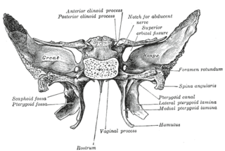
The pterygoid fossa is an anatomical term for the fossa formed by the divergence of the lateral pterygoid plate and the medial pterygoid plate of the sphenoid bone.
The great wings, or alae-sphenoids, are two strong processes of bone, which arise from the sides of the body, and are curved upward, lateralward, and backward; the posterior part of each projects as a triangular process which fits into the angle between the squama and the petrous portion of the temporal bone and presents at its apex a downwardly directed process, the spina angularis. It serves as the origin for the sphenomandibular ligament.

The petrotympanic fissure is a fissure in the temporal bone that runs from the temporomandibular joint to the tympanic cavity.
The superior surface of the body of the sphenoid bone presents in front a prominent spine, the ethmoidal spine, for articulation with the cribriform plate of the ethmoid; behind this is a smooth surface slightly raised in the middle line, and grooved on either side for the olfactory lobes of the brain.

The body of the sphenoid bone, more or less cubical in shape, is hollowed out in its interior to form two large cavities, the sphenoidal sinuses, which are separated from each other by a septum.

The clivus is a bony part of the cranium at the skull base, a shallow depression behind the dorsum sellæ that slopes obliquely backward. It forms a gradual sloping process at the anterior most portion of the basilar occipital bone at its junction with the sphenoid bone. On axial planes, it sits just posterior to the sphenoid sinuses. Just lateral to the clivus bilaterally is the foramen lacerum, proximal to its anastomosis with the Circle of Willis. Posterior to the clivus is the basilar artery.

The base of skull, also known as the cranial base or the cranial floor, is the most inferior area of the skull. It is composed of the endocranium and the lower parts of the skull roof.

The ligaments of malleus are three ligaments that attach the malleus in the middle ear. They are the anterior, lateral and superior ligaments.