Rasmussen's encephalitis

Last updated

Rasmussen's encephalitis is a rare inflammatory neurological disease, characterized by frequent and severe seizures, loss of motor skills and speech, hemiparesis (weakness on one side of the body), encephalitis (inflammation of the brain), and dementia. The illness affects a single cerebral hemisphere and generally occurs in children under the age of 15.

Contents

Signs and symptoms

The condition mostly affects children, with an average age of 6 years. However, one in ten people with the condition develops it in adulthood.[ citation needed ]

There are two main stages, sometimes preceded by a 'prodromal stage' of a few months. In the acute stage, lasting four to eight months, the inflammation is active and the symptoms become progressively worse. These include weakness of one side of the body (hemiparesis), loss of vision for one side of the visual field (hemianopia), and cognitive difficulties (affecting learning, memory or language, for example). Epileptic seizures are also a major part of the illness, although these are often partial. Focal motor seizures or epilepsia partialis continua are particularly common, and may be very difficult to control with drugs.[ citation needed ]

In the chronic or residual stage, the inflammation is no longer active, but the affected individual is left with some or all of the symptoms because of the damage that the inflammation has caused. In the long term, most patients are left with some epilepsy, paralysis and cognitive problems, but the severity varies considerably. [1]

Pathophysiology

In Rasmussen's encephalitis, there is chronic inflammation of the brain, with infiltration of T lymphocytes into the brain tissue. In most cases, this affects only one cerebral hemisphere, either the left or the right. This inflammation causes permanent damage to the cells of the brain, leading to atrophy of the hemisphere; the epilepsy that this causes may itself contribute to the brain damage. The epilepsy might derive from a disturbed GABA release, [2] the main inhibitory neurotransmitter of the mammalian brain.

The cause of the inflammation is not known: infection by a virus has been suggested, but the evidence for this is inconclusive. [1] In the 1990s it was suggested that auto-antibodies against the glutamate receptor GluR3 were important in causing the disease, [3] but this is no longer thought to be the case. [4] However, more recent studies report the presence of autoantibodies against the NMDA-type glutamate receptor subunit GluRepsilon2 (anti-NR2A antibodies) in a subset of patients with Rasmussen's encephalitis. [5] There has also been some evidence that patients with RE express auto-antibodies against alpha 7 subunit of the nicotinic acetylcholine receptor. [6] By sequencing T cell receptors from various compartments it could be shown that RE patients present with peripheral CD8+ T-cell expansion which in some cases have been proven for years after disease onset. [7]

Rasmussen's encephalitis has been recorded with a neurovisceral porphyria, and acute intermittent porphyria. [8]

Diagnosis

Brain biopsy in Rasmussen's encephalitis showing lymphocytic infiltrates staining for CD8 on immunohistochemistry Rasmussen Brainbiopsy CD8.jpg
Brain biopsy in Rasmussen's encephalitis showing lymphocytic infiltrates staining for CD8 on immunohistochemistry

The diagnosis may be made on the clinical features alone, along with tests to rule out other possible causes. An EEG will usually show the electrical features of epilepsy and slowing of brain activity in the affected hemisphere, and MRI brain scans will show gradual shrinkage of the affected hemisphere with signs of inflammation or scarring. [9]

Brain biopsy can provide very strong confirmation of the diagnosis, but this is not always necessary. [9] [10]

Treatment

During the acute stage, treatment is aimed at reducing the inflammation. As in other inflammatory diseases, steroids may be used first of all, either as a short course of high-dose treatment, or in a lower dose for long-term treatment. Intravenous immunoglobulin is also effective both in the short term and in the long term, particularly in adults where it has been proposed as first-line treatment. [11] Other similar treatments include plasmapheresis and tacrolimus, though there is less evidence for these. None of these treatments can prevent permanent disability from developing. [9] [12]

During the residual stage of the illness when there is no longer active inflammation, treatment is aimed at improving the remaining symptoms. Standard anti-epileptic drugs are usually ineffective in controlling seizures, and it may be necessary to surgically remove or disconnect the affected cerebral hemisphere, in an operation called hemispherectomy or via a corpus callosotomy. This usually results in further weakness, hemianopsia and cognitive problems, but the other side of the brain may be able to take over some of the function, particularly in young children. The operation may not be advisable if the left hemisphere is affected, since this hemisphere contains most of the parts of the brain that control language. However, hemispherectomy is often very effective in reducing seizures. [1] [9]

History

It is named for the neurosurgeon Theodore Rasmussen (1910–2002), who succeeded Wilder Penfield as head of the Montreal Neurological Institute, and served as Neurosurgeon-in-Chief at the Royal Victoria Hospital. [13] [14]

Society

The Hemispherectomy Foundation was formed in 2008 to assist families with children who have Rasmussen's encephalitis and other conditions that require hemispherectomy. [15]

The RE Children's Project was founded in 2010 to increase awareness of Rasmussen's encephalitis. Its primary purpose is to support scientific research directed toward finding a cure for this disease.[ citation needed ]

Related Research Articles

Encephalomyelitis is inflammation of the brain and spinal cord. Various types of encephalomyelitis include:

Hemispherectomy is a neurosurgical procedure in which a cerebral hemisphere is removed or disconnected. It is used to treat a variety of refractory or drug-resistant seizure disorders (epilepsy).

Epilepsia partialis continua is a rare type of brain disorder in which a patient experiences recurrent motor epileptic seizures that are focal, and recur every few seconds or minutes for extended periods. It is sometimes called Kozhevnikov's epilepsia named after Russian psychiatrist Aleksei Yakovlevich Kozhevnikov who first described this type of epilepsy.

<span class="mw-page-title-main">Glutamate receptor</span> Cell-surface proteins that bind glutamate and trigger changes which influence the behavior of cells

Glutamate receptors are synaptic and non synaptic receptors located primarily on the membranes of neuronal and glial cells. Glutamate is abundant in the human body, but particularly in the nervous system and especially prominent in the human brain where it is the body's most prominent neurotransmitter, the brain's main excitatory neurotransmitter, and also the precursor for GABA, the brain's main inhibitory neurotransmitter. Glutamate receptors are responsible for the glutamate-mediated postsynaptic excitation of neural cells, and are important for neural communication, memory formation, learning, and regulation.

<span class="mw-page-title-main">Temporal lobe epilepsy</span> Chronic focal seizure disorder

In the field of neurology, temporal lobe epilepsy is an enduring brain disorder that causes unprovoked seizures from the temporal lobe. Temporal lobe epilepsy is the most common type of focal onset epilepsy among adults. Seizure symptoms and behavior distinguish seizures arising from the medial temporal lobe from seizures arising from the lateral (neocortical) temporal lobe. Memory and psychiatric comorbidities may occur. Diagnosis relies on electroencephalographic (EEG) and neuroimaging studies. Anticonvulsant medications, epilepsy surgery and dietary treatments may improve seizure control.

Juvenile myoclonic epilepsy (JME), also known as Janz syndrome or impulsive petit mal, is a form of hereditary, idiopathic generalized epilepsy, representing 5–10% of all epilepsy cases. Typically it first presents between the ages of 12 and 18 with myoclonic seizures. These events typically occur after awakening from sleep, during the evening or when sleep-deprived. JME is also characterized by generalized tonic–clonic seizures, and a minority of patients have absence seizures. It was first described by Théodore Herpin in 1857. Understanding of the genetics of JME has been rapidly evolving since the 1990s, and over 20 chromosomal loci and multiple genes have been identified. Given the genetic and clinical heterogeneity of JME some authors have suggested that it should be thought of as a spectrum disorder.

Epilepsy surgery involves a neurosurgical procedure where an area of the brain involved in seizures is either resected, ablated, disconnected or stimulated. The goal is to eliminate seizures or significantly reduce seizure burden. Approximately 60% of all people with epilepsy have focal epilepsy syndromes. In 15% to 20% of these patients, the condition is not adequately controlled with anticonvulsive drugs. Such patients are potential candidates for surgical epilepsy treatment.

<span class="mw-page-title-main">Limbic encephalitis</span> Inflammation involving the limbic system in the brain

Limbic encephalitis is a form of encephalitis, a disease characterized by inflammation of the brain. Limbic encephalitis is caused by autoimmunity: an abnormal state where the body produces antibodies against itself. Some cases are associated with cancer and some are not. Although the disease is known as "limbic" encephalitis, it is seldom limited to the limbic system and post-mortem studies usually show involvement of other parts of the brain. The disease was first described by Brierley and others in 1960 as a series of three cases. The link to cancer was first noted in 1968 and confirmed by later investigators.

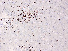
Anti-glutamate receptor antibodies are autoantibodies detected in serum and/or cerebrospinal fluid samples of a variety of disorders such as encephalitis, epilepsy and ataxia. Clinical and experimental studies starting around the year 2000 suggest that these antibodies are not simply epiphenomena and are involved in autoimmune disease pathogenesis.

<span class="mw-page-title-main">GRIK2</span> Protein-coding gene in the species Homo sapiens

Glutamate ionotropic receptor kainate type subunit 2, also known as ionotropic glutamate receptor 6 or GluR6, is a protein that in humans is encoded by the GRIK2 gene.

<span class="mw-page-title-main">LGI1</span> Protein-coding gene in the species Homo sapiens

Leucine-rich, glioma inactivated 1, also known as LGI1, is a protein which in humans is encoded by the LGI1 gene. It may be a metastasis suppressor.

<span class="mw-page-title-main">Anti-NMDA receptor encephalitis</span> Rare disease which results in brain inflammation

Anti-NMDA receptor encephalitis is a type of brain inflammation caused by antibodies. Early symptoms may include fever, headache, and feeling tired. This is then typically followed by psychosis which presents with false beliefs (delusions) and seeing or hearing things that others do not see or hear (hallucinations). People are also often agitated or confused. Over time, seizures, decreased breathing, and blood pressure and heart rate variability typically occur. In some cases, patients may develop catatonia.

Febrile infection-related epilepsy syndrome (FIRES), is onset of severe seizures following a febrile illness in someone who was previously healthy. The seizures may initially be focal; however, often become tonic-clonic. Complications often include intellectual disability, behavioral problems, and ongoing seizures.

James O. McNamara is an American neurologist and neuroscientist, known for his research of epileptogenesis, the process underlying development and progression of epilepsy. He is the Duke School of Medicine Professor of Neuroscience in the Departments of Neurobiology, Neurology, and Pharmacology and Cancer Biology at Duke University. He served as chair of the Department of Neurobiology at Duke from 2002 to 2011

People with epilepsy may be classified into different syndromes based on specific clinical features. These features include the age at which seizures begin, the seizure types, and EEG findings, among others. Identifying an epilepsy syndrome is useful as it helps determine the underlying causes as well as deciding what anti-seizure medication should be tried. Epilepsy syndromes are more commonly diagnosed in infants and children. Some examples of epilepsy syndromes include benign rolandic epilepsy, childhood absence epilepsy and juvenile myoclonic epilepsy. Severe syndromes with diffuse brain dysfunction caused, at least partly, by some aspect of epilepsy, are also referred to as epileptic encephalopathies. These are associated with frequent seizures that are resistant to treatment and severe cognitive dysfunction, for instance Lennox-Gastaut syndrome and West syndrome.

<span class="mw-page-title-main">Autoimmune encephalitis</span> Type of encephalitis

Autoimmune encephalitis (AIE) is a type of encephalitis, and one of the most common causes of noninfectious encephalitis. It can be triggered by tumors, infections, or it may be cryptogenic. The neurological manifestations can be either acute or subacute and usually develop within six weeks. The clinical manifestations include behavioral and psychiatric symptoms, autonomic disturbances, movement disorders, and seizures.

Anti-VGKC-complex encephalitis are caused by antibodies against the voltage gated potassium channel-complex (VGKC-complex) and are implicated in several autoimmune conditions including limbic encephalitis, epilepsy and neuromyotonia.

<span class="mw-page-title-main">Svetlana Dambinova</span> Russian neuroscientist

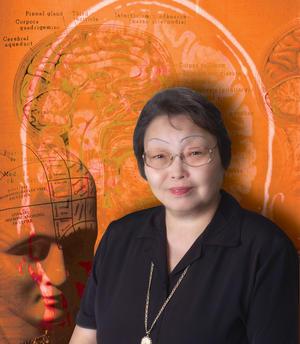
Svetlana Dambinova is a Russian neuroscientist, Doctor of Biological Sciences, Distinguished Professor at Laboratory of biomarkers at Medical Center "DeKalb", Atlanta, USA. Dambinova was awarded as Honored Worker of Science of the Republic of Buryatia (1996) and Russian Federation (1998). Known in the world for research of glutamate receptors. The Head of the project of the International Department of Neurology SPBGMU them. Acad. Pavlov's.

Drug-resistant epilepsy (DRE), also known as refractory epilepsy, intractable epilepsy, or pharmacoresistant epilepsy, is diagnosed following a failure of adequate trials of two tolerated and appropriately chosen and used antiepileptic drugs (AEDs) to achieve sustained seizure freedom. The probability that the next medication will achieve seizure freedom drops with every failed AED. For example, after two failed AEDs, the probability that the third will achieve seizure freedom is around 4%. Drug-resistant epilepsy is commonly diagnosed after several years of uncontrolled seizures, however, in most cases, it is evident much earlier. Approximately 30% of people with epilepsy have a drug-resistant form.

Anti-IgLON5 disease is a neurodegenerative autoimmune disease. It is marked by parasomnias and chorea - an involuntary movement disorder.

References

  1. 1 2 3 Bien, CG; et al. (2005). "Pathogenesis, diagnosis and treatment of Rasmussen encephalitis: a European consensus statement". Brain. 128 (Pt 3): 454–471. doi: 10.1093/brain/awh415 . PMID   15689357.
  2. Rassner, Michael P.; van Velthoven-Wurster, Vera; Ramantani, Georgia; Feuerstein, Thomas J. (March 2013). "Altered transporter-mediated neocortical GABA release in Rasmussen encephalitis". Epilepsia. 54 (3): e41–e44. doi:10.1111/epi.12093. PMID   23360283.
  3. Rogers SW, Andrews PI, Gahring LC, et al. (1994). "Autoantibodies to glutamate receptor GluR3 in Rasmussen's encephalitis". Science. 265 (5172): 648–51. doi:10.1126/science.8036512. PMID   8036512.
  4. Watson R, Jiang Y, Bermudez I, et al. (2004). "Absence of antibodies to glutamate receptor type 3 (GluR3) in Rasmussen encephalitis". Neurology. 63 (1): 43–50. doi:10.1212/01.WNL.0000132651.66689.0F. PMID   15249609. S2CID   30712041.
  5. Takahashi Y, Mori H, Mishina M, et al. (2005). "Autoantibodies and cell-mediated autoimmunity to NMDA-type GluRepsilon2 in patients with Rasmussen's encephalitis (RE) and chronic progressive epilepsia partialis continua". Epilepsia. 46 (Suppl 5): 152–158. doi: 10.1111/j.1528-1167.2005.01024.x . PMID   15987271. S2CID   19120589.
  6. Watson, R; Jepson, JE; Bermudez, I; Alexander, S; Hart, Y; McKnight, K; Roubertie, A; Fecto, F; Valmier, J; Sattelle, DB; Beeson, D; Vincent, A; Lang, B (Dec 13, 2005). "Alpha7-acetylcholine receptor antibodies in two patients with Rasmussen encephalitis". Neurology. 65 (11): 1802–4. doi:10.1212/01.wnl.0000191566.86977.04. PMID   16344526. S2CID   20681278.
  7. Schneider-Hohendorf T, Mohan H, Bien CG, Breuer J, Becker A, Görlich D, Kuhlmann T, Widman G, Herich S, Elpers C, Melzer N, Dornmair K, Kurlemann G, Wiendl H, Schwab N (2016). "CD8(+) T-cell pathogenicity in Rasmussen encephalitis elucidated by large-scale T-cell receptor sequencing". Nat Commun. 7: 11153. doi:10.1038/ncomms11153. PMC   4822013 . PMID   27040081.
  8. Tziperman B, Garty BZ, Schoenfeld N, Hoffer V, Watemberg N, Lev D, Ganor Y, Levite M, Lerman-Sagie T (2007). "Acute intermittent porphyria, Rasmussen encephalitis, or both?". J. Child Neurol. 22 (1): 99–105. doi:10.1177/0883073807299962. PMID   17608316. S2CID   23773750.
  9. 1 2 3 4 Varadkar S, Bien CG, Kruse CA, Jensen FE, Bauer J, Pardo CA, Vincent A, Mathern GW, Cross JH (2014). "Rasmussen's encephalitis: clinical features, pathobiology, and treatment advances". Lancet Neurol. 13 (2): 195–205. doi:10.1016/S1474-4422(13)70260-6. PMC   4005780 . PMID   24457189.
  10. Owens GC, Chang JW, Huynh MN, Chirwa T, Vinters HV, Mathern GW (2016). "Evidence for Resident Memory T Cells in Rasmussen Encephalitis". Front Immunol. 7: 64. doi: 10.3389/fimmu.2016.00064 . PMC   4763066 . PMID   26941743.
  11. Hart, YM; Cortez, Andermann; Hwang, Fish (1994). "Medical treatment of Rasmussen syndrome (chronic encephalitis and epilepsy): effect of high-dose steroids or immunoglobulins in 19 patients". Neurology. 44 (6): 1030–1036. doi:10.1212/WNL.44.6.1030. PMID   8208394. S2CID   32279177. 8208394.
  12. Takahashi Y, Yamazaki E, Mine J, Kubota Y, Imai K, Mogami Y, Baba K, Matsuda K, Oguni H, Sugai K, Ohtsuka Y, Fujiwara T, Inoue Y (2013). "Immunomodulatory therapy versus surgery for Rasmussen syndrome in early childhood". Brain Dev. 35 (8): 778–85. doi:10.1016/j.braindev.2013.01.010. PMID   23433490. S2CID   34514886.
  13. Rasmussen's encephalitis at Who Named It?
  14. Rasmussen T, Olszewski J, Lloyd-Smith D (1958). "Focal seizures due to chronic localized encephalitis". Neurology. 8 (6): 435–45. doi: 10.1212/WNL.8.6.435 . PMID   13566382.
  15. "The Community News". Archived from the original on March 29, 2009. Retrieved 2009-02-25.