Rathke's pouch

Last updated
Rathke's pouch
Gray982.png
Sketches in profile of two stages in the development of the human digestive tube.
Details
Carnegie stage 10
Gives rise to Anterior pituitary
Identifiers
Latin fovea adenohypophysialis
Anatomical terminology

In embryogenesis, Rathke's pouch is an evagination at the roof of the developing mouth in front of the buccopharyngeal membrane. It gives rise to the anterior pituitary (adenohypophysis), a part of the endocrine system.

Contents

Development

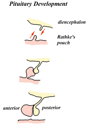
Rathke's pouch, and therefore the anterior pituitary, is derived from ectoderm.

The pouch eventually loses its connection with the pharynx giving rise to the anterior pituitary. The anterior wall of Rathke's pouch proliferates, filling most of the pouch to form pars distalis and pars tuberalis . The posterior wall forms pars intermedia .

In some organisms, the proliferating anterior wall does not fully occupy Rathke's pouch, leaving a remnant (Rathke's cleft) between the pars distalis and pars intermedia. This remnant may develop into a large cyst, the Rathke's cleft cyst. This cyst typically doesn't cause symptoms, but, if large enough, it may cause vision loss, bitemporal hemianopsia, blurry vision, and dulled color vision. [1]

Clinical significance

Rathke's pouch may develop benign cysts. Craniopharyngioma is a neoplasm which can arise from the epithelium within the cleft.

Eponym

It is named for Martin Rathke. [2] [3]

See also

Related Research Articles

<span class="mw-page-title-main">Endocrine system</span> Hormone-producing glands of a body

The endocrine system is a messenger system in an organism comprising feedback loops of hormones that are released by internal glands directly into the circulatory system and that target and regulate distant organs. In vertebrates, the hypothalamus is the neural control center for all endocrine systems.

<span class="mw-page-title-main">Pituitary gland</span> Endocrine gland at the base of the brain

The pituitary gland is an endocrine gland in vertebrates. In humans, the pituitary gland is located at the base of the brain, protruding off the bottom of the hypothalamus. The human pituitary gland is oval shaped, about the size of a chickpea, and weighs 0.5 grams (0.018 oz) on average.

<span class="mw-page-title-main">Anterior pituitary</span> Anterior lobe of the pituitary gland

A major organ of the endocrine system, the anterior pituitary is the glandular, anterior lobe that together with the posterior lobe makes up the pituitary gland (hypophysis) which, in humans, is located at the base of the brain, protruding off the bottom of the hypothalamus.

<span class="mw-page-title-main">Posterior pituitary</span> Posterior lobe of the pituitary gland

The posterior pituitary is the posterior lobe of the pituitary gland which is part of the endocrine system. The posterior pituitary is not glandular as is the anterior pituitary. Instead, it is largely a collection of axonal projections from the hypothalamus that terminate behind the anterior pituitary, and serve as a site for the secretion of neurohypophysial hormones directly into the blood. The hypothalamic–neurohypophyseal system is composed of the hypothalamus, posterior pituitary, and these axonal projections.

<span class="mw-page-title-main">Scalp</span> Anatomical area bordered by the face at the front, and by the neck at the sides and back

The scalp is the area of the head where head hair grows. It is made up of skin, layers of connective and fibrous tissues, and the membrane of the skull. Anatomically, the scalp is part of the epicranium, a collection of structures covering the cranium. The scalp is bordered by the face at the front, and by the neck at the sides and back. The scientific study of hair and scalp is called trichology.

<span class="mw-page-title-main">Hypopituitarism</span> Medical condition

Hypopituitarism is the decreased (hypo) secretion of one or more of the eight hormones normally produced by the pituitary gland at the base of the brain. If there is decreased secretion of one specific pituitary hormone, the condition is known as selective hypopituitarism. If there is decreased secretion of most or all pituitary hormones, the term panhypopituitarism is used.

<span class="mw-page-title-main">Pars intermedia</span> Boundary between the anterior and posterior lobes of the pituitary

Pars intermedia is the boundary between the anterior and posterior lobes of the pituitary. It contains colloid-filled cysts and two types of cells - basophils and chromophobes. The cysts are the remainder of Rathke's pouch. As technically part of the anterior pituitary, it separates the posterior pituitary and pars distalis. It is composed of large, pale cells that encompass the aforementioned colloid-filled follicles.

<span class="mw-page-title-main">Dental lamina</span>

The dental lamina is a band of epithelial tissue seen in histologic sections of a developing tooth. The dental lamina is first evidence of tooth development and begins at the sixth week in utero or three weeks after the rupture of the buccopharyngeal membrane. It is formed when cells of the oral ectoderm proliferate faster than cells of other areas. Best described as an in-growth of oral ectoderm, the dental lamina is frequently distinguished from the vestibular lamina, which develops concurrently. This dividing tissue is surrounded by and, some would argue, stimulated by ectomesenchymal growth. When it is present, the dental lamina connects the developing tooth bud to the epithelium of the oral cavity. Eventually, the dental lamina disintegrates into small clusters of epithelium and is resorbed. In situations when the clusters are not resorbed, eruption cysts are formed over the developing tooth and delay its eruption into the oral cavity. This invagination of ectodermal tissues is the progenitor to the later ameloblasts and enamel while the ectomesenchyme is responsible for the dental papilla and later odontoblasts.

<span class="mw-page-title-main">Craniopharyngioma</span> Medical condition

A craniopharyngioma is a rare type of brain tumor derived from pituitary gland embryonic tissue that occurs most commonly in children, but also affects adults. It may present at any age, even in the prenatal and neonatal periods, but peak incidence rates are childhood-onset at 5–14 years and adult-onset at 50–74 years. People may present with bitemporal inferior quadrantanopia leading to bitemporal hemianopsia, as the tumor may compress the optic chiasm. It has a point prevalence around two per 1,000,000. Craniopharyngiomas are distinct from Rathke's cleft tumours and intrasellar arachnoid cysts.

<span class="mw-page-title-main">Pharyngeal arch</span> Embryonic precursor structures in vertebrates

The pharyngeal arches, also known as visceral arches, are structures seen in the embryonic development of vertebrates that are recognisable precursors for many structures. In fish, the arches are known as the branchial arches, or gill arches.

Neuroepithelial cells, or neuroectodermal cells, form the wall of the closed neural tube in early embryonic development. The neuroepithelial cells span the thickness of the tube's wall, connecting with the pial surface and with the ventricular or lumenal surface. They are joined at the lumen of the tube by junctional complexes, where they form a pseudostratified layer of epithelium called neuroepithelium.

<span class="mw-page-title-main">Urachus</span> Structure of the urinary system

The urachus is a fibrous remnant of the allantois, a canal that drains the urinary bladder of the fetus that joins and runs within the umbilical cord. The fibrous remnant lies in the space of Retzius, between the transverse fascia anteriorly and the peritoneum posteriorly.

Pituitary apoplexy is bleeding into or impaired blood supply of the pituitary gland. This usually occurs in the presence of a tumor of the pituitary, although in 80% of cases this has not been diagnosed previously. The most common initial symptom is a sudden headache, often associated with a rapidly worsening visual field defect or double vision caused by compression of nerves surrounding the gland. This is often followed by acute symptoms caused by lack of secretion of essential hormones, predominantly adrenal insufficiency.

<span class="mw-page-title-main">Chiasmal syndrome</span> Set of signs and symptoms that are associated with lesions of the optic chiasm

Chiasmal syndrome is the set of signs and symptoms that are associated with lesions of the optic chiasm, manifesting as various impairments of the affected's visual field according to the location of the lesion along the optic nerve. Pituitary adenomas are the most common cause; however, chiasmal syndrome may be caused by cancer, or associated with other medical conditions such as multiple sclerosis and neurofibromatosis.

<span class="mw-page-title-main">Branchial cleft cyst</span> Medical condition

A branchial cleft cyst or simply branchial cyst is a cyst as a swelling in the upper part of neck anterior to sternocleidomastoid. It can, but does not necessarily, have an opening to the skin surface, called a fistula. The cause is usually a developmental abnormality arising in the early prenatal period, typically failure of obliteration of the second, third, and fourth branchial cleft, i.e. failure of fusion of the second branchial arches and epicardial ridge in lower part of the neck. Branchial cleft cysts account for almost 20% of neck masses in children. Less commonly, the cysts can develop from the first, third, or fourth clefts, and their location and the location of associated fistulas differs accordingly.

<span class="mw-page-title-main">Rathke's cleft cyst</span> Benign cystic growth on the pituitary gland

A Rathke's cleft cyst is a benign growth on the pituitary gland in the brain, specifically a mucin-filled cyst in the posterior portion of the anterior pituitary gland. It occurs when the Rathke's pouch does not develop properly and ranges in size from 2 to 40 mm in diameter.

A cyst is a pathological epithelial lined cavity that fills with fluid or soft material and usually grows from internal pressure generated by fluid being drawn into the cavity from osmosis. The bones of the jaws, the mandible and maxilla, are the bones with the highest prevalence of cysts in the human body. This is due to the abundant amount of epithelial remnants that can be left in the bones of the jaws. The enamel of teeth is formed from ectoderm, and so remnants of epithelium can be left in the bone during odontogenesis. The bones of the jaws develop from embryologic processes which fuse, and ectodermal tissue may be trapped along the lines of this fusion. This "resting" epithelium is usually dormant or undergoes atrophy, but, when stimulated, may form a cyst. The reasons why resting epithelium may proliferate and undergo cystic transformation are generally unknown, but inflammation is thought to be a major factor. The high prevalence of tooth impactions and dental infections that occur in the bones of the jaws is also significant to explain why cysts are more common at these sites.

The fetal endocrine system is one of the first systems to develop during prenatal development of a human individual. The endocrine system arises from all three embryonic germ layers. The endocrine glands that produce the steroid hormones, such as the gonads and adrenal cortex, arise from the mesoderm. In contrast, endocrine glands that arise from the endoderm and ectoderm produce the amine, peptide, and protein hormones.

<span class="mw-page-title-main">Pituitary pars intermedia dysfunction</span>

Pituitary pars intermedia dysfunction (PPID), or equine Cushing's disease, is an endocrine disease affecting the pituitary gland of horses. It is most commonly seen in older animals, and is classically associated with the formation of a long, wavy coat (hirsutism) and chronic laminitis.

A cervical thymic cyst, also called thymopharyngeal duct cyst, is a fluid-filled mass that occurs when the thymopharyngeal duct, an embryonic structure connecting the nascent thymus with the embryonic pharynx, fails to close and disappear. A thymic cyst is typically a solitary mass on one side of the neck, and is usually found near the carotid sheath. Some cervical thymic cysts may extend into the mediastinum. It is usually asymptomatic. The diagnostic process includes differentiating between other causes of neck masses in infants and children, including branchial cleft cysts and cystic hygromas. The treatment is surgical excision. On histologic examination, the wall of the cyst includes thymic tissue, and may include parathyroid gland tissue because of the parathyroid gland's common embryonic origin with the thymus gland in the third pharyngeal pouch. Fewer than 100 cases of cervical thymic cysts have been reported in the medical literature.

References

  1. "Rathke's Cleft Cyst - Pituitary & Skull Base Tumor | UCLA Health". UCLA Health. Retrieved 9 February 2023.
  2. synd/3564 at Who Named It?
  3. M. H. Rathke. Entwicklungsgeschichte der Natter (Coluber natrix). Königsberg, Bornträger, 1839.