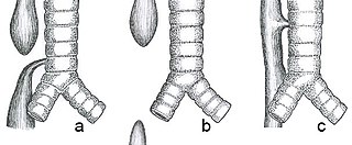
A saber-sheath trachea also known as scabbard trachea is a trachea that has an abnormal shape. The posterior area of the trachea increases in diameter while the lateral measurement decreases.

A saber-sheath trachea also known as scabbard trachea is a trachea that has an abnormal shape. The posterior area of the trachea increases in diameter while the lateral measurement decreases.
It can occur in chronic obstructive pulmonary disease or prolonged bilateral compression on it as in goitre.

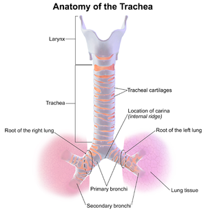
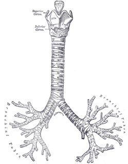
The trachea, also known as the windpipe, is a cartilaginous tube that connects the larynx to the bronchi of the lungs, allowing the passage of air, and so is present in almost all air-breathing animals with lungs. The trachea extends from the larynx and branches into the two primary bronchi. At the top of the trachea the cricoid cartilage attaches it to the larynx. The trachea is formed by a number of horseshoe-shaped rings, joined together vertically by overlying ligaments, and by the trachealis muscle at their ends. The epiglottis closes the opening to the larynx during swallowing.

In vertebrate anatomy, the throat is the front part of the neck, internally positioned in front of the vertebrae. It contains the pharynx and larynx. An important section of it is the epiglottis, separating the esophagus from the trachea (windpipe), preventing food and drinks being inhaled into the lungs. The throat contains various blood vessels, pharyngeal muscles, the nasopharyngeal tonsil, the tonsils, the palatine uvula, the trachea, the esophagus, and the vocal cords. Mammal throats consist of two bones, the hyoid bone and the clavicle. The "throat" is sometimes thought to be synonymous for the fauces.

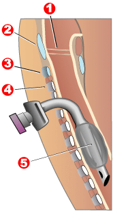
Tracheal intubation, usually simply referred to as intubation, is the placement of a flexible plastic tube into the trachea (windpipe) to maintain an open airway or to serve as a conduit through which to administer certain drugs. It is frequently performed in critically injured, ill, or anesthetized patients to facilitate ventilation of the lungs, including mechanical ventilation, and to prevent the possibility of asphyxiation or airway obstruction.

Esophageal atresia is a congenital medical condition that affects the alimentary tract. It causes the esophagus to end in a blind-ended pouch rather than connecting normally to the stomach. It comprises a variety of congenital anatomic defects that are caused by an abnormal embryological development of the esophagus. It is characterized anatomically by a congenital obstruction of the esophagus with interruption of the continuity of the esophageal wall.

Tracheotomy, or tracheostomy, is a surgical procedure which consists of making an incision (cut) on the anterior aspect (front) of the neck and opening a direct airway through an incision in the trachea (windpipe). The resulting stoma (hole) can serve independently as an airway or as a site for a tracheal tube or tracheostomy tube to be inserted; this tube allows a person to breathe without the use of the nose or mouth.

The respiratory tract is the subdivision of the respiratory system involved with the process of respiration in mammals. The respiratory tract is lined with respiratory mucosa or respiratory epithelium.

A bronchus is a passage or airway in the lower respiratory tract that conducts air into the lungs. The first or primary bronchi pronounced (BRAN-KAI) to branch from the trachea at the carina are the right main bronchus and the left main bronchus. These are the widest bronchi, and enter the right lung, and the left lung at each hilum. The main bronchi branch into narrower secondary bronchi or lobar bronchi, and these branch into narrower tertiary bronchi or segmental bronchi. Further divisions of the segmental bronchi are known as 4th order, 5th order, and 6th order segmental bronchi, or grouped together as subsegmental bronchi. The bronchi when too narrow to be supported by cartilage are known as bronchioles. No gas exchange takes place in the bronchi.
Stridor is a high-pitched extra-thoracic breath sound resulting from turbulent air flow in the larynx or lower in the bronchial tree. It is different from a stertor which is a noise originating in the pharynx. Stridor is a physical sign which is caused by a narrowed or obstructed airway. It can be inspiratory, expiratory or biphasic, although it is usually heard during inspiration. Inspiratory stridor often occurs in children with croup. It may be indicative of serious airway obstruction from severe conditions such as epiglottitis, a foreign body lodged in the airway, or a laryngeal tumor. Stridor should always command attention to establish its cause. Visualization of the airway by medical experts equipped to control the airway may be needed.

The hyperpyron was a Byzantine coin in use during the late Middle Ages, replacing the solidus as the Byzantine Empire's gold coinage.

Tracheitis is an inflammation of the trachea. Although the trachea is usually considered part of the lower respiratory tract, in ICD-10 tracheitis is classified under "acute upper respiratory infections".

The syrinx is the vocal organ of birds. Located at the base of a bird's trachea, it produces sounds without the vocal folds of mammals. The sound is produced by vibrations of some or all of the membrana tympaniformis and the pessulus, caused by air flowing through the syrinx. This sets up a self-oscillating system that modulates the airflow creating the sound. The muscles modulate the sound shape by changing the tension of the membranes and the bronchial openings. The syrinx enables some species of birds to mimic human speech. Unlike the larynx in mammals, the syrinx is located where the trachea forks into the lungs. Thus, lateralization is possible, with muscles on the left and right branch modulating vibrations independently so that some songbirds can produce more than one sound at a time. Some species of birds, such as New World vultures, lack a syrinx and communicate through throaty hisses. Birds do have a larynx, but unlike in mammals, it does not vocalize.
Oliver's sign, or the tracheal tug sign, is an abnormal downward movement of the trachea during systole that can indicate a dilation or aneurysm of the aortic arch.

In anatomy, the carina is a ridge of cartilage in the trachea that occurs between the division of the two main bronchi.

A gapeworm, also known as a red worm and forked worm, is a parasitic nematode worm that infects the tracheas of certain birds. The resulting disease, known as "gape" occurs when the worms clog and obstruct the airway. The worms are also known as "red worms" or "forked worms" due to their red color and the permanent procreative conjunction of males and females. Gapeworms are common in young, domesticated chickens and turkeys.

The tracheobronchial lymph nodes are lymph nodes that are located around the division of trachea and main bronchi.

The pretracheal fascia is a portion of the structure of the human neck. It extends medially in front of the carotid vessels and assists in forming the carotid sheath.
The trachealis muscle is a sheet of smooth muscle in the trachea.

Tracheobronchial injury is damage to the tracheobronchial tree. It can result from blunt or penetrating trauma to the neck or chest, inhalation of harmful fumes or smoke, or aspiration of liquids or objects.
Paolo Macchiarini is a Swiss-born Italian thoracic surgeon and former regenerative medicine researcher who became known for research fraud and manipulative behavior.

A spiracle or stigma is the opening in the exoskeletons of insects and some spiders to allow air to enter the trachea. In the respiratory system of insects, the tracheal tubes primarily deliver oxygen directly into the animals' tissues. The spiracles can be opened and closed in an efficient manner to reduce water loss. This is done by contracting closer muscles surrounding the spiracle. In order to open, the muscle relaxes. The closer muscle is controlled by the central nervous system, but can also react to localized chemical stimuli. Several aquatic insects have similar or alternative closing methods to prevent water from entering the trachea. The timing and duration of spiracle closures can affect the respiratory rates of the organism. Spiracles may also be surrounded by hairs to minimize bulk air movement around the opening, and thus minimize water loss.