A sex organ is any part of an animal's body that is involved in sexual reproduction. The reproductive organs together constitute the reproductive system. The testis in the male, and the ovary in the female, are called the primary sex organs. The external sex organs – the genitals or genitalia, visible at birth in both sexes, and the internal sex organs are called the secondary sex organs.
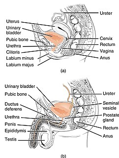
In anatomy, the urethra is a tube that connects the urinary bladder to the urinary meatus for the removal of urine from the body. In males, the urethra travels through the penis and also carries semen. In human females and other primates, the urethra connects to the urinary meatus above the vagina, whereas in non-primates, the female's urethra empties into the urogenital sinus.

The bulbospongiosus muscle is one of the superficial muscles of the perineum. It has a slightly different origin, insertion and function in males and females. In males, it covers the bulb of the penis. In females, it covers the vestibular bulb.
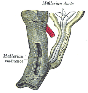
Paramesonephric ducts are paired ducts of the embryo that run down the lateral sides of the urogenital ridge and terminate at the sinus tubercle in the primitive urogenital sinus. In the female, they will develop to form the uterine tubes, uterus, cervix, and the upper two third of the vagina. Lower 1/3 of vagina is derived from sinovaginal bulb derived from urogenital sinus; in the male, they are lost. These ducts are made of tissue of mesodermal origin.
Pseudovaginal perineoscrotal hypospadias (PPSH) refers to a configuration of the external genitalia of an infant. In a sense, this configuration is roughly midway between phenotypical human male genitalia, and phenotypical human female genitalia, in structure and appearance. It is a relatively common form of genital ambiguity caused by undervirilization of genetic males due to several different intersex conditions.
The development of the urinary system begins as a part of prenatal development, and relates to the development of both the urinary system and the sex organs. It continues as a part of sexual differentiation.

The male reproductive system consists of a number of sex organs that play a role in the process of human reproduction. These organs are located on the outside of the body and within the pelvis.

Vaginal atresia is a condition in which the vagina is abnormally closed or absent. The main causes can either be complete vaginal hypoplasia, or a vaginal obstruction, often caused by an imperforate hymen or, less commonly, a transverse vaginal septum. It results in uterovaginal outflow tract obstruction. This condition does not usually occur by itself within an individual, but coupled with other developmental disorders within the female. The disorders that are usually coupled with a female who has vaginal atresia are Rokitansky-Mayer- Küster-Hauser syndrome, Bardet-Biedl syndrome, or Fraser syndrome. One out of every 5,000 women have this abnormality.

The vulval vestibule is a part of the vulva between the labia minora into which the urinary meatus and the vaginal opening open. Its edge is marked by Hart's line. It represents the distal end of the urogenital sinus of the embryo.

The urogenital triangle is the anterior part of the perineum. In female mammals, it contains the vagina and associated parts of the external genitalia.

Sexual differentiation in humans is the process of development of sex differences in humans. It is defined as the development of phenotypic structures consequent to the action of hormones produced following gonadal determination. Sexual differentiation includes development of different genitalia and the internal genital tracts, breasts, body hair, and plays a role in gender identification.
The development of the reproductive system is a part of prenatal development, and concerns the sex organs. It is a part of the stages of sexual differentiation. Because its location, to a large extent, overlaps the urinary system, the development of them can also be described together as the development of the urinary and reproductive organs.
A vaginal septum is a vaginal anomaly that is partition within the vagina; such a septum could be either longitudinal or transverse. In some affected women, the septum is partial or does not extend the length or width of the vagina. Pain during intercourse can be a symptom. A longitudinal vaginal septum develops during embryogenesis when there is an incomplete fusion of the lower parts of the two Müllerian ducts. As a result, there may appear to be two openings to the vagina. There may be associated duplications of the more cranial parts of the Müllerian derivatives, a double cervix, and either a uterine septum or uterus didelphys. A transverse septum forms during embryogenesis when the Müllerian ducts do not fuse to the urogenital sinus. A complete transverse septum can occur across the vagina at different levels. Menstrual flow can be blocked. and is a cause of primary amenorrhea. The accumulation of menstrual debris behind the septum is termed cryptomenorrhea. Some transverse septa are incomplete and may lead to dyspareunia or obstruction in labour.

Diphallia, penile duplication (PD), diphallic terata, or diphallasparatus, is an extremely rare developmental abnormality in which a male is born with two penises. The first reported case was by Johannes Jacob Wecker in 1609. Its occurrence is 1 in 5.5 million boys in the United States.
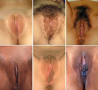
The vulva consists of the external female sex organs. The vulva includes the mons pubis, labia majora, labia minora, clitoris, vestibular bulbs, vulval vestibule, urinary meatus, the vaginal opening, and Bartholin's and Skene's vestibular glands. The urinary meatus is also included as it opens into the vulval vestibule. Other features of the vulva include the pudendal cleft, sebaceous glands, the urogenital triangle, and pubic hair. The vulva includes the entrance to the vagina, which leads to the uterus, and provides a double layer of protection for this by the folds of the outer and inner labia. Pelvic floor muscles support the structures of the vulva. Other muscles of the urogenital triangle also give support.
The Prader scale or Prader staging, named after Dr. Andrea Prader, is a coarse rating system for the measurement of the degree of virilization of the genitalia of the human body and is similar to the Quigley scale. It primarily relates to virilization of the female genitalia in cases of congenital adrenal hyperplasia (CAH) and identifies five distinct stages, but in recent times has been used to describe the range of differentiation of genitalia, with normal infant presentation being shown on either end of the scale, female on the left (0) and male on the right (6).

