A. James Hudspeth

Last updated
A. James Hudspeth
KavliPrize-7273 (42704615130).jpg
Alma mater
Awards Kavli Prize in Neuroscience (2018)
Scientific career
Institutions

A. James Hudspeth is the F.M. Kirby Professor at Rockefeller University in New York City, where he is director of the F.M. Kirby Center for Sensory Neuroscience. His laboratory studies the physiological basis of hearing.

Contents

Early life and education

As a teenager, James Hudspeth spent his summers working as a technician in the lab of neurophysiologist Peter Kellaway at Baylor College of Medicine. [1] Hudspeth was expelled from high school for mixing dangerous chemicals and other mischief. [1]

Hudspeth graduated from Harvard College in 1967, and received his master's degree from Harvard University in 1968. He enrolled in a graduate program in neurobiology to avoid being drafted into the military, but a year later the policy was changed, requiring him to enter medical school for exemption. He studied under Nobel prize winners Torsten Wiesel and David Hubel. He completed both programs and received his PhD in 1973 and MD in 1974, both from Harvard University. [1] [2]

He began a postdoctoral fellowship with Åke Flock at the Karolinska Institute, but left early without much success to return to Harvard Medical School. [1] [2]

Career

Following his postdoctoral training, Hudspeth was a professor at Caltech from 1975 to 1983. [2] He then moved to the UCSF School of Medicine where he was a professor from 1983 to 1989. He directed the neuroscience program at University of Texas Southwestern Medical Center from 1989 until 1995, when the department was closed. [1] In 1995, he was recruited to the Rockefeller University. [1] [3]

Hudspeth has been an HHMI investigator since 1993. [4]

Research

Hudspeth's research is focused on sensorineural hearing loss, and the deterioration of the hair cells, the sensory cells of the cochlea. [5] Hudspeth's bold interpretation of the data obtained in his careful experimental research combined with biophysical modelling lead him to propose for the first time that the sense of hearing depends on a channel that is opened by mechanical force: [6] The hair cells located in the inner ear perceive sound when their apical end -consisting of a bundle of filaments- bends in response to the movement caused by this sound. The activated hair cell rapidly fills with calcium entering from the outside of the cell, which in turn activates the release of neurotransmitters that start a signal to the brain. Hudspeth proposed the existence of a "gating spring" opened by direct mechanical force that would open an hypothetical channel responsible for the entry of calcium ions. The hypothesis was based on the following evidence: [7] 1) Part of the energy needed to bend the filament bundle was mysteriously lost, but could be explained if it was used to opening this gating spring, 2) The entry of calcium ions was microseconds long, this is so fast that only direct opening -without a cascade of chemical reactions- could account for it and 3) Hudspeth tested a model analogue to the opening of a door with a string attached to the door knob and demonstrated that a similar process was taking place when the filaments of the hair cell moved. Furthermore, microscopic evidence showed the existence of such a string-like structure tethering the tip of one filament to the side of and adjacent filament that could be the elusive gating spring; [7] this string—called the tip link—would tense if the filament bundle was bend and then open the channel. Although the precise identity of the proteins forming the tip link [8] and the mechanosensitive channel [9] is still controversial 30 years later. Hudspeth's hypothesis was correct and fundamental for the understanding of the sense of hearing.

Noted publications

Awards

Related Research Articles

<span class="mw-page-title-main">Inner ear</span> Innermost part of the vertebrate ear

The inner ear is the innermost part of the vertebrate ear. In vertebrates, the inner ear is mainly responsible for sound detection and balance. In mammals, it consists of the bony labyrinth, a hollow cavity in the temporal bone of the skull with a system of passages comprising two main functional parts:

<span class="mw-page-title-main">Cochlea</span> Snail-shaped part of inner ear involved in hearing

The cochlea is the part of the inner ear involved in hearing. It is a spiral-shaped cavity in the bony labyrinth, in humans making 2.75 turns around its axis, the modiolus. A core component of the cochlea is the Organ of Corti, the sensory organ of hearing, which is distributed along the partition separating the fluid chambers in the coiled tapered tube of the cochlea.

<span class="mw-page-title-main">Basilar membrane</span> Stiff structural element within the cochlea of the inner ear which separates two liquid-filled tubes

The basilar membrane is a stiff structural element within the cochlea of the inner ear which separates two liquid-filled tubes that run along the coil of the cochlea, the scala media and the scala tympani. The basilar membrane moves up and down in response to incoming sound waves, which are converted to traveling waves on the basilar membrane.

<span class="mw-page-title-main">Organ of Corti</span> Receptor organ for hearing

The organ of Corti, or spiral organ, is the receptor organ for hearing and is located in the mammalian cochlea. This highly varied strip of epithelial cells allows for transduction of auditory signals into nerve impulses' action potential. Transduction occurs through vibrations of structures in the inner ear causing displacement of cochlear fluid and movement of hair cells at the organ of Corti to produce electrochemical signals.

<span class="mw-page-title-main">Auditory system</span> Sensory system used for hearing

The auditory system is the sensory system for the sense of hearing. It includes both the sensory organs and the auditory parts of the sensory system.

<span class="mw-page-title-main">Hair cell</span> Auditory sensory receptor nerve cells

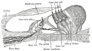
Hair cells are the sensory receptors of both the auditory system and the vestibular system in the ears of all vertebrates, and in the lateral line organ of fishes. Through mechanotransduction, hair cells detect movement in their environment.

In physiology, tonotopy is the spatial arrangement of where sounds of different frequency are processed in the brain. Tones close to each other in terms of frequency are represented in topologically neighbouring regions in the brain. Tonotopic maps are a particular case of topographic organization, similar to retinotopy in the visual system.

Presbycusis, or age-related hearing loss, is the cumulative effect of aging on hearing. It is a progressive and irreversible bilateral symmetrical age-related sensorineural hearing loss resulting from degeneration of the cochlea or associated structures of the inner ear or auditory nerves. The hearing loss is most marked at higher frequencies. Hearing loss that accumulates with age but is caused by factors other than normal aging is not presbycusis, although differentiating the individual effects of distinct causes of hearing loss can be difficult.

<span class="mw-page-title-main">Stereocilia (inner ear)</span>

In the inner ear, stereocilia are the mechanosensing organelles of hair cells, which respond to fluid motion in numerous types of animals for various functions, including hearing and balance. They are about 10–50 micrometers in length and share some similar features of microvilli. The hair cells turn the fluid pressure and other mechanical stimuli into electric stimuli via the many microvilli that make up stereocilia rods. Stereocilia exist in the auditory and vestibular systems.

<span class="mw-page-title-main">Superior olivary complex</span> Collection of brainstem nuclei related to hearing

The superior olivary complex (SOC) or superior olive is a collection of brainstem nuclei that functions in multiple aspects of hearing and is an important component of the ascending and descending auditory pathways of the auditory system. The SOC is intimately related to the trapezoid body: most of the cell groups of the SOC are dorsal to this axon bundle while a number of cell groups are embedded in the trapezoid body. Overall, the SOC displays a significant interspecies variation, being largest in bats and rodents and smaller in primates.

<span class="mw-page-title-main">Tectorial membrane</span>

The tectoria membrane (TM) is one of two acellular membranes in the cochlea of the inner ear, the other being the basilar membrane (BM). "Tectorial" in anatomy means forming a cover. The TM is located above the spiral limbus and the spiral organ of Corti and extends along the longitudinal length of the cochlea parallel to the BM. Radially the TM is divided into three zones, the limbal, middle and marginal zones. Of these the limbal zone is the thinnest (transversally) and overlies the auditory teeth of Huschke with its inside edge attached to the spiral limbus. The marginal zone is the thickest (transversally) and is divided from the middle zone by Hensen's Stripe. It overlies the sensory inner hair cells and electrically-motile outer hair cells of the organ of Corti and during acoustic stimulation stimulates the inner hair cells through fluid coupling, and the outer hair cells via direct connection to their tallest stereocilia.

A kinocilium is a special type of cilium on the apex of hair cells located in the sensory epithelium of the vertebrate inner ear.

Binaural fusion or binaural integration is a cognitive process that involves the combination of different auditory information presented binaurally, or to each ear. In humans, this process is essential in understanding speech as one ear may pick up more information about the speech stimuli than the other.

The olivocochlear system is a component of the auditory system involved with the descending control of the cochlea. Its nerve fibres, the olivocochlear bundle (OCB), form part of the vestibulocochlear nerve, and project from the superior olivary complex in the brainstem (pons) to the cochlea.

The ribbon synapse is a type of neuronal synapse characterized by the presence of an electron-dense structure, the synaptic ribbon, that holds vesicles close to the active zone. It is characterized by a tight vesicle-calcium channel coupling that promotes rapid neurotransmitter release and sustained signal transmission. Ribbon synapses undergo a cycle of exocytosis and endocytosis in response to graded changes of membrane potential. It has been proposed that most ribbon synapses undergo a special type of exocytosis based on coordinated multivesicular release. This interpretation has recently been questioned at the inner hair cell ribbon synapse, where it has been instead proposed that exocytosis is described by uniquantal release shaped by a flickering vesicle fusion pore.

The cochlear amplifier is a positive feedback mechanism within the cochlea that provides acute sensitivity in the mammalian auditory system. The main component of the cochlear amplifier is the outer hair cell (OHC) which increases the amplitude and frequency selectivity of sound vibrations using electromechanical feedback.

The neural encoding of sound is the representation of auditory sensation and perception in the nervous system. The complexities of contemporary neuroscience are continually redefined. Thus what is known of the auditory system has been continually changing. The encoding of sounds includes the transduction of sound waves into electrical impulses along auditory nerve fibers, and further processing in the brain.

Electrocochleography is a technique of recording electrical potentials generated in the inner ear and auditory nerve in response to sound stimulation, using an electrode placed in the ear canal or tympanic membrane. The test is performed by an otologist or audiologist with specialized training, and is used for detection of elevated inner ear pressure or for the testing and monitoring of inner ear and auditory nerve function during surgery.

<span class="mw-page-title-main">Tip link</span>

Tip links are extracellular filaments that connect stereocilia to each other or to the kinocilium in the hair cells of the inner ear. Mechanotransduction is thought to occur at the site of the tip links, which connect to spring-gated ion channels. These channels are cation-selective transduction channels that allow potassium and calcium ions to enter the hair cell from the endolymph that bathes its apical end. When the hair cells are deflected toward the kinocilium, depolarization occurs; when deflection is away from the kinocilium, hyperpolarization occurs. The tip link is made of two different cadherin molecules, protocadherin 15 and cadherin 23. It has been found that the tip links are relatively stiff, so it is thought that there has to be something else in the hair cells that is stretchy which allows the stereocilia to move back and forth.

Edwin Rubel is an American academic and Developmental Neurobiologist holding the position of emeritus professor at the University of Washington. He was the Founding Director and first Virginia Merrill Bloedel Chair in Basic Hearing Research from 1989 to 2017.

References

  1. 1 2 3 4 5 6 "The Ears Have It". The Scientist.
  2. 1 2 3 4 "A. James Hudspeth – Our Scientists". Our Scientists.
  3. "The Rockefeller University » Scientists & Research". www2.rockefeller.edu.
  4. "A. James Hudspeth, MD, PhD | HHMI.org". HHMI.org.
  5. "James Hudspeth, MD, PhD | Duke Neurobiology". www.neuro.duke.edu. Archived from the original on 2016-03-09. Retrieved 2018-05-23.
  6. 1 2 Hudspeth, A. J.; Corey, D. P. (May 1983). "Corey, D.P., Hudspeth, A.J. (1983) Kinetics of the receptor current in bullfrog saccular hair cells J Neuro 3 (5): 962–976 : In this paper, the direct mechanical opening necessary for the sense of hearing is stated for the first time". Journal of Neuroscience. 3 (5): 962–976. doi: 10.1523/JNEUROSCI.03-05-00962.1983 . PMC   6564517 . PMID   6601694.
  7. 1 2 Hudspeth, A. J. (1989). "AJ Hudspeth (1989) How the ear's works work Nature 341 page 397-404". Nature. 341 (6241): 397–404. doi:10.1038/341397a0. PMID   2677742. S2CID   33117543.
  8. Bartsch, T. F.; Hengel, F. E.; Oswald, A.; Dionne, G.; Chipendo, I. V.; Mangat, S. S.; El Shatanofy, M.; Shapiro, L.; Müller, U.; Hudspeth, A. J. (2019). "Bartsch TF & Hudspeth AJ. et al (2019) Elasticity of individual protocadherin 15 molecules implicates tip links as the gating springs for hearing. Proc Natl Acad Sci U S A. 28; 116(22):11048-11056". Proceedings of the National Academy of Sciences of the United States of America. 116 (22): 11048–11056. doi: 10.1073/pnas.1902163116 . PMC   6561218 . PMID   31072932.
  9. Qiu, X.; Müller, U. (2018). "Qiu, X., & Müller, U. (2018). Mechanically Gated Ion Channels in Mammalian Hair Cells. Frontiers in cellular neuroscience, 12, 100". Frontiers in Cellular Neuroscience. 12: 100. doi: 10.3389/fncel.2018.00100 . PMC   5932396 . PMID   29755320.
  10. Holton, T.; Hudspeth, A. J. (1983). "In this study from 1983, quantitative measurements were made of the motion of individual hair bundles in an excised preparation of the cochlea stimulated at auditory frequencies. The angular displacement of hair bundles is frequency selective and tonotopically organized, demonstrating the existence of a micromechanical tuning mechanism". Science. 222 (4623): 508–10. doi:10.1126/science.6623089. PMID   6623089.
  11. Rosenblatt, K. P.; Sun, Z. P.; Heller, S.; Hudspeth, A. J. (1997). "This landmark research has been featured in the textbook "Molecular Cell Biology" by JE Darnell". Neuron. 19 (5): 1061–75. doi: 10.1016/S0896-6273(00)80397-9 . PMID   9390519. S2CID   18165145.
  12. Miranda-Rottmann, S.; Kozlov, A. S.; Hudspeth, A. J. (2010). "Revisiting how a molecular gradient of a potassium channel allows the chicken cochlea to sense progressively lower tones along its structure". Molecular and Cellular Biology. 30 (14): 3646–60. doi:10.1128/MCB.00073-10. PMC   2897565 . PMID   20479127.
  13. Hudspeth, A. J. (November 1997). "In this review AJ Hudspeth explains the biophysics of the hearing in the light of his own vast contribution to the field". Neuron. 19 (5): 947–950. doi: 10.1016/S0896-6273(00)80385-2 . PMID   9390507. S2CID   16020028.
  14. López-Schier, H.; Starr, C. J.; Kappler, J. A.; Kollmar, R.; Hudspeth, A. J. (2004). "This research shows the embryonic development of the har cells necessary for the zebrafish directional movement in the water". Developmental Cell. 7 (3): 401–12. doi: 10.1016/j.devcel.2004.07.018 . PMID   15363414.
  15. Chan, D. K.; Hudspeth, A. J. (2005). "These results suggest that the Ca2+ current drives the cochlear active process, and they support the hypothesis that active hair-bundle motility underlies cochlear amplification". Nature Neuroscience. 8 (2): 149–55. doi:10.1038/nn1385. PMC   2151387 . PMID   15643426.
  16. Kozlov, A. S.; Risler, T.; Hudspeth, A. J. (2007). "Research showing the coordinated movement of the entire hair cell filament bundle". Nature Neuroscience. 10 (1): 87–92. doi:10.1038/nn1818. PMC   2174432 . PMID   17173047.
  17. Hudspeth, A. J.; Versteegh, Corstiaen P. C.; Risler, Thomas; Baumgart, Johannes; Kozlov, Andrei S. (June 2011). "A combination of high-resolution experiments and detailed numerical modelling of fluid-structure interactions reveals the physical principles behind the basic structural features of hair bundles and shows quantitatively how these organelles are adapted to the needs of sensitive mechanotransduction". Nature. 474 (7351): 376–379. doi:10.1038/nature10073. PMC   3150833 . PMID   21602823.
  18. Fisher, J. A.; Nin, F.; Reichenbach, T.; Uthaiah, R. C.; Hudspeth, A. J. (2012). "The spatial pattern of cochlear amplification note: featured as a cover of this journal issue". Neuron. 76 (5): 989–97. doi:10.1016/j.neuron.2012.09.031. PMC   3721062 . PMID   23217746.
  19. "APS Member History". search.amphilsoc.org. Retrieved 2021-02-22.
  20. Louisa Gross Horwitz Prize 2020