
In vertebrate anatomy, the pituitary gland, or hypophysis, is an endocrine gland, about the size of a pea and weighing 0.5 grams (0.018 oz) in humans. It is a protrusion off the bottom of the hypothalamus at the base of the brain. The hypophysis rests upon the hypophysial fossa of the sphenoid bone in the center of the middle cranial fossa and is surrounded by a small bony cavity covered by a dural fold. The anterior pituitary is a lobe of the gland that regulates several physiological processes. The intermediate lobe synthesizes and secretes melanocyte-stimulating hormone. The posterior pituitary is a lobe of the gland that is functionally connected to the hypothalamus by the median eminence via a small tube called the pituitary stalk.
Peptide hormones or protein hormones are hormones whose molecules are peptides or proteins, respectively. The latter have longer amino acid chain lengths than the former. These hormones have an effect on the endocrine system of animals, including humans. Most hormones can be classified as either amino acid–based hormones or steroid hormones. The former are water-soluble and act on the surface of target cells via second messengers; the latter, being lipid-soluble, move through the plasma membranes of target cells to act within their nuclei.
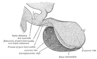
A major organ of the endocrine system, the anterior pituitary is the glandular, anterior lobe that together with the posterior lobe makes up the pituitary gland (hypophysis). The anterior pituitary regulates several physiological processes, including stress, growth, reproduction, and lactation. Proper functioning of the anterior pituitary and of the organs it regulates can often be ascertained via blood tests that measure hormone levels.

Leydig cells, also known as interstitial cells of Leydig, are found adjacent to the seminiferous tubules in the testicle. They produce testosterone in the presence of luteinizing hormone (LH). Leydig cells are polyhedral in shape, and have a large prominent nucleus, an eosinophilic cytoplasm and numerous lipid-filled vesicles.
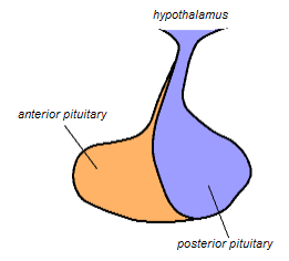
The posterior pituitary is the posterior lobe of the pituitary gland which is part of the endocrine system. The posterior pituitary is not glandular as is the anterior pituitary. Instead, it is largely a collection of axonal projections from the hypothalamus that terminate behind the anterior pituitary, and serve as a site for the secretion of neurohypophysial hormones directly into the blood. The hypothalamic–neurohypophyseal system is composed of the hypothalamus, posterior pituitary, and these axonal projections.

Seminiferous tubules are located within the testes, and are the specific location of meiosis, and the subsequent creation of male gametes, namely spermatozoa.

Chromaffin cells, also pheochromocytes, are neuroendocrine cells found mostly in the medulla of the adrenal glands in mammals. These cells serve a variety of functions such as serving as a response to stress, monitoring carbon dioxide and oxygen concentrations in the body, maintenance of respiration and the regulation of blood pressure. They are in close proximity to pre-synaptic sympathetic ganglia of the sympathetic nervous system, with which they communicate, and structurally they are similar to post-synaptic sympathetic neurons. In order to activate chromaffin cells, the splanchnic nerve of the sympathetic nervous system releases acetylcholine, which then binds to nicotinic acetylcholine receptors on the adrenal medulla. This causes the release of catecholamines. The chromaffin cells release catecholamines: ~80% of adrenaline (epinephrine) and ~20% of noradrenaline (norepinephrine) into systemic circulation for systemic effects on multiple organs, and can also send paracrine signals. Hence they are called neuroendocrine cells.

Thyroid follicular cells are the major cell type in the thyroid gland, and are responsible for the production and secretion of the thyroid hormones thyroxine (T4) and triiodothyronine (T3). They form the single layer of cuboidal epithelium that makes up the outer structure of the almost spherical thyroid follicle.

Parafollicular cells, also called C cells, are neuroendocrine cells in the thyroid. The primary function of these cells is to secrete calcitonin. They are located adjacent to the thyroid follicles and reside in the connective tissue. These cells are large and have a pale stain compared with the follicular cells. In teleost and avian species these cells occupy a structure outside the thyroid gland named the ultimobranchial body.

Parathyroid chief cells are one of the two cell types of the parathyroid glands, along with oxyphil cells. The chief cells are much more prevalent in the parathyroid gland than the oxyphil cells. It is perceived that oxyphil cells may be derived from chief cells at puberty, as they are not present at birth like chief cells.

Enterocytes, or intestinal absorptive cells, are simple columnar epithelial cells which line the inner surface of the small and large intestines. A glycocalyx surface coat contains digestive enzymes. Microvilli on the apical surface increase its surface area. This facilitates transport of numerous small molecules into the enterocyte from the intestinal lumen. These include broken down proteins, fats, and sugars, as well as water, electrolytes, vitamins, and bile salts. Enterocytes also have an endocrine role, secreting hormones such as leptin.
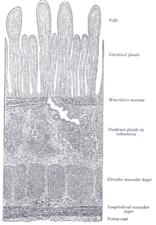
Brunner's glands are compound tubular submucosal glands found in that portion of the duodenum which is above the hepatopancreatic sphincter. The main function of these glands is to produce a mucus-rich alkaline secretion i.e. mucous in order to:
Neuroendocrine cells are cells that receive neuronal input and, as a consequence of this input, release message molecules (hormones) into the blood. In this way they bring about an integration between the nervous system and the endocrine system, a process known as neuroendocrine integration. An example of a neuroendocrine cell is a cell of the adrenal medulla, which releases adrenaline to the blood. The adrenal medullary cells are controlled by the sympathetic division of the autonomic nervous system. These cells are modified postganglionic neurons. Autonomic nerve fibers lead directly to them from the central nervous system. The adrenal medullary hormones are kept in vesicles much in the same way neurotransmitters are kept in neuronal vesicles. Hormonal effects can last up to ten times longer than those of neurotransmitters. Sympathetic nerve fiber impulses stimulate the release of adrenal medullary hormones. In this way the sympathetic division of the autonomic nervous system and the medullary secretions function together.
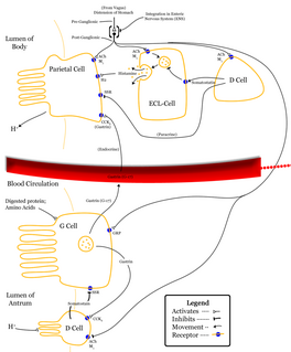
Enterochromaffin-like cells or ECL cells are a type of neuroendocrine cell found in the gastric glands of the gastric mucosa beneath the epithelium, in particular in the vicinity of parietal cells, that aid in the production of gastric acid via the release of histamine. They are also considered a type of enteroendocrine cell.

The juxtaglomerular cells are cells in the kidney that synthesize, store, and secrete the enzyme renin. They are specialized smooth muscle cells mainly in the walls of the afferent arterioles that deliver blood to the glomerulus. In synthesizing renin, they play a critical role in the renin–angiotensin system and thus in autoregulation of the kidney.

In histology, an intestinal gland(also crypt of Lieberkühn and intestinal crypt) is a gland found in between villi in the intestinal epithelium lining of the small intestine and large intestine. The glands and intestinal villi are covered by epithelium, which contains multiple types of cells: enterocytes, goblet cells, enteroendocrine cells, cup cells, tuft cells, and at the base of the gland, Paneth cells and stem cells.
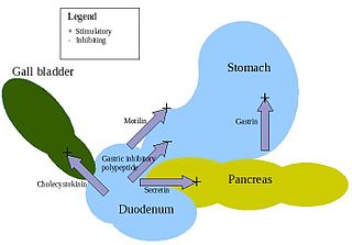
Enteroendocrine cells are specialized cells of the gastrointestinal tract and pancreas with endocrine function. They produce gastrointestinal hormones or peptides in response to various stimuli and release them into the bloodstream for systemic effect, diffuse them as local messengers, or transmit them to the enteric nervous system to activate nervous responses. Enteroendocrine cells of the intestine are the most numerous endocrine cells of the body. They constitute an enteric endocrine system as a subset of the endocrine system just as the enteric nervous system is a subset of the nervous system. In a sense they are known to act as chemoreceptors, initiating digestive actions and detecting harmful substances and initiating protective responses. Enteroendocrine cells are located in the stomach, in the intestine and in the pancreas.
Acidophile is a term used by histologists to describe a particular staining pattern of cells and tissues when using haematoxylin and eosin stains. Specifically, the name refers to structures which "love" acid, and take it up readily. More specifically, acidophilia can be described by cationic groups of most often proteins in the cell readily reacting with acidic stains.

A chromophobe is a histological structure that does not stain readily, and thus appears relatively pale under the microscope.

Foveolar cells or surface mucous cells are mucus-producing cells which cover the inside of the stomach, protecting it from the corrosive nature of gastric acid. These cells line the gastric mucosa. The mucus-secreting cells of the stomach can be distinguished histologically from the intestinal goblet cells, another type of mucus-secreting cell.
