Bioluminescence imaging

Last updated
Imaging of engineered E. coli Nissle 1917 in the mouse gut Imaging of engineered E. coli Nissle 1917 in the mouse gut.pdf
Imaging of engineered E. coli Nissle 1917 in the mouse gut

Bioluminescence imaging (BLI) is a technology developed over the past decades (1990's and onward). [1] [2] [3] [ when? ] that allows for the noninvasive study of ongoing biological processes [4] [1] [5] [6] [7] Recently, bioluminescence tomography (BLT) has become possible and several systems have become commercially available. In 2011, PerkinElmer acquired one of the most popular lines of optical imaging systems with bioluminescence from Caliper Life Sciences. [8]

Contents

Background

Bioluminescence is the process of light emission in living organisms. Bioluminescence imaging utilizes native light emission from one of several organisms which bioluminesce, also known as luciferase enzymes. [9] [10] [11] The three main sources are the North American firefly, the sea pansy (and related marine organisms), and bacteria like Photorhabdus luminescens and Vibrio fischeri . The DNA encoding the luminescent protein is incorporated into the laboratory animal either via a viral vector or by creating a transgenic animal. Rodent models of cancer spread can be studied through bioluminescence imaging.for e.g.Mouse models of breast cancer metastasis.

Systems derived from the three groups above differ in key ways:

While the total amount of light emitted from bioluminescence is typically small and not detected by the human eye, an ultra-sensitive CCD camera can image bioluminescence from an external vantage point.

Applications

Common applications of BLI include in vivo studies of infection [14] (with bioluminescent pathogens), cancer progression (using a bioluminescent cancer cell line), and reconstitution kinetics (using bioluminescent stem cells). [15]

Researchers at UT Southwestern Medical Center have shown that bioluminescence imaging can be used to determine the effectiveness of cancer drugs that choke off a tumor's blood supply. The technique requires luciferin to be added to the bloodstream, which carries it to cells throughout the body. When luciferin reaches cells that have been altered to carry the firefly gene, those cells emit light. [16]

The BLT inverse problem of 3D reconstruction of the distribution of bioluminescent molecules from data measured on the animal surface is inherently ill-posed. The first small animal study using BLT was conducted by researchers at the University of Southern California, Los Angeles, USA in 2005. Following this development, many research groups in USA and China have built systems that enable BLT.

Mustard plants have had the gene that makes fireflies' tails glow added to them so that the plants glow when touched. The effect lasts for an hour, but an utra-sensitive camera is needed to see the glow. [17]

Autoluminograph

An autoluminograph is a photograph produced by placing a light emitting object directly on a piece of film. A famous example is an autoluminograph published in Science magazine in 1986 [18] of a glowing transgenic tobacco plant bearing the luciferase gene of fireflies placed on Kodak Ektachrome 200 film.

Induced metabolic bioluminescence imaging

Induced metabolic bioluminescence imaging (imBI) is used to obtain a metabolic snapshot of biological tissues. [19] Metabolites that may be quantified through imBI include glucose, lactate, pyruvate, ATP, glucose-6-phosphate, or D2-hydroxygluturate. [20] imBI can be used to determine the lactate concentration of tumors or to measure the metabolism of the brain. [20] [19]

Related Research Articles

Biophotons are photons of light in the ultraviolet and low visible light range that are produced by a biological system. They are non-thermal in origin, and the emission of biophotons is technically a type of bioluminescence, though bioluminescence is generally reserved for higher luminance luciferin/luciferase systems. The term biophoton used in this narrow sense should not be confused with the broader field of biophotonics, which studies the general interaction of light with biological systems.

<span class="mw-page-title-main">Bioluminescence</span> Emission of light by a living organism

Bioluminescence is the production and emission of light by living organisms. It is a form of chemiluminescence. Bioluminescence occurs widely in marine vertebrates and invertebrates, as well as in some fungi, microorganisms including some bioluminescent bacteria, and terrestrial arthropods such as fireflies. In some animals, the light is bacteriogenic, produced by symbiotic bacteria such as those from the genus Vibrio; in others, it is autogenic, produced by the animals themselves.

<span class="mw-page-title-main">Chemiluminescence</span> Emission of light as a result of a chemical reaction

Chemiluminescence is the emission of light (luminescence) as the result of a chemical reaction, i.e. a chemical reaction results in a flash or glow of light. A standard example of chemiluminescence in the laboratory setting is the luminol test. Here, blood is indicated by luminescence upon contact with iron in hemoglobin. When chemiluminescence takes place in living organisms, the phenomenon is called bioluminescence. A light stick emits light by chemiluminescence.

<span class="mw-page-title-main">Luciferase</span> Enzyme family

Luciferase is a generic term for the class of oxidative enzymes that produce bioluminescence, and is usually distinguished from a photoprotein. The name was first used by Raphaël Dubois who invented the words luciferin and luciferase, for the substrate and enzyme, respectively. Both words are derived from the Latin word lucifer, meaning "lightbearer", which in turn is derived from the Latin words for "light" (lux) and "to bring or carry" (ferre).

<span class="mw-page-title-main">Luciferin</span> Class of light-emitting chemical compounds

Luciferin is a generic term for the light-emitting compound found in organisms that generate bioluminescence. Luciferins typically undergo an enzyme-catalyzed reaction with molecular oxygen. The resulting transformation, which usually involves breaking off a molecular fragment, produces an excited state intermediate that emits light upon decaying to its ground state. The term may refer to molecules that are substrates for both luciferases and photoproteins.

<i>Aliivibrio fischeri</i> Species of bacterium

Aliivibrio fischeri is a Gram-negative, rod-shaped bacterium found globally in marine environments. This species has bioluminescent properties, and is found predominantly in symbiosis with various marine animals, such as the Hawaiian bobtail squid. It is heterotrophic, oxidase-positive, and motile by means of a single polar flagella. Free-living A. fischeri cells survive on decaying organic matter. The bacterium is a key research organism for examination of microbial bioluminescence, quorum sensing, and bacterial-animal symbiosis. It is named after Bernhard Fischer, a German microbiologist.

<i>Noctiluca scintillans</i> Bioluminescent, marine dinoflagellate

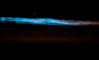
Noctiluca scintillans is a marine species of dinoflagellate that can exist in a green or red form, depending on the pigmentation in its vacuoles. It can be found worldwide, but its geographical distribution varies depending on whether it is green or red. This unicellular microorganism is known for its ability to bioluminesce, giving the water a bright blue glow seen at night. However, blooms of this species can be responsible for environmental hazards, such as toxic red tides. They may also be an indicator of anthropogenic eutrophication.

<span class="mw-page-title-main">Firefly luciferase</span>

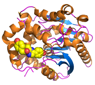
Firefly luciferase is the light-emitting enzyme responsible for the bioluminescence of fireflies and click beetles. The enzyme catalyses the oxidation of firefly luciferin, requiring oxygen and ATP. Because of the requirement of ATP, firefly luciferases have been used extensively in biotechnology.

<span class="mw-page-title-main">Firefly luciferin</span> Chemical compound

Firefly luciferin is the luciferin, or light-emitting compound, used for the firefly (Lampyridae), railroad worm (Phengodidae), starworm (Rhagophthalmidae), and click-beetle (Pyrophorini) bioluminescent systems. It is the substrate of luciferase, which is responsible for the characteristic yellow light emission from many firefly species.

<span class="mw-page-title-main">Renilla-luciferin 2-monooxygenase</span>

Renilla-luciferin 2-monooxygenase, Renilla luciferase, or RLuc, is a bioluminescent enzyme found in Renilla reniformis, belonging to a group of coelenterazine luciferases. Of this group of enzymes, the luciferase from Renilla reniformis has been the most extensively studied, and due to its bioluminescence requiring only molecular oxygen, has a wide range of applications, with uses as a reporter gene probe in cell culture, in vivo imaging, and various other areas of biological research. Recently, chimeras of RLuc have been developed and demonstrated to be the brightest luminescent proteins to date, and have proved effective in both noninvasive single-cell and whole body imaging.

<span class="mw-page-title-main">Bioreporter</span> Genetically engineered microbial cells

Bioreporters are intact, living microbial cells that have been genetically engineered to produce a measurable signal in response to a specific chemical or physical agent in their environment. Bioreporters contain two essential genetic elements, a promoter gene and a reporter gene. The promoter gene is turned on (transcribed) when the target agent is present in the cell’s environment. The promoter gene in a normal bacterial cell is linked to other genes that are then likewise transcribed and then translated into proteins that help the cell in either combating or adapting to the agent to which it has been exposed. In the case of a bioreporter, these genes, or portions thereof, have been removed and replaced with a reporter gene. Consequently, turning on the promoter gene now causes the reporter gene to be turned on. Activation of the reporter gene leads to production of reporter proteins that ultimately generate some type of a detectable signal. Therefore, the presence of a signal indicates that the bioreporter has sensed a particular target agent in its environment.

<span class="mw-page-title-main">Coelenterazine</span> Chemical compound

Coelenterazine is a luciferin, a molecule that emits light after reaction with oxygen, found in many aquatic organisms across eight phyla. It is the substrate of many luciferases such as Renilla reniformis luciferase (Rluc), Gaussia luciferase (Gluc), and photoproteins, including aequorin, and obelin. All these proteins catalyze the oxidation of this substance, a reaction catalogued EC 1.13.12.5.

<span class="mw-page-title-main">Vargulin</span> Chemical compound

Vargulin, also called Cypridinid luciferin, Cypridina luciferin, or Vargula luciferin, is the luciferin found in the ostracod Cypridina hilgendorfii, also named Vargula hilgendorfii. These bottom dwelling ostracods emit a light stream into water when disturbed presumably to deter predation. Vargulin is also used by the midshipman fish, Porichthys.

<i>Vargula hilgendorfii</i> Species of seed shrimp

Vargula hilgendorfii, sometimes called the sea-firefly and one of three bioluminescent species known in Japan as umi-hotaru (海蛍), is a species of ostracod crustacean. It is the only member of genus Vargula to inhabit Japanese waters; all other members of its genus inhabit the Gulf of Mexico, the Caribbean Sea, and waters off the coast of California. V. hilgendorfii was formerly more common, but its numbers have fallen significantly.

<span class="mw-page-title-main">John Woodland Hastings</span>

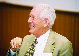
John Woodland "Woody" Hastings, was a leader in the field of photobiology, especially bioluminescence, and was one of the founders of the field of circadian biology. He was the Paul C. Mangelsdorf Professor of Natural Sciences and Professor of Molecular and Cellular Biology at Harvard University. He published over 400 papers and co-edited three books.

<span class="mw-page-title-main">Dinoflagellate luciferase</span>

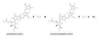
Dinoflagellate luciferase (EC 1.13.12.18, Gonyaulax luciferase) is a specific luciferase, an enzyme with systematic name dinoflagellate-luciferin:oxygen 132-oxidoreductase.

<span class="mw-page-title-main">Bioluminescent bacteria</span>

Bioluminescent bacteria are light-producing bacteria that are predominantly present in sea water, marine sediments, the surface of decomposing fish and in the gut of marine animals. While not as common, bacterial bioluminescence is also found in terrestrial and freshwater bacteria. These bacteria may be free living or in symbiosis with animals such as the Hawaiian Bobtail squid or terrestrial nematodes. The host organisms provide these bacteria a safe home and sufficient nutrition. In exchange, the hosts use the light produced by the bacteria for camouflage, prey and/or mate attraction. Bioluminescent bacteria have evolved symbiotic relationships with other organisms in which both participants benefit close to equally. Another possible reason bacteria use luminescence reaction is for quorum sensing, an ability to regulate gene expression in response to bacterial cell density.

Breast cancer metastatic mouse models are experimental approaches in which mice are genetically manipulated to develop a mammary tumor leading to distant focal lesions of mammary epithelium created by metastasis. Mammary cancers in mice can be caused by genetic mutations that have been identified in human cancer. This means models can be generated based upon molecular lesions consistent with the human disease.

<span class="mw-page-title-main">Scintillon</span>

Scintillons are small structures in cytoplasm that produce light. Among bioluminescent organisms, only dinoflagellates have scintillons.

Howard Harold Seliger was a physicist, biochemist, and biology professor, known for his research on bioluminescence.

References

  1. 1 2 Zambito G, Chawda C, Mezzanotte L (August 2021). "Emerging tools for bioluminescence imaging". Current Opinion in Chemical Biology. Chemical Genetics and Epigenetics * Molecular Imaging. 63: 86–94. doi: 10.1016/j.cbpa.2021.02.005 . PMID   33770744. S2CID   232377256.
  2. Contag CH, Bachmann MH (2002-08-01). "Advances in in vivo bioluminescence imaging of gene expression". Annual Review of Biomedical Engineering. 4 (1): 235–260. doi:10.1146/annurev.bioeng.4.111901.093336. PMID   12117758.
  3. Mezzanotte L, van 't Root M, Karatas H, Goun EA, Löwik CW (July 2017). "In Vivo Molecular Bioluminescence Imaging: New Tools and Applications". Trends in Biotechnology. 35 (7): 640–652. doi:10.1016/j.tibtech.2017.03.012. PMID   28501458.
  4. Brennan CK, Ornelas MY, Yao ZW, Prescher JA (August 2021). "Multicomponent Bioluminescence Imaging with Naphthylamino Luciferins". ChemBioChem. 22 (16): 2650–2654. doi:10.1002/cbic.202100202. PMC   8496354 . PMID   34139065.
  5. Syed AJ, Anderson JC (May 2021). "Applications of bioluminescence in biotechnology and beyond". Chemical Society Reviews. 50 (9): 5668–5705. doi: 10.1039/D0CS01492C . PMID   33735357. S2CID   232304401.
  6. Thorne N, Inglese J, Auld DS (June 2010). "Illuminating insights into firefly luciferase and other bioluminescent reporters used in chemical biology". Chemistry & Biology. 17 (6): 646–657. doi:10.1016/j.chembiol.2010.05.012. PMC   2925662 . PMID   20609414.
  7. Yeh HW, Ai HW (June 2019). "Development and Applications of Bioluminescent and Chemiluminescent Reporters and Biosensors". Annual Review of Analytical Chemistry. 12 (1): 129–150. Bibcode:2019ARAC...12..129Y. doi:10.1146/annurev-anchem-061318-115027. PMC   6565457 . PMID   30786216.
  8. "PerkinElmer to Acquire Caliper Life Sciences for $600M in Cash". Genetic Engineering & Biotechnology News (GEN). Mary Ann Liebert, Inc. 8 September 2011. Retrieved 2016-06-10.
  9. Prescher JA, Contag CH (February 2010). "Guided by the light: visualizing biomolecular processes in living animals with bioluminescence". Current Opinion in Chemical Biology. 14 (1): 80–89. doi:10.1016/j.cbpa.2009.11.001. PMID   19962933.
  10. Branchini BR, Behney CE, Southworth TL, Fontaine DM, Gulick AM, Vinyard DJ, Brudvig GW (June 2015). "Experimental Support for a Single Electron-Transfer Oxidation Mechanism in Firefly Bioluminescence". Journal of the American Chemical Society. 137 (24): 7592–7595. doi:10.1021/jacs.5b03820. PMID   26057379.
  11. Rathbun CM, Porterfield WB, Jones KA, Sagoe MJ, Reyes MR, Hua CT, Prescher JA (December 2017). "Parallel Screening for Rapid Identification of Orthogonal Bioluminescent Tools". ACS Central Science. 3 (12): 1254–1261. doi:10.1021/acscentsci.7b00394. PMC   5746862 . PMID   29296665.
  12. Close DM, Patterson SS, Ripp S, Baek SJ, Sanseverino J, Sayler GS (August 2010). Pan X (ed.). "Autonomous bioluminescent expression of the bacterial luciferase gene cassette (lux) in a mammalian cell line". PLOS ONE. 5 (8): e12441. Bibcode:2010PLoSO...512441C. doi: 10.1371/journal.pone.0012441 . PMC   2929204 . PMID   20805991.
  13. Close DM, Hahn RE, Patterson SS, Baek SJ, Ripp SA, Sayler GS (April 2011). "Comparison of human optimized bacterial luciferase, firefly luciferase, and green fluorescent protein for continuous imaging of cell culture and animal models". Journal of Biomedical Optics. 16 (4): 047003–047003–10. Bibcode:2011JBO....16d7003C. doi:10.1117/1.3564910. PMC   3094131 . PMID   21529093.
  14. Xiong YQ, Willard J, Kadurugamuwa JL, Yu J, Francis KP, Bayer AS (January 2005). "Real-time in vivo bioluminescent imaging for evaluating the efficacy of antibiotics in a rat Staphylococcus aureus endocarditis model". Antimicrobial Agents and Chemotherapy. 49 (1): 380–387. doi:10.1128/AAC.49.1.380-387.2005. PMC   538900 . PMID   15616318.
  15. Di Rocco G, Gentile A, Antonini A, Truffa S, Piaggio G, Capogrossi MC, Toietta G (1 September 2012). "Analysis of biodistribution and engraftment into the liver of genetically modified mesenchymal stromal cells derived from adipose tissue". Cell Transplantation. 21 (9): 1997–2008. doi: 10.3727/096368911X637452 . PMID   22469297. S2CID   21603693.
  16. Zhao D, Richer E, Antich PP, Mason RP (July 2008). "Antivascular effects of combretastatin A4 phosphate in breast cancer xenograft assessed using dynamic bioluminescence imaging and confirmed by MRI". FASEB Journal. 22 (7): 2445–2451. doi: 10.1096/fj.07-103713 . PMC   4426986 . PMID   18263704.
  17. Riley C (17 May 2000). "Glowing plants reveal touch sensitivity". BBC News.
  18. Ow DW, DE Wet JR, Helinski DR, Howell SH, Wood KV, Deluca M (November 1986). "Transient and stable expression of the firefly luciferase gene in plant cells and transgenic plants". Science. 234 (4778). American Association for the Advancement of Science: 856–859. Bibcode:1986Sci...234..856O. doi:10.1126/science.234.4778.856. PMID   17758108. S2CID   32603977.
  19. 1 2 Walenta S, Voelxen NF, Sattler UG, Mueller-Klieser W (2014). "Localizing and Quantifying Metabolites in Situ with Luminometry: Induced Metabolic Bioluminescence Imaging (ImBI)". Brain Energy Metabolism. Neuromethods. Vol. 90. pp. 195–216. doi:10.1007/978-1-4939-1059-5_9. ISBN   978-1-4939-1058-8.
  20. 1 2 Parks SK, Mueller-Klieser W, Pouysségur J (2020). "Lactate and Acidity in the Cancer Microenvironment". Annual Review of Cancer Biology. 4: 141–158. doi: 10.1146/annurev-cancerbio-030419-033556 .

Further reading