Carcinoid syndrome

Last updated
Carcinoid syndrome
Specialty Endocrinology, oncology

Carcinoid syndrome is a paraneoplastic syndrome comprising the signs and symptoms that occur secondary to neuroendocrine tumors (formerly known as carcinoid tumors). [1] The syndrome is caused by neuroendocrine tumors most often found in the gut releasing biologically active substances into the blood causing symptoms such as flushing and diarrhea, and less frequently, heart failure, vomiting and bronchoconstriction. [2] [1]

Contents

Signs and symptoms

Possible symptoms of the carcinoid syndrome Carcinoid syndrome presentation.svg
Possible symptoms of the carcinoid syndrome

The carcinoid syndrome occurs in approximately 10% of all neuroendocrine tumors [1] or about 30–40% of more advanced/well developed neuroendocrine tumors. [2] The biologically active substances that are released by the tumors cause the symptoms of the carcinoid syndrome. [4] [2] [1] These substances act on the vessels to produce the symptoms of the carcinoid syndrome. [2] [1]

Less common symptoms include malabsorption (leading to pellagra), fatigue, muscle loss, and cognitive impairment. [1] Late complications may include mesenteric and retroperitoneal fibroses as well. [2]

Pathophysiology

Serotonin Serotonin (5-HT).svg
Serotonin
Tryptophan Tryptophan - Tryptophan.svg
Tryptophan

The carcinoid syndrome occurs secondary to neuroendocrine tumors. [1] [2] These tumors occur mostly in the gut and less commonly in the lungs, but may also occur in other places in the body such as the pancreas, kidneys, and other organs. [1] [2] [5] [6] [7] Neuroendocrine tumors produce several biologically active substances, mainly amines and peptides. [1] There are over 40 substances known to be secreted by these tumors but the exact effect of each and their contribution to the carcinoid syndrome is unknown. [6] The most common substances found to be released and contribute to the syndrome include serotonin, histamine, tachykinins, kallikrein, and prostaglandins with the greatest contribution appearing to be from serotonin. [6] [2] [1] The symptoms of the carcinoid syndrome result from the action of these substances largely on the blood vessels. [1] These biologic substances are often metabolized and inactivated by the liver in a process known as first pass metabolism. This is why carcinoid syndrome most often occurs in patients whom the neuroendocrine tumor has metastasized to the liver, which allows the substances to bypass the first pass metabolism. [1] [6] [7] Neuroendocrine tumors arising in the bronchi may be associated with manifestations of carcinoid syndrome without liver metastases because their biologically active products reach the systemic circulation before passing through the liver and being metabolized.

Tryptophan metabolism is altered in the carcinoid syndrome. With neuroendocrine tumors, there is a shift in conversion of tryptophan to serotonin from the normal 1% to as high as 70%. [1] [8] Increased amounts of serotonin lead to increased gut motility causing the diarrhea seen in carcinoid syndrome. [1] [6] [8] Increased amounts of serotonin can also cause the flushing seen as the main symptom of carcinoid syndrome. [2] Tryptophan is also needed for niacin synthesis which can be a cause for pellagra associated with carcinoid syndrome. [1] In the pulmonary neuroendocrine tumors or metastases, histamine release and kallikrein metabolism are the vasoactive mediators of flushing and the other symptoms of carcinoid syndrome. [2] [1]

Carcinoid crisis

Carcinoid crisis is an extreme exacerbation of the carcinoid syndrome. This results from excessive release of amines by the neuroendocrine tumors. It is largely a result of stressful procedures such as anesthesia, surgery, or radiation treatment. Symptoms of carcinoid crisis include flushing, hypotension, arrhythmia and bronchospasm. [2] [9]

Carcinoid heart disease

Carcinoid heart disease is the result of valvular damage related to the vasoactive substances released by the neuroendocrine tumor reaching the right side of the heart. [5] This mainly affects the right side of the heart unless there is anomalous circulation (i.e. patent foramen ovale) because the lungs will metabolize the substances released by the tumor similar to how the liver will. [5] After initial tissue injury around the valves, plaque will develop and fibrosis will occur, possibly mediated by excess serotonin. [5]

Diagnosis

With a certain degree of clinical suspicion, the most useful initial test is the 24-hour urine levels of 5-HIAA (5-hydroxyindoleacetic acid), the end product of serotonin metabolism. Chromogramin A, a glycoprotein released by neuroendocrine tumors, can be used to detect non-secreting tumors. [1] [10]

Imaging

Imaging studies should be largely focused on the abdomen and pelvis because the neuroendocrine tumors causing the carcinoid syndrome largely arise in the gut. [10] CT and MRI that utilize radioactive somatostatin analogues such as indium-111 pentetreotide are used to localize the tumor. [10] PET scan can also be used to find the primary tumor site. [7] Bronchoscopy with biopsy can performed if there is evidence of a pulmonary tumor. [1] For patients with serotonin elevated 5x the upper limit of normal or more, an echocardiogram is recommended for evaluation of carcinoid heart disease. [1]

Differential diagnosis

Other conditions similar to the carcinoid syndrome that should be considered include: [1]

Treatment

Treatment of the carcinoid syndrome is focused on controlling the proliferation of the primary tumor and symptomatic control of the symptoms with somatostatin analogues octreotide or lanreotide. [2] [1] [11] These analogues can help control the growth of the tumor itself and the associated symptoms of the carcinoid syndrome. [11] In patients whose symptoms are refractory to initial doses, increasing the dose or switching to another analogue pasireotide may be effective. [11] In patients who continue to be refractory, mTOR inhibitors such as everolimus. [11] The TPH inhibitor telotristat ethyl may be useful in controlling diarrhea associated with the carcinoid syndrome.

[11] Peptide directed radiotherapy (PRRT) is another alternative treatment for patients who failed somatostain analogue therapy. [11] This method uses radioactive somatostatin analogues such as 177Lu-Dotatate or 90Y-Edotreotide to target tumor directly. [11] These therapies are effective for metastatic disease but studies have been limited to about 6-month time periods. [11]

Cytoreductive surgery performed chemically with 131metaiodobenzylguanidine (131I-MIBG) may also control symptoms starting around 6–15 months post procedure and lasting as long as 39 months. [11] There are also procedures that target the liver directly such as radiofrequency ablation or radioembolization that deliver targeted therapy directly to the liver through special catheters. [11] This is especially useful for patients with liver metastases. [11] [2] [7]

Carcinoid heart disease

The most important aspect of treating carcinoid heart disease is detecting its presence with echocardiography, likely with color doppler. [11] Treatment consists of the same treatment as patients with heart failure with definitive treatment being surgical valve repair or replacement. [11]

Uncertainties

Disease progression is difficult to ascertain because the disease can metastasize anywhere in the body and can be too small to identify with any current technology. Markers of the condition such as chromogranin-A are imperfect indicators of disease progression. [12]

Epidemiology

The incidence of neuroendocrine tumors in the US lies somewhere from 2.7 to 4.3 per 100,000 people and appears to be increasing over time. [1] [9] The incidence of the carcinoid syndrome is about 0.27 per 100,000 people in the US, [9] about 10% of all people with neuroendocrine tumors. [1] There does not appear to be any variance by gender however patients of African American ethnicity appear to be affected by the carcinoid syndrome more often. [1] [9]

Nonhuman animals

The carcinoid syndrome can affect other animals similarly to humans. [13] Similarly to humans, the carcinoid syndrome is due to neuroendocrine tumors that arise mainly from the bowel but also from other organs. [13] Common signs in animals include vomiting, diarrhea, and weight loss but other symptoms that are more common in humans such as flushing, hypotension and diarrhea can also occur. [13] Similar to humans, the cause of the carcinoid syndrome is the release of bioactive substances such as serotonin and histamine. [13]

See also

Related Research Articles

<span class="mw-page-title-main">Pheochromocytoma</span> Type of neuroendocrine tumor

Pheochromocytoma is a rare tumor of the adrenal medulla composed of chromaffin cells, also known as pheochromocytes. When a tumor composed of the same cells as a pheochromocytoma develops outside the adrenal gland, it is referred to as a paraganglioma. These neuroendocrine tumors typically release massive amounts of catecholamines which result in the most common symptoms, including hypertension, tachycardia, and sweating. Rarely, some tumors may secrete little to no catecholamines, making diagnosis difficult. While tumors of the head and neck are parasympathetic, their sympathetic counterparts are predominantly located in the abdomen and pelvis, particularly concentrated at the organ of Zuckerkandl.

<span class="mw-page-title-main">Zollinger–Ellison syndrome</span> Condition in which tumours stimulate excessive gastric acid production

Zollinger–Ellison syndrome is rare disease in which tumors cause the stomach to produce too much acid, resulting in peptic ulcers. Symptoms include abdominal pain and diarrhea.

<span class="mw-page-title-main">Octreotide</span> Octapeptide that mimics natural somatostatin pharmacologically

Octreotide, sold under the brand name Sandostatin among others, is an octapeptide that mimics natural somatostatin pharmacologically, though it is a more potent inhibitor of growth hormone, glucagon, and insulin than the natural hormone. It was first synthesized in 1979 by the chemist Wilfried Bauer, and binds predominantly to the somatostatin receptors SSTR2 and SSTR5.

<span class="mw-page-title-main">Enterochromaffin cell</span> Cell type

Enterochromaffin (EC) cells are a type of enteroendocrine cell, and neuroendocrine cell. They reside alongside the epithelium lining the lumen of the digestive tract and play a crucial role in gastrointestinal regulation, particularly intestinal motility and secretion. They were discovered by Nikolai Kulchitsky.

<span class="mw-page-title-main">Carcinoid</span> Slow-growing type of neuroendocrine tumor

A carcinoid is a slow-growing type of neuroendocrine tumor originating in the cells of the neuroendocrine system. In some cases, metastasis may occur. Carcinoid tumors of the midgut are associated with carcinoid syndrome.

<span class="mw-page-title-main">Multiple endocrine neoplasia type 1</span> Medical condition

Multiple endocrine neoplasia type 1 (MEN-1) is one of a group of disorders, the multiple endocrine neoplasias, that affect the endocrine system through development of neoplastic lesions in pituitary, parathyroid gland and pancreas. Individuals suffering from this disorder are prone to developing multiple endocrine and nonendocrine tumors. It was first described by Paul Wermer in 1954.

A VIPoma or vipoma is a rare endocrine tumor that overproduces vasoactive intestinal peptide. The incidence is about 1 per 10,000,000 per year. VIPomas usually originate from the non-β islet cells of the pancreas. They are sometimes associated with multiple endocrine neoplasia type 1. Roughly 50–75% of VIPomas are malignant, but even when they are benign, they are problematic because they tend to cause a specific syndrome: the massive amounts of VIP cause a syndrome of profound and chronic watery diarrhea and resultant dehydration, hypokalemia, achlorhydria, acidosis, flushing and hypotension, hypercalcemia, and hyperglycemia. This syndrome is called Verner–Morrison syndrome (VMS), WDHA syndrome, or pancreatic cholera syndrome (PCS). The eponym reflects the physicians who first described the syndrome.

<span class="mw-page-title-main">Gastrinoma</span> Medical condition

Gastrinomas are neuroendocrine tumors (NETs), usually located in the duodenum or pancreas, that secrete gastrin and cause a clinical syndrome known as Zollinger–Ellison syndrome (ZES). A large number of gastrinomas develop in the pancreas or duodenum, with near-equal frequency, and approximately 10% arise as primary neoplasms in lymph nodes of the pancreaticoduodenal region.

<span class="mw-page-title-main">5-Hydroxyindoleacetic acid</span> Chemical compound

5-Hydroxyindoleacetic acid (5-HIAA) is the main metabolite of serotonin. In chemical analysis of urine samples, 5-HIAA is used to determine serotonin levels in the body.

<span class="mw-page-title-main">Neuroendocrine tumor</span> Medical condition

Neuroendocrine tumors (NETs) are neoplasms that arise from cells of the endocrine (hormonal) and nervous systems. They most commonly occur in the intestine, where they are often called carcinoid tumors, but they are also found in the pancreas, lung, and the rest of the body.

<span class="mw-page-title-main">Enteroendocrine cell</span>

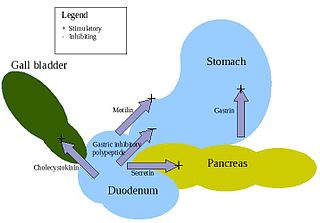
Enteroendocrine cells are specialized cells of the gastrointestinal tract and pancreas with endocrine function. They produce gastrointestinal hormones or peptides in response to various stimuli and release them into the bloodstream for systemic effect, diffuse them as local messengers, or transmit them to the enteric nervous system to activate nervous responses. Enteroendocrine cells of the intestine are the most numerous endocrine cells of the body. They constitute an enteric endocrine system as a subset of the endocrine system just as the enteric nervous system is a subset of the nervous system. In a sense they are known to act as chemoreceptors, initiating digestive actions and detecting harmful substances and initiating protective responses. Enteroendocrine cells are located in the stomach, in the intestine and in the pancreas. Microbiota play key roles in the intestinal immune and metabolic responses in these enteroendocrine cells via their fermentation product, acetate.

Cardiac fibrosis commonly refers to the excess deposition of extracellular matrix in the cardiac muscle, but the term may also refer to an abnormal thickening of the heart valves due to inappropriate proliferation of cardiac fibroblasts. Fibrotic cardiac muscle is stiffer and less compliant and is seen in the progression to heart failure. The description below focuses on a specific mechanism of valvular pathology but there are other causes of valve pathology and fibrosis of the cardiac muscle.

<span class="mw-page-title-main">Lanreotide</span> Pharmaceutical drug

Lanreotide, sold under the brand name Somatuline among others, is a medication used in the management of acromegaly and symptoms caused by neuroendocrine tumors, most notably carcinoid syndrome. It is a long-acting analogue of somatostatin, like octreotide.

<span class="mw-page-title-main">Medullary thyroid cancer</span> Malignant thyroid neoplasm originating from C-cells

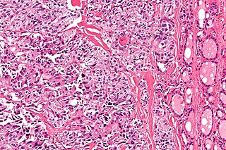
Medullary thyroid cancer is a form of thyroid carcinoma which originates from the parafollicular cells, which produce the hormone calcitonin. Medullary tumors are the third most common of all thyroid cancers and together make up about 3% of all thyroid cancer cases. MTC was first characterized in 1959.

<span class="mw-page-title-main">Acromegaly</span> Human disease that results in excess growth of certain parts of the body

Acromegaly is a disorder that results in excess growth of certain parts of the human body. It is caused by excess growth hormone (GH) after the growth plates have closed. The initial symptom is typically enlargement of the hands and feet. There may also be an enlargement of the forehead, jaw, and nose. Other symptoms may include joint pain, thicker skin, deepening of the voice, headaches, and problems with vision. Complications of the disease may include type 2 diabetes, sleep apnea, and high blood pressure.

<span class="mw-page-title-main">Pancreatic neuroendocrine tumor</span> Medical condition

Pancreatic neuroendocrine tumours, often referred to as "islet cell tumours", or "pancreatic endocrine tumours" are neuroendocrine neoplasms that arise from cells of the endocrine (hormonal) and nervous system within the pancreas.

Hepatic artery embolization, also known as trans-arterial embolization (TAE), is one of the several therapeutic methods to treat primary liver tumors or metastases to the liver. The embolization therapy can reduce the size of the tumor, and decrease the tumor's impact such its hormone production, effectively decreasing symptoms. The treatment was initially developed in the early 1970s. The several types of hepatic artery treatments are based on the observation that tumor cells get nearly all their nutrients from the hepatic artery, while the normal cells of the liver get about 70-80 percent of their nutrients and 50% their oxygen supply from the portal vein, and thus can survive with the hepatic artery effectively blocked. In practice, hepatic artery embolization occludes the blood flow to the tumors, achieving significant tumor shrinkage in over 80% of people. Shrinkage rates vary.

Diffuse idiopathic pulmonary neuroendocrine cell hyperplasia (DIPNECH) is a diffuse parenchymal lung disease which often presents with symptoms of cough and shortness of breath. The pathological definition published by the World Health Organization is “a generalized proliferation of scattered single cells, small nodules, or linear proliferations of pulmonary neuroendocrine (PNE) cells that may be confined to the bronchial and bronchiolar epithelium.” The true prevalence of this disease is not known. To date, just under 200 cases have been reported in the literature. However, with an increase in recognition of this disease by radiologists and pulmonologists, the number of cases has been increasing. DIPNECH predominantly affects middle-aged women with slowly progressive lung obstruction. DIPNECH is usually discovered in one of two ways: 1) as an unexpected finding following a lung surgery; or 2) by evaluation of a patient in a pulmonary clinic with longstanding, unexplained symptoms.

<span class="mw-page-title-main">Telotristat ethyl</span> Chemical compound

Telotristat ethyl is a prodrug of telotristat, which is an inhibitor of tryptophan hydroxylase. It is formulated as telotristat etiprate — a hippurate salt of telotristat ethyl.

A small intestine neuroendocrine tumor is a carcinoid in the distal small intestine or the proximal large intestine. It is a relatively rare cancer and is diagnosed in approximately 1/100000 people every year. In recent decades the incidence has increased. The prognosis is comparatively good with a median survival of more than 8 years. The disease was named by Siegfried Oberndorfer, a German pathologist, in 1907.

References

  1. 1 2 3 4 5 6 7 8 9 10 11 12 13 14 15 16 17 18 19 20 21 22 23 24 25 26 27 28 Pandit, Sudha; Annamaraju, Pavan; Bhusal, Kamal (2022), "Carcinoid Syndrome", StatPearls, Treasure Island (FL): StatPearls Publishing, PMID   28846309 , retrieved 2023-01-16
  2. 1 2 3 4 5 6 7 8 9 10 11 12 13 Rubin de Celis Ferrari AC; Glasberg, J.; Riechelmann, R. P. (2018). "Carcinoid syndrome: Update on the pathophysiology and treatment". Clinics (Sao Paulo, Brazil). 73 (suppl 1): e490s. doi:10.6061/clinics/2018/e490s. PMC   6096975 . PMID   30133565.
  3. Mitchell, Richard Sheppard; Kumar, Vinay; Abbas, Abul K.; Fausto, Nelson Robbins Basic Pathology, Philadelphia: Saunders ISBN: 1-4160-2973-7. Table 15-4. 8th edition.
  4. Kumar, Vinay (2023). Robbins & Kumar Basic Pathology (11e ed.). Web: Elsevier. pp. 483–532. ISBN   978-0-323-79018-5.
  5. 1 2 3 4 5 Ram, P.; Penalver, J. L.; Lo KBU; Rangaswami, J.; Pressman, G. S. (2019). "Carcinoid Heart Disease: Review of Current Knowledge". Texas Heart Institute Journal. 46 (1): 21–27. doi:10.14503/THIJ-17-6562. PMC   6378997 . PMID   30833833.
  6. 1 2 3 4 5 Guilmette, J.; Nosé, V. (2019). "Paraneoplastic syndromes and other systemic disorders associated with neuroendocrine neoplasms". Seminars in Diagnostic Pathology. 36 (4): 229–239. doi:10.1053/j.semdp.2019.03.002. PMID   30910348. S2CID   85514650.
  7. 1 2 3 4 Tsoli, M.; Chatzellis, E.; Koumarianou, A.; Kolomodi, D.; Kaltsas, G. (2019). "Current best practice in the management of neuroendocrine tumors". Therapeutic Advances in Endocrinology and Metabolism. 10. doi:10.1177/2042018818804698. PMC   6378464 . PMID   30800264.
  8. 1 2 Strosberg, Jonathan (November 16, 2021). "Clinical Features of Carcinoid Syndrome". Uptodate.
  9. 1 2 3 4 Gade AK, Olariu E, Douthit NT (March 2020). "Carcinoid Syndrome: A Review". Cureus. 12 (3): e7186. doi: 10.7759/cureus.7186 . PMC   7124884 . PMID   32257725.
  10. 1 2 3 Strosberg, Jonathan (April 20, 2022). "Diagnosis of Carcinoid Syndrome and Tumor Localization". Uptodate.
  11. 1 2 3 4 5 6 7 8 9 10 11 12 13 Ito, T.; Lee, L.; Jensen, R. T. (2018). "Carcinoid-syndrome: Recent advances, current status and controversies". Current Opinion in Endocrinology, Diabetes, and Obesity. 25 (1): 22–35. doi:10.1097/MED.0000000000000376. PMC   5747542 . PMID   29120923.
  12. Nobels FR, Kwekkeboom DJ, Bouillon R, Lamberts SW (June 1998). "Chromogranin A: its clinical value as marker of neuroendocrine tumours". European Journal of Clinical Investigation. 28 (6): 431–440. doi:10.1046/j.1365-2362.1998.00305.x. PMID   9693933. S2CID   37911197.
  13. 1 2 3 4 Kemppainen, Robert (April 2019). "Neuroendocrine Tumors in Animals - Endocrine System". Merck Veterinary Manual. Retrieved 2023-01-25.

Further reading