In vitro toxicity testing is the scientific analysis of the toxic effects of chemical substances on cultured bacteria or mammalian cells. In vitro testing methods are employed primarily to identify potentially hazardous chemicals and/or to confirm the lack of certain toxic properties in the early stages of the development of potentially useful new substances such as therapeutic drugs, agricultural chemicals and food additives.
Cytotoxicity is the quality of being toxic to cells. Examples of toxic agents are an immune cell or some types of venom, e.g. from the puff adder or brown recluse spider.
An assay is an investigative (analytic) procedure in laboratory medicine, mining, pharmacology, environmental biology and molecular biology for qualitatively assessing or quantitatively measuring the presence, amount, or functional activity of a target entity. The measured entity is often called the analyte, the measurand, or the target of the assay. The analyte can be a drug, biochemical substance, chemical element or compound, or cell in an organism or organic sample. An assay usually aims to measure an analyte's intensive property and express it in the relevant measurement unit.
Genotoxicity is the property of chemical agents that damage the genetic information within a cell causing mutations, which may lead to cancer. While genotoxicity is often confused with mutagenicity, all mutagens are genotoxic, but some genotoxic substances are not mutagenic. The alteration can have direct or indirect effects on the DNA: the induction of mutations, mistimed event activation, and direct DNA damage leading to mutations. The permanent, heritable changes can affect either somatic cells of the organism or germ cells to be passed on to future generations. Cells prevent expression of the genotoxic mutation by either DNA repair or apoptosis; however, the damage may not always be fixed leading to mutagenesis.

The MTT assay is a colorimetric assay for assessing cell metabolic activity. NAD(P)H-dependent cellular oxidoreductase enzymes may, under defined conditions, reflect the number of viable cells present. These enzymes are capable of reducing the tetrazolium dye MTT, which is chemically 3-(4,5-dimethylthiazol-2-yl)-2,5-diphenyltetrazolium bromide, to its insoluble formazan, which has a purple color. Other closely related tetrazolium dyes including XTT, MTS and the WSTs, are used in conjunction with the intermediate electron acceptor, 1-methoxy phenazine methosulfate (PMS). With WST-1, which is cell-impermeable, reduction occurs outside the cell via plasma membrane electron transport. However, this traditionally assumed explanation is currently contended as proof has also been found of MTT reduction to formazan in lipidic cellular structures without apparent involvement of oxidoreductases.

Neutral red is a eurhodin dye used for staining in histology. It stains lysosomes red. It is used as a general stain in histology, as a counterstain in combination with other dyes, and for many staining methods. Together with Janus Green B, it is used to stain embryonal tissues and supravital staining of blood. Can be used for staining Golgi apparatus in cells and Nissl granules in neurons.

Antibody-dependent cellular cytotoxicity (ADCC), also referred to as antibody-dependent cell-mediated cytotoxicity, is a mechanism of cell-mediated immune defense whereby an effector cell of the immune system actively lyses a target cell, whose membrane-surface antigens have been bound by specific antibodies. It is one of the mechanisms through which antibodies, as part of the humoral immune response, can act to limit and contain infection.
A neurosphere is a culture system composed of free-floating clusters of neural stem cells. Neurospheres provide a method to investigate neural precursor cells in vitro. Putative neural stem cells are suspended in a medium lacking adherent substrates but containing necessary growth factors, such as epidermal growth factor and fibroblast growth factor. This allows the neural stem cells to form into the characteristic 3-D clusters. However, neurospheres are not identical to stem cells; rather, they only contain a small percent of neural stem cells.

Carboxyfluorescein succinimidyl ester (CFSE) is a fluorescent cell staining dye. CFSE is cell permeable and covalently couples, via its succinimidyl group, to intracellular molecules, notably, to intracellular lysine residues and other amine sources. Due to this covalent coupling reaction, fluorescent CFSE can be retained within cells for extremely long periods. Also, due to this stable linkage, once incorporated within cells, the dye is not transferred to adjacent cells.

Toxicology testing, also known as safety assessment, or toxicity testing, is the process of determining the degree to which a substance of interest negatively impacts the normal biological functions of an organism, given a certain exposure duration, route of exposure, and substance concentration. Toxicology testing is often conducted by researchers who follow established toxicology test protocol for a certain substance, mode of exposure, exposure environment, duration of exposure, or for a particular organism of interest, or for a particular developmental stage of interest. Toxicology testing is commonly conducted during preclinical development for a substance intended for human exposure. Stages of in silico, in vitro and in vivo research are conducted to determine safe exposure doses in model organisms. If necessary, the next phase of research involves human toxicology testing during a first-in-man study. Toxicology testing may be conducted by the pharmaceutical industry, biotechnology companies, contract research organizations, or environmental scientists.

Resazurin is a phenoxazine dye that is weakly fluorescent, nontoxic, cell-permeable, and redox‐sensitive. Resazurin has a blue to purple color and is used in microbiological, cellular, and enzymatic assays because it can be irreversibly reduced to the pink-colored and highly fluorescent resorufin (7-Hydroxy-3H-phenoxazin-3-one). At circum-neutral pH, resorufin can be detected by visual observation of its pink color or by fluorimetry, with an excitation maximum at 530-570 nm and an emission maximum at 580-590 nm.

5-Ethynyl-2′-deoxyuridine (EdU) is a thymidine analogue which is incorporated into the DNA of dividing cells. EdU is used to assay DNA synthesis in cell culture and detect cells in embryonic, neonatal and adult animals which have undergone DNA synthesis. Whilst at high doses it can be cytotoxic, this molecule is now widely used to track proliferating cells in multiple biological systems.

A viability assay is an assay that is created to determine the ability of organs, cells or tissues to maintain or recover a state of survival. Viability can be distinguished from the all-or-nothing states of life and death by the use of a quantifiable index that ranges between the integers of 0 and 1 or, if more easily understood, the range of 0% and 100%. Viability can be observed through the physical properties of cells, tissues, and organs. Some of these include mechanical activity, motility, such as with spermatozoa and granulocytes, the contraction of muscle tissue or cells, mitotic activity in cellular functions, and more. Viability assays provide a more precise basis for measurement of an organism's level of vitality.
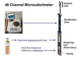
Isothermal microcalorimetry (IMC) is a laboratory method for real-time monitoring and dynamic analysis of chemical, physical and biological processes. Over a period of hours or days, IMC determines the onset, rate, extent and energetics of such processes for specimens in small ampoules at a constant set temperature.
A 3D cell culture is an artificially created environment in which biological cells are permitted to grow or interact with their surroundings in all three dimensions. Unlike 2D environments, a 3D cell culture allows cells in vitro to grow in all directions, similar to how they would in vivo. These three-dimensional cultures are usually grown in bioreactors, small capsules in which the cells can grow into spheroids, or 3D cell colonies. Approximately 300 spheroids are usually cultured per bioreactor.
Albert P. Li is president and CEO of In Vitro ADMET Laboratories (IVAL), Columbia, Maryland, and Malden, Massachusetts. For the past three decades, Li has devoted his scientific career to the advancement of scientific concepts and technologies to accurately predict human drug properties. His research is focused on the development and application of human-based in vitro experimental systems in drug discovery and development. He is a pioneer in the isolation, cryopreservation, and culturing of human hepatocytes and their application in the evaluation of drug metabolism, drug-drug interactions, and drug toxicity.
Complement-dependent cytotoxicity (CDC) is an effector function of IgG and IgM antibodies. When they are bound to surface antigen on target cell, the classical complement pathway is triggered by bonding protein C1q to these antibodies, resulting in formation of a membrane attack complex (MAC) and target cell lysis.

Bisphenol F is an organic compound with the chemical formula (HOC
6H
4)
2CH
2. It is structurally related to bisphenol A (BPA), a popular precursor for forming plastics, as both belong to the category of molecules known as bisphenols, which feature two phenol groups connected via a linking group. In BPF, the two aromatic rings are linked by a methylene connecting group. In response to concern about the health effects of BPA, BPF is increasingly used as a substitute for BPA.
A bioassay is an analytical method to determine the concentration or potency of a substance by its effect on living animals or plants, or on living cells or tissues(in vitro). A bioassay can be either quantal or quantitative, direct or indirect. If the measured response is binary, the assay is quantal, if not, it is quantitative.
Cycloheximide chase assays are an experimental technique used in molecular and cellular biology to measure steady state protein stability. Cycloheximide is a drug that inhibits the elongation step in eukaryotic protein translation, thereby preventing protein synthesis. The addition of cycloheximide to cultured cells followed by protein lysis at multiple timepoints is conducted to observe protein degradation over time and can be used to determine a protein's half-life. These assays are often followed by western blotting to assess protein abundance and can be analyzed using quantitative tools such as ImageJ.