Related Research Articles

In mammals, the vagina is the elastic, muscular part of the female genital tract. In humans, it extends from the vulva to the cervix. The outer vaginal opening is normally partly covered by a membrane called the hymen. At the deep end, the cervix bulges into the vagina. The vagina allows for sexual intercourse and birth. It also channels menstrual flow (menses), which occurs in humans and closely related primates as part of the monthly menstrual cycle.
Obstetrics and gynaecology or obstetrics and gynecology is the medical specialty that encompasses the two subspecialties of obstetrics and gynecology. It is commonly abbreviated as OB-GYN or OB/GYN in US English, and as obs and gynae or O&G in British English.
Gynaecology or gynecology is the medical practice dealing with the health of the female reproductive system. Outside medicine, the term means "the science of women". Its counterpart is andrology, which deals with medical issues specific to the male reproductive system.

Hysterectomy is the surgical removal of the uterus. It may also involve removal of the cervix, ovaries (oophorectomy), Fallopian tubes (salpingectomy), and other surrounding structures.

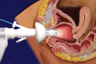
A pessary is a prosthetic device inserted into the vagina for structural and pharmaceutical purposes. It is most commonly used to treat stress urinary incontinence to stop urinary leakage, and pelvic organ prolapse to maintain the location of organs in the pelvic region. It can also be used to administer medications locally in the vagina or as a method of contraception. Pessaries come in different shapes and sizes, so it is important that individuals be fitted for them by health care professionals to avoid any complications.However, there are a few instances and circumstances that allow individuals to purchase pessaries from a store without a prescription or without seeking help from a health care professional. Some side effects may occur if pessaries are not sized properly or regularly maintained, but with the appropriate care, pessaries are generally safe and well tolerated.
Müllerian agenesis, also known as Mayer–Rokitansky–Küster–Hauser syndrome (MRKH) or vaginal agenesis, is a congenital malformation characterized by a failure of the Müllerian duct to develop, resulting in a missing uterus and variable degrees of vaginal hypoplasia of its upper portion. Müllerian agenesis is the cause in 15% of cases of primary amenorrhoea. Because most of the vagina does not develop from the Müllerian duct, instead developing from the urogenital sinus, along with the bladder and urethra, it is present even when the Müllerian duct is completely absent. Because ovaries do not develop from the Müllerian ducts, affected people might have normal secondary sexual characteristics but are infertile due to the lack of a functional uterus. However, parenthood is possible through use of gestational surrogates.
Vaginal bleeding is any bleeding from the vagina. This bleeding may originate from the uterus, vaginal wall, or cervix. Generally, it is either part of a normal menstrual cycle or is caused by hormonal or other problems of the reproductive system, such as abnormal uterine bleeding.

A uterine malformation is a type of female genital malformation resulting from an abnormal development of the Müllerian duct(s) during embryogenesis. Symptoms range from amenorrhea, infertility, recurrent pregnancy loss, and pain, to normal functioning depending on the nature of the defect.
An imperforate hymen is a congenital disorder where a hymen without an opening completely obstructs the vagina. It is caused by a failure of the hymen to perforate during fetal development. It is most often diagnosed in adolescent girls when menstrual blood accumulates in the vagina and sometimes also in the uterus. It is treated by surgical incision of the hymen.
Vaginal atresia is a condition in which the vagina is abnormally closed or absent. The main causes can either be complete vaginal hypoplasia, or a vaginal obstruction, often caused by an imperforate hymen or, less commonly, a transverse vaginal septum. It results in uterovaginal outflow tract obstruction. This condition does not usually occur by itself within an individual, but coupled with other developmental disorders within the female. The disorders that are usually coupled with a female who has vaginal atresia are Rokitansky-Mayer- Küster-Hauser syndrome, Bardet-Biedl syndrome, or Fraser syndrome. One out of every 5,000 women have this abnormality.

Uterus didelphys represents a uterine malformation where the uterus is present as a paired organ when the embryogenetic fusion of the Müllerian ducts fails to occur. As a result, there is a double uterus with two separate cervices, and possibly a double vagina as well. Each uterus has a single horn linked to the ipsilateral fallopian tube that faces its ovary.
'Hematocolpos' is a medical condition in which the vagina is pooled with menstrual blood due to multiple factors leading to the blockage of menstrual blood flow. The medical definition of Hematocolpos is 'an accumulation of blood within the vagina'. It is often caused by the combination of menstruation with an imperforate hymen. It is sometimes seen in Robinow syndrome, uterus didelphys, or other vaginal anomalies.
Vaginal hypoplasia is the underdevelopment or incomplete development of the vagina. It is a birth defect or congenital abnormality of the female genitourinary system.

A pelvic examination is the physical examination of the external and internal female pelvic organs. It is frequently used in gynecology for the evaluation of symptoms affecting the female reproductive and urinary tract, such as pain, bleeding, discharge, urinary incontinence, or trauma. It can also be used assess a patient's anatomy in preparation for procedures. The exam can be done awake in the clinic and emergency department, or under anesthesia in the operating room. The most commonly performed components of the exam are 1) the external exam, to evaluate the external genitalia 2) the internal exam with palpation to examine the uterus, ovaries, and fallopian tubes, and 3) the internal exam using the speculum to visualize the vaginal walls and cervix. During the pelvic exam, sample of cells and fluids may be collected to screen for sexually transmitted infections or cancer.

Hematometra is a medical condition involving collection or retention of blood in the uterus. It is most commonly caused by an imperforate hymen or a transverse vaginal septum.
A vaginal disease is a pathological condition that affects part or all of the vagina.
Neuroendocrine carcinoma of the cervix is best defined separately:Neuroendocrine: Of, relating to, or involving the interaction between the nervous system and the hormones of the endocrine glands.Carcinoma: An invasive malignant tumor derived from epithelial tissue that tends to metastasize to other areas of the body.
Postcoital bleeding is bleeding from the vagina in women after sexual intercourse and may or may not be associated with pain. The bleeding can be from the uterus, cervix, vagina and other tissue or organs located near the vagina. Postcoital bleeding can be one of the first indications of cervical cancer. There are other reasons why a woman may bleed after intercourse. Some women will bleed after intercourse for the first time but others will not. The hymen may bleed if it is stretched since it is thin tissue. Other activities may have an effect on the vagina such as sports and tampon use. Postcoital bleeding may stop without treatment. In some instances, postcoital bleeding may resemble menstrual irregularities. Postcoital bleeding may occur throughout pregnancy. The presence of cervical polyps may result in postcoital bleeding during pregnancy because the tissue of the polyps is more easily damaged. Postcoital bleeding can be due to trauma after consensual and non-consensual sexual intercourse.
Mullerian duct anomalies are those structural anomalies caused by errors in müllerian-duct development during embryonic morphogenesis. Factors that precipitate include genetics, and maternal exposure to teratogens.

Vaginal anomalies are abnormal structures that are formed during the prenatal development of the female reproductive system and are rare congenital defects that result in an abnormal or absent vagina. When present, they are often found with uterine, skeletal and urinary abnormalities. This is because these structures, like the vagina, are most susceptible to disruption during crucial times of organ-genesis. Many of these defects are classified under the broader term Müllerian duct anomalies. Müllerian duct anomalies are caused by a disturbance during the embryonic time of genitourinary development. The other isolated incidents of vaginal anomalies can occur with no apparent cause. Oftentimes vaginal anomalies are part of a cluster of defects or syndromes. In addition, inheritance can play a part as can prenatal exposure to some teratogens. Many vaginal anomalies are not detected at birth because the external genitalia appear to be normal. Other organs of the reproductive system may not be affected by an abnormality of the vagina. The uterus, fallopian tubes and ovaries can be functional despite the presence of a defect of the vagina and external genitalia. A vaginal anomaly may not affect fertility. Though it depends on the extent of the vaginal defect, it is possible for conception to occur. In instances where a functional ovary exists, IVF may be successful. Functioning ovaries in a woman with a vaginal defect allows the implantation of a fertilized ovum into the uterus of an unaffected gestational carrier. A successful conception and can occur. Vaginal length varies from 6.5 to 12.5 cm. Since this is slightly shorter than older descriptions, it may impact the diagnosis of women with vaginal agenesis or hypoplasia who may unnecessarily be encouraged to undergo treatment to increase the size of the vagina. Vaginal anomalies may cause difficulties in urination, conception, pregnancy, impair sex. Psychosocial effects can also exist.
References
- 1 2 3 4 Falcone, Tommaso; Hurd, William W. (2013). Clinical Reproductive Medicine and Surgery: A Practical Guide. Springer Science+Business Media. pp. 312–314. ISBN 9781461468370.
- 1 2 3 4 Sokol, Andrew I.; Sokol, Eric R. (2013). General Gynecology: The Requisites in Obstetrics and Gynecology. Elsevier. p. 217. ISBN 9780323032476.
- 1 2 3 4 Drutz, Harold P.; Herschorn, Sender; Diamant, Nicholas E. (2007). Female Pelvic Medicine and Reconstructive Pelvic Surgery. Springer Science+Business Media. p. 54. ISBN 9781846282386.
- 1 2 Arulkumaran, Sabaratnam, ed. (2011). Oxford Desk Reference: Obstetrics and Gynaecology. Oxford University Press. p. 533. ISBN 9780199552214.