Chaperonin

Last updated
TCP-1/cpn60 chaperonin family
PDB 1grl EBI.jpg
Structure of the bacterial chaperonin GroEL. [1]
Identifiers
SymbolCpn60_TCP1
Pfam PF00118
InterPro IPR002423
PROSITE PDOC00610
CATH 5GW5
SCOP2 1grl / SCOPe / SUPFAM
CDD cd00309
Available protein structures:
Pfam   structures / ECOD  
PDB RCSB PDB; PDBe; PDBj
PDBsum structure summary
PDB 1sx3 K:23-525 1kpo Z:23-525 1fya A:190-375

1gru H:23-525 1xck F:23-525 1kp8 E:23-525 1pcq J:23-524 1aon J:23-524 1mnf I:23-525 1svt J:23-524 2c7d K:23-525 1dkd C:190-335 1j4z L:23-525 1oel E:23-524 2c7c H:23-525 1gr5 H:23-525 1sx4 E:23-524 1kid :190-375 1gr6 F:23-525 1ss8 B:23-524 1fy9 A:190-375 1dk7 A:190-335 1jon :190-335 1la1 A:187-378 1iok A:23-526 1wf4 e:22-526 1we3 E:22-526 1sjp B:42-522 1srv A:1-143 1a6d B:33-521 1a6e B:33-521 1e0r B:215-366 1ass :214-364

Contents

1asx :214-364 1gn1 H:210-380 1gml B:210-380

HSP60, also known as chaperonins (Cpn), is a family of heat shock proteins originally sorted by their 60kDa molecular mass. They prevent misfolding of proteins during stressful situations such as high heat, by assisting protein folding. HSP60 belong to a large class of molecules that assist protein folding, called molecular chaperones. [2] [3]

Newly made proteins usually must fold from a linear chain of amino acids into a three-dimensional tertiary structure. The energy to fold proteins is supplied by non-covalent interactions between the amino acid side chains of each protein, and by solvent effects. Most proteins spontaneously fold into their most stable three-dimensional conformation, which is usually also their functional conformation, but occasionally proteins mis-fold. Molecular chaperones catalyze protein refolding by accelerating partial unfolding of misfolded proteins, aided by energy supplied by the hydrolysis of adenosine triphosphate (ATP). Chaperonin proteins may also tag misfolded proteins to be degraded. [3]

Structure

The structure of these chaperonins resemble two donuts stacked on top of one another to create a barrel. Each ring is composed of either 7, 8 or 9 subunits depending on the organism in which the chaperonin is found. Each ~60kDa peptide chain can be divided into three domains, apical, intermediate, and equatorial. [4]

The original chaperonin is proposed to have evolved from a peroxiredoxin. [5]

Classification

Group I

GroES/GroEL complex (side) GroES-GroEL.png
GroES/GroEL complex (side)

Group I chaperonins (Cpn60) [lower-alpha 1] are found in bacteria as well as organelles of endosymbiotic origin: chloroplasts and mitochondria.

The GroEL/GroES complex in E. coli is a Group I chaperonin and the best characterized large (~ 1 MDa) chaperonin complex.

GroEL/GroES may not be able to undo protein aggregates, but kinetically it competes in the pathway of misfolding and aggregation, thereby preventing aggregate formation. [6]

The Cpn60 subfamily was discovered in 1988. [7] It was sequenced in 1992. The cpn10 and cpn60 oligomers also require Mg2+-ATP in order to interact to form a functional complex. [8] The binding of cpn10 to cpn60 inhibits the weak ATPase activity of cpn60. [9]

The RuBisCO subunit binding protein is a member of this family. [10] The crystal structure of Escherichia coli GroEL has been resolved to 2.8 Å. [11]

Some bacteria use multiple copies of this chaperonin, probably for different peptides. [4]

Group II

Structure of Saccharomyces cerevisiae TRiC in the AMP-PNP bound state (PDB: 5GW5 ). PDB-5GW5-TRiC-AMP-PNP.png
Structure of Saccharomyces cerevisiae TRiC in the AMP-PNP bound state ( PDB: 5GW5 ).

Group II chaperonins (TCP-1), found in the eukaryotic cytosol and in archaea, are more poorly characterized.

Methanococcus maripaludis chaperonin (Mm cpn) is composed of sixteen identical subunits (eight per ring). It has been shown to fold the mitochondrial protein rhodanese; however, no natural substrates have yet been identified. [13]

Group II chaperonins are not thought to utilize a GroES-type cofactor to fold their substrates. They instead contain a "built-in" lid that closes in an ATP-dependent manner to encapsulate its substrates, a process that is required for optimal protein folding activity. They also interact with a co-chaperone, prefoldin, that helps move the substrate in. [3]

Other families

Group III includes some bacterial Cpns that are related to Group II. They have a lid, but the lid opening is noncooperative in them. They are thought to be an ancient relative of Group II. [3] [4]

A Group I chaperonin gp146 from phage EL does not use a lid, and its donut interface is more similar to Group II. It might represent another ancient type of chaperonin. [14]

Mechanism of action

Chaperonins undergo large conformational changes during a folding reaction as a function of the enzymatic hydrolysis of ATP as well as binding of substrate proteins and cochaperonins, such as GroES. These conformational changes allow the chaperonin to bind an unfolded or misfolded protein, encapsulate that protein within one of the cavities formed by the two rings, and release the protein back into solution. Upon release, the substrate protein will either be folded or will require further rounds of folding, in which case it can again be bound by a chaperonin.

The exact mechanism by which chaperonins facilitate folding of substrate proteins is unknown. According to recent analyses by different experimental techniques, GroEL-bound substrate proteins populate an ensemble of compact and locally expanded states that lack stable tertiary interactions. [15] A number of models of chaperonin action have been proposed, which generally focus on two (not mutually exclusive) roles of chaperonin interior: passive and active. Passive models treat the chaperonin cage as an inert form, exerting influence by reducing the conformational space accessible to a protein substrate or preventing intermolecular interactions e.g. by aggregation prevention. [16] The active chaperonin role is in turn involved with specific chaperonin–substrate interactions that may be coupled to conformational rearrangements of the chaperonin. [17] [18] [19]

Probably the most popular model of the chaperonin active role is the iterative annealing mechanism (IAM), which focuses on the effect of iterative, and hydrophobic in nature, binding of the protein substrate to the chaperonin. According to computational simulation studies, the IAM leads to more productive folding by unfolding the substrate from misfolded conformations [19] or by prevention from protein misfolding through changing the folding pathway. [17]

Conservation of structural and functional homology

As mentioned, all cells contain chaperonins.

These protein complexes appear to be essential for life in E. coli, Saccharomyces cerevisiae and higher eukaryotes. While there are differences between eukaryotic, bacterial and archaeal chaperonins, the general structure and mechanism are conserved. [3]

Bacteriophage T4 morphogenesis

The gene product 31 (gp31) of bacteriophage T4 is a protein required for bacteriophage morphogenesis that acts catalytically rather than being incorporated into the bacteriophage structure. [20] The bacterium E. coli is the host for bacteriophage T4. The bacteriophage encoded gp31 protein appears to be homologous to the E. coli cochaperonin protein GroES and is able to substitute for it in the assembly of phage T4 virions during infection. [21] Like GroES, gp31 forms a stable complex with GroEL chaperonin that is absolutely necessary for the folding and assembly in vivo of the bacteriophage T4 major capsid protein gp23. [21]

The main reason for the phage to need its own GroES homolog is that the gp23 protein is too large to fit into a conventional GroES cage. gp31 has longer loops that create a taller container. [22]

Clinical significance

Human GroEL is the immunodominant antigen of patients with Legionnaire's disease, [10] and is thought to play a role in the protection of the Legionella bacteria from oxygen radicals within macrophages. This hypothesis is based on the finding that the cpn60 gene is upregulated in response to hydrogen peroxide, a source of oxygen radicals. Cpn60 has also been found to display strong antigenicity in many bacterial species [23] and has the potential for inducing immune protection against unrelated bacterial infections.

Examples

Human genes encoding proteins containing this domain include:

See also

Notes

  1. The GroEL family is referred to, by InterPro, as Cpn60. However, CDD uses Cpn60 to refer to the Group II proteins in archaea.
  2. Some archaeons have evolved to use, like eukaryotes, different subunits. Methanosarcina acetivorans is known to have five types of subunits. [3] The ancestor to eukarotic TriC is thought to have two. [5]

Related Research Articles

<span class="mw-page-title-main">Capsid</span> Protein shell of a virus

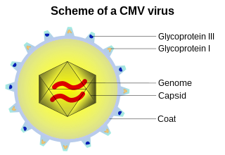
A capsid is the protein shell of a virus, enclosing its genetic material. It consists of several oligomeric (repeating) structural subunits made of protein called protomers. The observable 3-dimensional morphological subunits, which may or may not correspond to individual proteins, are called capsomeres. The proteins making up the capsid are called capsid proteins or viral coat proteins (VCP). The capsid and inner genome is called the nucleocapsid.

<span class="mw-page-title-main">Chaperone (protein)</span> Proteins assisting in protein folding

In molecular biology, molecular chaperones are proteins that assist the conformational folding or unfolding of large proteins or macromolecular protein complexes. There are a number of classes of molecular chaperones, all of which function to assist large proteins in proper protein folding during or after synthesis, and after partial denaturation. Chaperones are also involved in the translocation of proteins for proteolysis.

<span class="mw-page-title-main">Hsp90</span> Heat shock proteins with a molecular mass around 90kDa

Hsp90 is a chaperone protein that assists other proteins to fold properly, stabilizes proteins against heat stress, and aids in protein degradation. It also stabilizes a number of proteins required for tumor growth, which is why Hsp90 inhibitors are investigated as anti-cancer drugs.

DNA gyrase, or simply gyrase, is an enzyme within the class of topoisomerase and is a subclass of Type II topoisomerases that reduces topological strain in an ATP dependent manner while double-stranded DNA is being unwound by elongating RNA-polymerase or by helicase in front of the progressing replication fork. It is the only known enzyme to actively contribute negative supercoiling to DNA, while it also is capable of relaxing positive supercoils. It does so by looping the template to form a crossing, then cutting one of the double helices and passing the other through it before releasing the break, changing the linking number by two in each enzymatic step. This process occurs in bacteria, whose single circular DNA is cut by DNA gyrase and the two ends are then twisted around each other to form supercoils. Gyrase is also found in eukaryotic plastids: it has been found in the apicoplast of the malarial parasite Plasmodium falciparum and in chloroplasts of several plants. Bacterial DNA gyrase is the target of many antibiotics, including nalidixic acid, novobiocin, albicidin, and ciprofloxacin.

<span class="mw-page-title-main">GroEL</span> Protein-coding gene in the species Homo sapiens

GroEL is a protein which belongs to the chaperonin family of molecular chaperones, and is found in many bacteria. It is required for the proper folding of many proteins. To function properly, GroEL requires the lid-like cochaperonin protein complex GroES. In eukaryotes the organellar proteins Hsp60 and Hsp10 are structurally and functionally nearly identical to GroEL and GroES, respectively, due to their endosymbiotic origin.

<span class="mw-page-title-main">GroES</span> Protein-coding gene in the species Homo sapiens

Heat shock 10 kDa protein 1 (Hsp10), also known as chaperonin 10 (cpn10) or early-pregnancy factor (EPF), is a protein that in humans is encoded by the HSPE1 gene. The homolog in E. coli is GroES that is a chaperonin which usually works in conjunction with GroEL.

<span class="mw-page-title-main">Heat shock response</span> Type of cellular stress response

The heat shock response (HSR) is a cell stress response that increases the number of molecular chaperones to combat the negative effects on proteins caused by stressors such as increased temperatures, oxidative stress, and heavy metals. In a normal cell, proteostasis must be maintained because proteins are the main functional units of the cell. Many proteins take on a defined configuration in a process known as protein folding in order to perform their biological functions. If these structures are altered, critical processes could be affected, leading to cell damage or death. The heat shock response can be employed under stress to induce the expression of heat shock proteins (HSP), many of which are molecular chaperones, that help prevent or reverse protein misfolding and provide an environment for proper folding.

<span class="mw-page-title-main">Endopeptidase Clp</span>

Endopeptidase Clp (EC 3.4.21.92, endopeptidase Ti, caseinolytic protease, protease Ti, ATP-dependent Clp protease, ClpP, Clp protease). This enzyme catalyses the following chemical reaction

<span class="mw-page-title-main">DNA clamp</span>

A DNA clamp, also known as a sliding clamp, is a protein complex that serves as a processivity-promoting factor in DNA replication. As a critical component of the DNA polymerase III holoenzyme, the clamp protein binds DNA polymerase and prevents this enzyme from dissociating from the template DNA strand. The clamp-polymerase protein–protein interactions are stronger and more specific than the direct interactions between the polymerase and the template DNA strand; because one of the rate-limiting steps in the DNA synthesis reaction is the association of the polymerase with the DNA template, the presence of the sliding clamp dramatically increases the number of nucleotides that the polymerase can add to the growing strand per association event. The presence of the DNA clamp can increase the rate of DNA synthesis up to 1,000-fold compared with a nonprocessive polymerase.

<span class="mw-page-title-main">Type II topoisomerase</span>

Type II topoisomerases are topoisomerases that cut both strands of the DNA helix simultaneously in order to manage DNA tangles and supercoils. They use the hydrolysis of ATP, unlike Type I topoisomerase. In this process, these enzymes change the linking number of circular DNA by ±2. Topoisomerases are ubiquitous enzymes, found in all living organisms.

<span class="mw-page-title-main">Thermosome</span>

A thermosome is a group II chaperonin protein complex that functions in archaea. It is the homolog of eukaryotic CCT. This group II chaperonin is an ATP-dependent chaperonin that is responsible for folding or refolding of incipient or denatured proteins. A thermosome has two rings, each consisting of eight subunits, stacked together to form a cylindrical shape with a large cavity at the center. The thermosome is also defined by its heterooligomeric nature. The complex consists of two subunits that alternate location within its two rings.

Co-chaperones are proteins that assist chaperones in protein folding and other functions. Co-chaperones are the non-client binding molecules that assist in protein folding mediated by Hsp70 and Hsp90. They are particularly essential in stimulation of the ATPase activity of these chaperone proteins. There are a great number of different co-chaperones however based on their domain structure most of them fall into two groups: J-domain proteins and tetratricopeptide repeats (TPR).

<span class="mw-page-title-main">Prefoldin</span>

Prefoldin (GimC) is a superfamily of proteins used in protein folding complexes. It is classified as a heterohexameric molecular chaperone in both archaea and eukarya, including humans. A prefoldin molecule works as a transfer protein in conjunction with a molecule of chaperonin to form a chaperone complex and correctly fold other nascent proteins. One of prefoldin's main uses in eukarya is the formation of molecules of actin for use in the eukaryotic cytoskeleton.

<span class="mw-page-title-main">Arthur L. Horwich</span> American biologist (born 1951)

Arthur L. Horwich is an American biologist and Sterling Professor of Genetics and Pediatrics at the Yale School of Medicine. Horwich has also been a Howard Hughes Medical Institute investigator since 1990. His research into protein folding uncovered the action of chaperonins, protein complexes that assist the folding of other proteins; Horwich first published this work in 1989.

<span class="mw-page-title-main">CCT2 (gene)</span> Protein-coding gene in humans

T-complex protein 1 subunit beta is a protein that in humans is encoded by the CCT2 gene.

Franz-Ulrich Hartl is a German biochemist and the current Executive Director of the Max Planck Institute of Biochemistry. He is known for his pioneering work in chaperone-mediated protein folding.

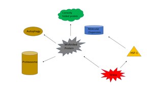
Proteostasis is the dynamic regulation of a balanced, functional proteome. The proteostasis network includes competing and integrated biological pathways within cells that control the biogenesis, folding, trafficking, and degradation of proteins present within and outside the cell. Loss of proteostasis is central to understanding the cause of diseases associated with excessive protein misfolding and degradation leading to loss-of-function phenotypes, as well as aggregation-associated degenerative disorders. Therapeutic restoration of proteostasis may treat or resolve these pathologies.

Chaperones, also called molecular chaperones, are proteins that assist other proteins in assuming their three-dimensional fold, which is necessary for protein function. However, the fold of a protein is sensitive to environmental conditions, such as temperature and pH, and thus chaperones are needed to keep proteins in their functional fold across various environmental conditions. Chaperones are an integral part of a cell's protein quality control network by assisting in protein folding and are ubiquitous across diverse biological taxa. Since protein folding, and therefore protein function, is susceptible to environmental conditions, chaperones could represent an important cellular aspect of biodiversity and environmental tolerance by organisms living in hazardous conditions. Chaperones also affect the evolution of proteins in general, as many proteins fundamentally require chaperones to fold or are naturally prone to misfolding, and therefore mitigates protein aggregation.

<span class="mw-page-title-main">GrpE</span> InterPro Family

GrpE is a bacterial nucleotide exchange factor that is important for regulation of protein folding machinery, as well as the heat shock response. It is a heat-inducible protein and during stress it prevents unfolded proteins from accumulating in the cytoplasm. Accumulation of unfolded proteins in the cytoplasm can lead to cell death.

<span class="mw-page-title-main">TRiC (complex)</span> Multiprotein complex used in cellular proteostasis

T-complex protein Ring Complex (TRiC), otherwise known as Chaperonin Containing TCP-1 (CCT), is a multiprotein complex and the chaperonin of eukaryotic cells. Like the bacterial GroEL, the TRiC complex aids in the folding of ~10% of the proteome, and actin and tubulin are some of its best known substrates. TRiC is an example of a biological machine that folds substrates within the central cavity of its barrel-like assembly using the energy from ATP hydrolysis.

References

  1. Braig K, Otwinowski Z, Hegde R, Boisvert DC, Joachimiak A, Horwich AL, Sigler PB (October 1994). "The crystal structure of the bacterial chaperonin GroEL at 2.8 A". Nature. 371 (6498): 578–86. Bibcode:1994Natur.371..578B. doi:10.1038/371578a0. PMID   7935790. S2CID   4341993.
  2. "Howard Hughes Investigators: Arthur L. Horwich, M.D." Archived from the original on 2019-07-26. Retrieved 2011-09-12.
  3. 1 2 3 4 5 6 Conway de Macario E, Yohda M, Macario AJ, Robb FT (2019-03-15). "Bridging human chaperonopathies and microbial chaperonins". Communications Biology. 2 (1): 103. doi:10.1038/s42003-019-0318-5. PMC   6420498 . PMID   30911678.
  4. 1 2 3 Ansari MY, Mande SC (2018). "A Glimpse Into the Structure and Function of Atypical Type I Chaperonins". Frontiers in Molecular Biosciences. 5: 31. doi: 10.3389/fmolb.2018.00031 . PMC   5904260 . PMID   29696145.
  5. 1 2 Willison, KR (5 October 2018). "The structure and evolution of eukaryotic chaperonin-containing TCP-1 and its mechanism that folds actin into a protein spring". The Biochemical Journal. 475 (19): 3009–3034. doi:10.1042/BCJ20170378. hdl: 10044/1/63924 . PMID   30291170. S2CID   52923821.
  6. Fenton WA, Horwich AL (May 2003). "Chaperonin-mediated protein folding: fate of substrate polypeptide". Quarterly Reviews of Biophysics. 36 (2): 229–56. doi:10.1017/S0033583503003883. PMID   14686103. S2CID   10328521.
  7. Hemmingsen SM, Woolford C, van der Vies SM, Tilly K, Dennis DT, Georgopoulos CP, et al. (May 1988). "Homologous plant and bacterial proteins chaperone oligomeric protein assembly". Nature. 333 (6171): 330–4. Bibcode:1988Natur.333..330H. doi:10.1038/333330a0. PMID   2897629. S2CID   4325057.
  8. Prasad TK, Stewart CR (March 1992). "cDNA clones encoding Arabidopsis thaliana and Zea mays mitochondrial chaperonin HSP60 and gene expression during seed germination and heat shock". Plant Molecular Biology. 18 (5): 873–85. doi:10.1007/BF00019202. PMID   1349837. S2CID   40768099.
  9. Schmidt A, Schiesswohl M, Völker U, Hecker M, Schumann W (June 1992). "Cloning, sequencing, mapping, and transcriptional analysis of the groESL operon from Bacillus subtilis". Journal of Bacteriology. 174 (12): 3993–9. doi:10.1128/jb.174.12.3993-3999.1992. PMC   206108 . PMID   1350777.
  10. 1 2 Hindersson P, Høiby N, Bangsborg J (January 1991). "Sequence analysis of the Legionella micdadei groELS operon". FEMS Microbiology Letters. 61 (1): 31–8. doi: 10.1111/j.1574-6968.1991.tb04317.x . PMID   1672279.
  11. Braig K, Otwinowski Z, Hegde R, Boisvert DC, Joachimiak A, Horwich AL, Sigler PB (October 1994). "The crystal structure of the bacterial chaperonin GroEL at 2.8 A". Nature. 371 (6498): 578–86. Bibcode:1994Natur.371..578B. doi:10.1038/371578a0. PMID   7935790. S2CID   4341993.
  12. Zang Y, Jin M, Wang H, Cui Z, Kong L, Liu C, Cong Y (December 2016). "Staggered ATP binding mechanism of eukaryotic chaperonin TRiC (CCT) revealed through high-resolution cryo-EM". Nature Structural & Molecular Biology. Springer Science and Business Media LLC. 23 (12): 1083–1091. doi:10.1038/nsmb.3309. PMID   27775711. S2CID   12001964.
  13. Kusmierczyk AR, Martin J (May 2003). "Nucleotide-dependent protein folding in the type II chaperonin from the mesophilic archaeon Methanococcus maripaludis". The Biochemical Journal. 371 (Pt 3): 669–73. doi:10.1042/BJ20030230. PMC   1223359 . PMID   12628000.
  14. Bracher A, Paul SS, Wang H, Wischnewski N, Hartl FU, Hayer-Hartl M (27 April 2020). "Structure and conformational cycle of a bacteriophage-encoded chaperonin". PLOS ONE. 15 (4): e0230090. Bibcode:2020PLoSO..1530090B. doi: 10.1371/journal.pone.0230090 . PMC   7185714 . PMID   32339190.
  15. Hartl FU, Hayer-Hartl M (June 2009). "Converging concepts of protein folding in vitro and in vivo". Nature Structural & Molecular Biology. 16 (6): 574–81. doi:10.1038/nsmb.1591. PMID   19491934. S2CID   205522841.
  16. Apetri AC, Horwich AL (November 2008). "Chaperonin chamber accelerates protein folding through passive action of preventing aggregation". Proceedings of the National Academy of Sciences of the United States of America. 105 (45): 17351–5. Bibcode:2008PNAS..10517351A. doi: 10.1073/pnas.0809794105 . PMC   2579888 . PMID   18987317.
  17. 1 2 Kmiecik S, Kolinski A (July 2011). "Simulation of chaperonin effect on protein folding: a shift from nucleation-condensation to framework mechanism". Journal of the American Chemical Society. 133 (26): 10283–9. doi:10.1021/ja203275f. PMC   3132998 . PMID   21618995.
  18. Chakraborty K, Chatila M, Sinha J, Shi Q, Poschner BC, Sikor M, et al. (July 2010). "Chaperonin-catalyzed rescue of kinetically trapped states in protein folding". Cell. 142 (1): 112–22. doi: 10.1016/j.cell.2010.05.027 . PMID   20603018. S2CID   3859016.
  19. 1 2 Todd MJ, Lorimer GH, Thirumalai D (April 1996). "Chaperonin-facilitated protein folding: optimization of rate and yield by an iterative annealing mechanism". Proceedings of the National Academy of Sciences of the United States of America. 93 (9): 4030–5. Bibcode:1996PNAS...93.4030T. doi: 10.1073/pnas.93.9.4030 . PMC   39481 . PMID   8633011.
  20. Snustad DP (August 1968). "Dominance interactions in Escherichia coli cells mixedly infected with bacteriophage T4D wild-type and amber mutants and their possible implications as to type of gene-product function: catalytic vs. stoichiometric". Virology. 35 (4): 550–63. doi:10.1016/0042-6822(68)90285-7. PMID   4878023.
  21. 1 2 Marusich EI, Kurochkina LP, Mesyanzhinov VV (April 1998). "Chaperones in bacteriophage T4 assembly". Biochemistry (Moscow). 63 (4): 399–406. PMID   9556522.
  22. Bukau B, Horwich AL (February 1998). "The Hsp70 and Hsp60 chaperone machines". Cell. 92 (3): 351–66. doi: 10.1016/S0092-8674(00)80928-9 . PMID   9476895. S2CID   16526409.
  23. Gor D, Mayfield JE (February 1992). "Cloning and nucleotide sequence of the Brucella abortus groE operon". Biochimica et Biophysica Acta (BBA) - Gene Structure and Expression. 1130 (1): 120–2. doi:10.1016/0167-4781(92)90476-g. PMID   1347461.
This article incorporates text from the public domain Pfam and InterPro: IPR002423