Development of the reproductive system

Last updated

The development of the reproductive system is the part of embryonic growth that results in the sex organs and contributes to sexual differentiation. Due to its large overlap with development of the urinary system, the two systems are typically described together as the genitourinary system.

Contents

The reproductive organs develop from the intermediate mesoderm and are preceded by more primitive structures that are superseded before birth. These embryonic structures are the mesonephric ducts (also known as Wolffian ducts) and the paramesonephric ducts, (also known as Müllerian ducts). The mesonephric duct gives rise to the male seminal vesicles, epididymides and vasa deferentia. The paramesonephric duct gives rise to the female fallopian tubes, uterus, cervix, and upper part of the vagina. [1]

Mesonephric ducts

The mesonephric duct originates from a part of the pronephric duct.

Origin

In the outer part of the intermediate mesoderm, immediately under the ectoderm, in the region from the fifth cervical segment to the third thoracic segment, a series of short evaginations from each segment grows dorsally and extends caudally, fusing successively from before backward to form the pronephric duct. This continues to grow caudally until it opens into the ventral part of the cloaca; beyond the pronephros it is termed the mesonephric duct. Thus, the mesonephric duct remains after the atrophy of the pronephros duct.

Development in male

In the male, the duct persists, and forms the tube of the epididymis, the vas deferens and the ejaculatory duct, while the seminal vesicle arises during the third month as a lateral diverticulum from its hinder end. A large part of the head end of the mesonephros atrophies and disappears; of the remainder the anterior tubules form the efferent ducts of the testicle; while the posterior tubules are represented by the ductuli aberrantes, and by the paradidymis, which is sometimes found in front of the spermatic cord above the head of the epididymis.

Atrophy in female

In the female, the mesonephric bodies and ducts atrophy. The nonfunctional remains of the mesonephric tubules are represented by the epoophoron, and the paroöphoron, two small collections of rudimentary blind tubules which are situated in the mesosalpinx.

Remnants

Section of the fold in the mesonephros of a chick embryo of the fourth day Gray1106.png
Section of the fold in the mesonephros of a chick embryo of the fourth day

The lower part of the mesonephric duct disappears, while the upper part persists as the longitudinal duct of the epoöphoron, called Gartner's duct.

There are also developments of other tissues from the mesonephric duct that persist, e.g. the development of the suspensory ligament of the ovary.

Paramesonephric ducts

A.--Diagram of the primitive urogenital organs in the embryo previous to sexual distinction. The common genital cord is labeled with gc. * 3. Ureter * 4. Urinary bladder * 5. Urachus * cl. Cloaca * cp. Elevation which becomes clitoris or penis * i. Lower part of the intestine * ls. Fold of integument from which the labia majora or scrotum are formed * m, m. Right and left paramesonephric ducts uniting together and running with the mesonephric ducts in gc, the common genital cord * ot. The gonadal ridge from which either the ovary or testis is formed * ug. Sinus urogenitalis * W. Left Wolffian body * w, w. Right and left mesonephric ducts. Gray1110-1.png
A.—Diagram of the primitive urogenital organs in the embryo previous to sexual distinction. The common genital cord is labeled with gc. * 3. Ureter * 4. Urinary bladder * 5. Urachus * cl. Cloaca * cp. Elevation which becomes clitoris or penis * i. Lower part of the intestine * ls. Fold of integument from which the labia majora or scrotum are formed * m, m. Right and left paramesonephric ducts uniting together and running with the mesonephric ducts in gc, the common genital cord * ot. The gonadal ridge from which either the ovary or testis is formed * ug. Sinus urogenitalis * W. Left Wolffian body * w, w. Right and left mesonephric ducts.

Shortly after the formation of the mesonephric ducts, a second pair of ducts is developed; these are the paramesonephric ducts. Each arises on the lateral aspect of the corresponding mesonephric duct as a tubular invagination of the cells lining the abdominal cavity. The orifice of the invagination remains open, and undergoes enlargement and modification to form the distal tubal opening (abdominal ostium) of the fallopian tube. The ducts pass backward lateral to the mesonephric ducts, but toward the posterior end of the embryo they cross to the medial side of these ducts, and thus come to lie side by side between and behind the latter—the four ducts forming what is termed the common genital cord[ citation needed ], to distinguish it from the genital cords of the germinal epithelium[ citation needed ] seen later in this article. The mesonephric ducts end in an epithelial elevation, the sinus tubercle, on the ventral part of the cloaca between the orifices of the mesonephric ducts. At a later stage the sinus tubercle opens in the middle, connecting the paramesonephric ducts with the cloaca.

Atrophy in males

In the male, the paramesonephric ducts atrophy (but traces of their anterior ends are represented by the appendix of testis of the male), while their terminal fused portions form the prostatic utricle in the floor of the prostatic urethra. This is due to the production of anti-Müllerian hormone by the Sertoli cells of the testes.

Development in females

In the female, the paramesonephric ducts persist and undergo further development. The portions which lie in the genital cord[ citation needed ] fuse to form the uterus and vagina. This fusion of the paramesonephric ducts begins in the third month, and the septum formed by their fused medial walls disappears from below upward.

The parts outside this cord remain separate, and each forms the corresponding fallopian tube. The ostium of the fallopian tube remains from the anterior extremity of the original tubular invagination from the abdominal cavity.

About the fifth month a ring-like constriction marks the position of the cervix of the uterus, and after the sixth month the walls of the uterus begin to thicken. For a time the vagina is represented by a solid rod of epithelial cells. A ring-like outgrowth of this epithelium occurs at the lower end of the uterus and marks the future vaginal fornix. At about the fifth or sixth month the lumen of the vagina is produced by the breaking down of the central cells of the epithelium. The hymen represents the remains of the sinus tubercle . [2]

Gonads

The gonads are the precursors of the testicles in males and ovaries in females. They initially develop from the mesothelial layer of the peritoneum.

Ovaries

The ovary is differentiated into a central part, the medulla of ovary, covered by a surface layer, the germinal epithelium. The immature ova originate from cells from the dorsal endoderm of the yolk sac. Once they have reached the gonadal ridge they are called oogonia. Development proceeds and the oogonia become fully surrounded by a layer of connective tissue cells (pre-granulosa cells). In this way, the rudiments of the ovarian follicles are formed. The embryological origin of granulosa cells, on the other hand, remains controversial. Just as in the male, there is a gubernaculum in the female, which pulls it downward, albeit not as much as in males. The gubernaculum later becomes the proper ovarian ligament and the round ligament of the uterus.

Testicles

The periphery of the testicles are converted into the tunica albuginea. Cords of the central mass run together and form a network which becomes the rete testis, and another network, which develops the seminiferous tubules. Via the rete testis, the seminiferous tubules become connected with outgrowths from the mesonephros, which form the efferent ducts of the testicle.

In short, the descent of the testicles consists of the opening of a connection from the testicle to its final location at the anterior abdominal wall, followed by the development of the gubernaculum, which subsequently pulls and translocates the testicle down into the developing scrotum. Ultimately, the passageway closes behind the testicle. A failure in this process can cause indirect inguinal hernia or an infantile hydrocoele.

Division of cloaca

Tail end of human embryo thirty-two to thirty-three days old. The endodermal cloaca is visible at center left, labeled in green. Gray1116.png
Tail end of human embryo thirty-two to thirty-three days old. The endodermal cloaca is visible at center left, labeled in green.

After the separation of the rectum from the dorsal part of the cloaca, the ventral part becomes the primary urogenital sinus. [3] The urogenital sinus, in turn, divides into the superficial definitive urogenital sinus and the deeper anterior vesico-urethral portion.

Definitive urogenital sinus

The definitive urogenital sinus consists of a caudal cephalic portion and an intermediate narrow channel, the pelvic portion.

Vesico-urethral portion

The vesico-urethral portion is the deepest portion, continuous with the allantois. It absorbs the ends of the mesonephric ducts and the associated ends of the renal diverticula, and these give rise to the trigone of urinary bladder and part of the prostatic urethra. The remainder of the vesico-urethral portion forms the body of the bladder and part of the prostatic urethra; its apex is prolonged to the umbilicus as a narrow canal, the urachus, which later is obliterated and becomes the median umbilical ligament of the adult.

Prostate

The prostate originally consists of two separate portions, each of which arises as a series of diverticular buds from the epithelial lining of the urogenital sinus and vesico-urethral part of the cloaca, between the third and fourth months. These buds become tubular, and form the glandular substance of the two lobes, which ultimately meet and fuse behind the urethra and also extend on to its ventral aspect. The median lobe of the prostate is formed as an extension of the lateral lobes between the common ejaculatory ducts and the bladder.

Skene's glands in the female urethra are regarded as the homologues of the prostatic glands.

The bulbourethral glands in the male, and Bartholin's gland in the female, also arise as diverticula from the epithelial lining of the urogenital sinus.

External genitalia

Human genitalia - development 1.png

Until about the ninth week of gestational age, [4] the external genitalia of human males and females look the same, and follow a common development. This includes the development of a genital tubercle and a membrane dorsally to it, covering the developing urogenital opening, and the development of the labioscrotal fold.

In mammals, the urogenital folds become the penile raphe or ventral aspect of the penis in the male and the labia minora (primates including humans)/labia vulvae (non-primates) in the female. [5] [6] [7]

Even after differentiation can be seen between the sexes, some stages are common, e.g. the disappearing of the membrane. On the other hand, sex-dependent development include further protrusion of the genital tubercle in the male to form the glans and shaft of the penis and in the female, the glans and body of the clitoris. [8] [9] The labioscrotal swellings evolve into the scrotum in males, and into the labia majora (dissipates in non-primates) in females. [8] [10]

Common development

Before differentiation

Urogenital membrane

There is initially a cloacal membrane, composed of ectoderm and endoderm, reaching from the umbilical cord to the tail, separating the cloaca from the exterior. After the separation of the rectum from the dorsal part of the cloaca, the ventral part of the cloacal membrane becomes the urogenital membrane.

Genital tubercle

Mesoderm extends to the midventral line for some distance behind the umbilical cord, and forms the lower part of the abdominal wall; it ends below in a prominent swelling, the cloacal tubercle, which after the separation of the rectum becomes the genital tubercle. Dorsally, to this tubercle the sides are not really fused. Rather, the urogenital part of the cloacal membrane separates the ingrowing sheets of mesoderm.

Phallus

The genital tubercle develops into the primordial phallus, the first rudiment of the penis or clitoris. [8]

The terminal part of the phallus, representing the future glans becomes solid. The remainder of the phallus, which remains hollow, is converted into a longitudinal groove by the absorption of the urogenital membrane.

The term genital tubercle still remains but only refers to the future glans [4]

Urogenital opening

In both sexes, the phallic portion of the urogenital sinus extends on to the under surface of the cloacal tubercle as far forward as the apex. At the apex, the walls of the phallic portion come together and fuse, obliterating the urogenital opening. Instead, a solid plate, the urethral plate, is formed. The remainder of the phallic portion is for a time tubular, and then, by the absorption of the urogenital membrane, it establishes a communication with the exterior. This opening is for a while the primitive urogenital opening, and it extends forward to the corona glandis.

After differentiation

Development of external genitalia. A: common development. C, E: male development. B, D, F: female development Development of external sexual organs in the male and female Gray1119.png
Development of external genitalia. A: common development. C, E: male development. B, D, F: female development

The following developments occur in both males and females, although a difference in the development between the sexes already can be seen:

  • The corpus cavernosum penis, and the corpus cavernosum of clitoris, and the corpus spongiosum penis arise from the mesodermal tissue in the phallus; they are at first dense structures, but later vascular spaces appear in them, and they gradually become cavernous. [8]
  • The prepuce in both sexes is formed by the growth of a solid plate of ectoderm into the superficial part of the phallus; on coronal section this plate presents the shape of a horseshoe. By the breaking down of its more centrally situated cells, the plate is split into two lamellæ. Thus, a cutaneous fold, the prepuce, is liberated and forms a hood over the glans.

Female

In the female, a deep groove forms around the phallus. The sides of it grow dorsalward as the labioscrotal folds, which ultimately form the labia majora in females. The labia minora, in contrast, arise by the continued growth of the lips of the groove on the under surface of the phallus; the remainder of the phallus forms the clitoral glans. [8]

Male

The labioscrotal folds extend around between the pelvic portion and the anus, and form a scrotal area. During the changes associated with the descent of the testes, this scrotal area is drawn out to form the scrotal sacs. The penis is developed from the phallus.

As in the female, the urogenital membrane undergoes absorption, forming a channel on the under surface of the phallus; this channel extends only as far forward as the corona glandis.

Urogenital opening

Later, this opening, which is located on the dorsal side of the penis, [11] closes from behind forward. Meanwhile, the urethral plate of the glans breaks down centrally to form a median groove continuous with the primitive ostium. This groove also closes from behind forward, leaving only a small pipe running in the middle of the penis. Thus, the urogenital opening is shifted forward to the end of the glans.

Diagram of internal differentiation

Diagrams to show the development of male and female generative organs from a common type Gray1110.png
Diagrams to show the development of male and female generative organs from a common type

A.—Diagram of the primitive urogenital organs in the embryo previous to sexual distinction.

B.—Diagram of the female type of sexual organs.

C.—Diagram of the male type of sexual organs.

Related Research Articles

<span class="mw-page-title-main">Labia majora</span> Folds of skin which contain and protect the vulvas other parts

In primates, and specifically in humans, the labia majora, also known as the outer lips or outer labia, are two prominent longitudinal skin folds that extend downward and backward from the mons pubis to the perineum. Together with the labia minora, they form the labia of the vulva.

<span class="mw-page-title-main">Sex organ</span> Biological part involved in sexual reproduction

A sex organ, also known as a reproductive organ, is a part of an organism that is involved in sexual reproduction. Sex organs constitute the primary sex characteristics of an organism. Sex organs are responsible for producing and transporting gametes, as well as facilitating fertilization and supporting the development and birth of offspring. Sex organs are found in many species of animals and plants, with their features varying depending on the species.

<span class="mw-page-title-main">Mesonephric duct</span> Paired organ in mammals

The mesonephric duct, also known as the Wolffian duct, archinephric duct, Leydig's duct or nephric duct, is a paired organ that develops in the early stages of embryonic development in humans and other mammals. It is an important structure that plays a critical role in the formation of male reproductive organs. The duct is named after Caspar Friedrich Wolff, a German physiologist and embryologist who first described it in 1759.

<span class="mw-page-title-main">Urogenital sinus</span> A part of the human body only present in the development of the urinary and reproductive organs

The urogenital sinus is a part of the human body only present in the development of the urinary and reproductive organs. It is the ventral part of the cloaca, formed after the cloaca separates from the anal canal during the fourth to seventh weeks of development.

<span class="mw-page-title-main">Corpus spongiosum (penis)</span> Spongy tissue within the human penis

The corpus spongiosum is the mass of spongy tissue surrounding the male urethra within the penis. It is also called the corpus cavernosum urethrae in older texts.

Gender-affirming surgery for male-to-female transgender women or transfeminine non-binary people describes a variety of surgical procedures that alter the body to provide physical traits more comfortable and affirming to an individual's gender identity and overall functioning.

<span class="mw-page-title-main">Paramesonephric duct</span> Paired ducts in the embryo in the primitive urogenital structures

The paramesonephric ducts are paired ducts of the embryo in the female reproductive system that run down the lateral sides of the genital ridge and terminate at the sinus tubercle in the primitive urogenital sinus. In the female, they will develop to form the fallopian tubes, uterus, cervix, and the upper one-third of the vagina.

<span class="mw-page-title-main">Genital tubercle</span> Body of tissue present in the development of the reproductive system

A genital tubercle or phallic tubercle is a body of tissue present in the development of the reproductive system. It forms in the ventral, caudal region of mammalian embryos of both sexes, and eventually develops into a primordial phallus. In the human fetus, the genital tubercle develops around week 4 of gestation, and by week 9 becomes recognizably either a clitoris or penis. This should not be confused with the sinus tubercle which is a proliferation of endoderm induced by paramesonephric ducts. Even after the phallus is developed, the term genital tubercle remains, but only as the terminal end of it, which develops into either the glans penis or the glans clitoridis.

The development of the urinary system begins during prenatal development, and relates to the development of the urogenital system – both the organs of the urinary system and the sex organs of the reproductive system. The development continues as a part of sexual differentiation.

<span class="mw-page-title-main">Male reproductive system</span> Reproductive system of the human male

The male reproductive system consists of a number of sex organs that play a role in the process of human reproduction. These organs are located on the outside of the body, and within the pelvis.

<span class="mw-page-title-main">Sexual differentiation in humans</span> Process of development of sex differences in humans

Sexual differentiation in humans is the process of development of sex differences in humans. It is defined as the development of phenotypic structures consequent to the action of hormones produced following gonadal determination. Sexual differentiation includes development of different genitalia and the internal genital tracts and body hair plays a role in sex identification.

Pseudohermaphroditism is a condition in which an individual has a matching chromosomal and gonadal tissue sex, but mismatching external genitalia.

<span class="mw-page-title-main">Urinary meatus</span> Orifice of the urethra

The urinary meatus, also known as the external urethral orifice, is the opening of the urethra. It is the point where urine exits the urethra in both sexes and where semen exits the urethra in males. The meatus has varying degrees of sensitivity to touch.

<span class="mw-page-title-main">Human penis</span> Human male external reproductive organ

In human anatomy, the penis is an external male sex organ that additionally serves as the urinary duct. The main parts are the root, body, the epithelium of the penis including the shaft skin, and the foreskin covering the glans. The body of the penis is made up of three columns of tissue: two corpora cavernosa on the dorsal side and corpus spongiosum between them on the ventral side. The human male urethra passes through the prostate gland, where it is joined by the ejaculatory duct, and then through the penis. The urethra traverses the corpus spongiosum, and its opening, the meatus, lies on the tip of the glans. It is a passage both for urination and ejaculation of semen.

The reproductive system of an organism, also known as the genital system, is the biological system made up of all the anatomical organs involved in sexual reproduction. Many non-living substances such as fluids, hormones, and pheromones are also important accessories to the reproductive system. Unlike most organ systems, the sexes of differentiated species often have significant differences. These differences allow for a combination of genetic material between two individuals, which allows for the possibility of greater genetic fitness of the offspring.

<span class="mw-page-title-main">Diphallia</span> Genital medical condition

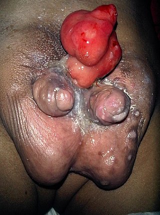
Diphallia, penile duplication (PD), diphallic terata, or diphallasparatus, is an extremely rare developmental abnormality in which a male is born with two penises. The first reported case was by Johannes Jacob Wecker in 1609. Its occurrence is 1 in 5.5 million boys in the United States.

<span class="mw-page-title-main">Sinus tubercle</span> Proliferation of endoderm induced by the paramesonephric ducts

Sinus tubercle is the proliferation of endoderm induced by the paramesonephric ducts. It is located in the developing fetus between the orifices of the mesonephric ducts on the urogenital sinus. The uterovaginal primoridium, which is a fusion of the caudal ends of paramesonephric ducts, contacts the dorsal wall of the urogenital sinus and, induces the formation of the sinus tubercle. This occurs in both sexes:

<span class="mw-page-title-main">Prader scale</span> Rating system for the degree of virilization of human genitalia

The Prader scale or Prader staging, named after Andrea Prader, is a coarse rating system for the measurement of the degree of virilization of the genitalia of the human body and is similar to the Quigley scale. It primarily relates to virilization of the female genitalia in cases of congenital adrenal hyperplasia (CAH) and identifies five distinct stages, but in recent times has been used to describe the range of differentiation of genitalia, with normal infant presentation being shown on either end of the scale, female on the left (0) and male on the right (6).

<span class="mw-page-title-main">Septum glandis</span> Part of the human glans penis

The septum glandis, also septum of the glans, refers to the fibrous partition of the ventral aspect of the glans penis that separates the two glans wings in the ventral midline. The septum extends from the urethral meatus through the glanular urethra and ends in the tunica albuginea of the human penis. Externally it is attached to the frenulum which extends lower on the neck of the penis.

References

In-line

  1. Carlson, Neil R. (2013). Physiology of behavior (11th ed.). Boston: Pearson. p.  329. ISBN   978-0205239399.
  2. 1918 Gray's Anatomy
  3. "Differentiation of the urogenital sinus in males". Embryology.ch. Archived from the original on 2018-02-15. Retrieved 2016-01-19.
  4. 1 2 "Embryo Images Online". Med.unc.edu. Retrieved 2016-01-19.
  5. Hodges, Frederick Mansfield S.; Denniston, George C.; Milos, Marilyn Fayre (2007). Male and Female Circumcision: Medical, Legal, and Ethical Considerations in Pediatric Practice. Springer US. p. 10. ISBN   978-0-58539-937-9 . Retrieved November 24, 2023.
  6. Martin, Richard J.; Fanaroff, Avory A.; Walsh, Michele C. (2014). Fanaroff and Martin's Neonatal-Perinatal Medicine E-Book: Diseases of the Fetus and Infant. Elsevier Health Sciences. p. 1522. ISBN   978-0-32329-537-6 . Retrieved November 24, 2023.
  7. McEntee, Mark (2012). Reproductive Pathology of Domestic Mammals. Elsevier Science. p. 192. ISBN   978-0-32313-804-8 . Retrieved November 19, 2023.
  8. 1 2 3 4 5 Keith L. Moore, T. V. N. Persaud, Mark G. Torchia, The Developing Human: Clinically Oriented Embryology 10th Ed. Elsevier Health Sciences, 2015 ISBN   978-0323313483, pp. 267–269
  9. Netter, Frank (2022). Netter Atlas of Human Anatomy: Classic Regional Approach - Ebook. Elsevier Health Sciences. p. 194. ISBN   978-0-32379-375-9 . Retrieved October 9, 2023.
  10. Kumar, M.S.A. (2015). Clinically Oriented Anatomy of the Dog & Cat. Linus Learning. p. 1254. ISBN   978-1-60797-552-6.
  11. "Embryo Images Online". Med.unc.edu. Retrieved 2016-01-19.

General