Fibromuscular dysplasia

Last updated
Fibromuscular dysplasia
Fibr.jpg
The "string-of-beads" feature in multi-focal fibromuscular dysplasia. The sign is caused by areas of relative stenoses alternating with small aneurysms.
Specialty Cardiology   OOjs UI icon edit-ltr-progressive.svg

Fibromuscular dysplasia (FMD) is a non-atherosclerotic, non-inflammatory disease of the blood vessels that causes abnormal growth within the wall of an artery. [1] FMD has been found in nearly every arterial bed in the body although the most common arteries affected are the renal and carotid arteries. [1] [2] [3]

Contents

There are various types of FMD, with multi-focal fibroplasia being the most common. Further, less common, forms of the disease include focal (previously known as intimal) and adventitial fibroplasia. [1] [2] [3] [4] FMD predominantly affects middle-aged women, but has been found in men and people of all ages. [1] Pediatric cases of FMD are vastly different from that of the adult population, and poorly studied. The prevalence of FMD is not known and, although the disease was initially thought to be rare, some studies have suggested that it may be underdiagnosed. [5]

Signs and symptoms

The symptoms expressed by FMD patients are largely dependent on the vascular bed(s) affected by the disease. Patients may also be entirely asymptomatic and have FMD discovered incidentally (e.g., when imaging studies are performed for other reasons). In a study from the United States Registry for Fibromuscular Dysplasia, the median age at first symptom was roughly 47 years. [2]

Renal arteries

The main symptoms associated with renal FMD are secondary hypertension and bruits that can be heard with a stethoscope over the abdomen or flanks. Complications such as aneurysms, dissections, or occlusion of the renal artery have been associated with renal artery FMD. [4]

Cerebrovascular regions

The carotid and vertebral arteries are most commonly affected. Middle and distal regions of the internal carotid arteries are frequently involved. [1] Patients with FMD in the carotid arteries typically present around 50 years of age. [3] Symptoms of craniocervical involvement include headaches (mostly migraine), pulsatile tinnitus, dizziness, and neck pain, although patients are often asymptomatic. On physical examination, one may detect neurological symptoms secondary to a stroke or transient ischemic attack (TIA), a bruit over an affected artery, and diminished distal pulses. Complications of cerebrovascular FMD include TIA, ischemic stroke, Horner syndrome, or subarachnoid hemorrhage. [1] [2] [3]

Other sites

Patients with mesenteric, or intestinal FMD, may experience abdominal pain after eating or weight loss. FMD within the extremities may cause claudication or may be detectable by bruits. [1] If the lower limb arteries are affected, the patient may present with cold legs or evidence of distal embolic disease. FMD present in the subclavian artery may cause arm weakness, paresthesia, claudication, and subclavial steal syndrome. [6]

Children

Children with FMD often report various non-specific symptoms or present with hypertension during routine physical examinations. Symptoms are commonly associated with the artery being affected. Symptoms may include headaches, insomnia, fatigue and chest or abdominal pain. FMD affecting the arteries of the head & neck is commonly recognized as a cause of childhood strokes. [7]

Detection may stem from a bruit being present over the affected vascular bed during a physical assessment. Yet, absence of a bruit does not rule out significant vascular disease. [8]

In children, renovascular disease accounts for approximately 10% of all causes of secondary hypertension. [8] Kidney failure is a common presentation in infants and children but is uncommon in adults, although it is occasionally the presenting problem in adults with focal disease. [3] “The presentation in infants and children younger than 4 years is especially likely to resemble vacuities.” [3]

The vascular subtype of Ehlers-Danlos Syndrome (type IV) has been associated with multi-focal FMD. This syndrome should be suspected in patients with multiple aneurysms and/or tears (dissections) in arteries in addition to the typical angiographic findings of FMD. There have been isolated reports of FMD associated with other disorders, including Alport syndrome, pheochromocytoma, Marfan syndrome, Moyamoya disease, and Takayasu's arteritis. [9]

Cause

While the cause of FMD remains unclear, current theory suggests that there may be a genetic predisposition as case reports have identified clusters of the disease and prevalence among twins. [7] In fact, according to the Cleveland Clinic approximately 10% of cases appear to be inherited and often coexists with other genetic abnormalities that affect the blood vessels. Approximately 10% of patients with FMD have an affected family member. [1] A study conducted from the patient registry at Michigan Cardiovascular Outcomes Research and Reporting Program (MCORRP) at the University of Michigan Health System reported a high prevalence of a family history of stroke (53.5%), aneurysm (23.5%), and sudden death (19.8%). [2] Even though FMD is a non-atherosclerotic disease family histories of hypertension and hyperlipidemia were also common among those diagnosed with FMD. It is believed that the cause of FMD is not a single identifier such as genetics but has multiple underlying factors. Theories of hormonal influence, mechanical stress from trauma and stress to the artery walls, and also the effect of loss of oxygen supply to the blood vessel wall caused by fibrous lesions. [7] It has been suggested that environmental factors, such as smoking and estrogen, may play a role in addition to genetic factors, however concerns for safety associated with exogenous female hormones in FMD remain theorectical. [10]

Pathophysiology

FMD can be found in almost every artery in the human body, but most often affects the carotid, vertebral, renal arteries and even those that supply the intestines, arms, and legs. [1] Patients may present with FMD in multiple vessels. FMD has been pathologically categorized into three types of classifications: Multi-focal, focal, and adventitial; referring to the particular layer of arterial wall being affected. [4]

Focal

In pediatrics, the most prevalent form of FMD is focal fibroplasia. Focal fibroplasia is described as long, narrow, irregular or smooth focal stenosis and can occur in any arterial bed. Whilst it is the most common type amongst children, it only accounts for approximately 10% of FMD cases overall. [8] It most often presents with ischemic symptoms, and frequently mistaken for Takayasu arteritis. [11] (previously known as intimal)

Multi-focal

The second type, multi-focal fibroplasia, involves thickening of the media and collagen formation. It is typically reported as having the appearance of a ‘string of beads’ on angiographic review. [4] "The 'bead' component is often larger than the normal arterial lumen, and in a subset of patients with FMD, aneurysms are present that may require treatment." [4] The multi-focal subtype of FMD accounts for nearly 80% to 90% of all FMD cases. [4] (previously known as medial)

Adventitial

The third classification is adventitial fibroplasia, in which collagen replaces the fibrous adventitia and extends beyond the artery. This form is considered rare but angiographic appearance may look similar to the focal subtype of FMD, making the distinction difficult.[ citation needed ]

Diagnosis

It is the lack of specific symptoms and their potential to appear anywhere that makes FMD a challenge to detect early on. The most accurate diagnosis comes from combining clinical presentation and angiographic imaging. According to the Michigan Cardiovascular Outcomes Research and Reporting Program (MCORRP, 2013) the length of time from a patient’s first signs or symptoms to diagnosis is commonly 5 years.[ citation needed ]

FMD is currently diagnosed through the use of both invasive and non-invasive tests. [10] Non-invasive testing includes duplex ultrasonography, magnetic resonance angiography (MRA), and computed tomography angiography (CTA). [4] Invasive testing through angiography is the gold standard. However, due to the higher risk of complications, this is typically not done early on. Occasionally, FMD is diagnosed asymptomatically after an unrelated x-ray presents the classic ‘string of beads’ appearance of the arteries, or when a practitioner investigates an unexpected bruit found during an exam. When a diagnosis of FMD is considered for a patient thorough medical history, family history, as well as vascular examination, should be completed.[ citation needed ]

A definitive diagnosis of FMD can only be made with imaging studies. Catheter-based angiography (with contrast) has proven to be the most accurate imaging technique: this test involves a catheter is inserted into a large artery and advanced until it reaches the vessel of question. [1] The catheter allows practitioners to view and measure the pressure of the artery aiding in the categorization and severity of the FMD diseased artery. According to Olin, “catheter-based angiography is the only imaging modality that can accurately identify the changes of FMD, aneurysm formation, and dissection in the branch vessels.” Practitioners believe it is important to utilize IVUS imaging because stenosis can sometimes only be detected through the methods of pressure gradient or IVUS imaging. [4] In addition, computed tomography angiography and magnetic resonance angiography are commonly used to evaluate arteries in the brain. Doppler ultrasound may be used in both the diagnosis and follow-up of FMD. [1]

Similar diseases

Children

The differentiating presentations are suggestive of FMD being a unique syndrome in respect to the pediatric population. Experienced FMD clinicians warn against relying on the “string of beads” angiography for a diagnosis. In fact, it is suggested that FMD may be both under and over-diagnosed in children with stroke. [7]

Treatment

There is no known cure for FMD. However, treatment focuses on relieving associated symptoms. Medical management is the most common form of treatment. The best approach to medically managing these patients is constantly being re-evaluated as more information is learned about the disease. [1]

Kidney treatment

Blood pressure control is the primary concern when treating patients with renal FMD since the ideal blood pressure target in patients with FMD is unknown. [10] In cases of renal artery stenosis and indications for intervention, percutaneous balloon angioplasty may be recommended. Many studies have assessed the success rate of percutaneous transluminal angioplasty (PTA) in these cases, and have found relief of hypertensive symptoms. [3] [12] Duplex ultrasonography should be performed soon after this procedure to ensure adequate renal velocities. [3]

Stents have a restenosis rate of 10-20%, and may make surgical revascularization more difficult. Surgical revascularization may be necessary if aneurysms develop within the affected artery or if PTA does not resolve the issue.[ citation needed ]

Ex vivo renal artery reconstruction is sometimes used for complex diseases where branches of the renal artery are affected. [13]

Cerebrovascular treatment

Patients with carotid or vertebral FMD should be medically managed to reduce the risk of a stroke. Aspirin 81 mg is typically prescribed for patients with carotid FMD. Antiplatelets and anticoagulants may be used to reduce the risk of blood clot formation. If a TIA or stroke are to occur, percutaneous angioplasty and antiplatelet therapy may be necessary. [3] Pulsatile tinnitus is recognized as a common manifestation in 32% of US cerebrovascular FMD patients and sound or cognitive behavioral therapy may be helpful for some patients with more severe symptoms. [10]

Treatment for FMD in other regions

Little information is known regarding the best treatment for FMD outside of the renal and extracranial regions. If claudication or limb ischemia is consequent to FMD in the extremities, angioplasty may be implemented.[ citation needed ]

Children

Pediatric FMD medical and surgical treatments or interventions are available. Treatment is determined by factors such as age and disease location but routinely involve controlling hypertension, re-establishing vascular flow, clot prevention, and improving lifestyle such as diet, exercise and smoking cessation. Medical therapy for pediatric population may involve the use of angiotensin-converting enzyme inhibitor (ACE inhibitors) and/or angiotensin II receptor blockers, multiple anti-hypertensive medications, diuretics, calcium channel blockers, and beta-blockers. Prevention of thrombosis of affected arteries may be taken through administration of an antiplatelet medication such as aspirin. [1]

Percutaneous transluminal renal angioplasty (PTRA) remains the gold standard for renal-artery FMD. This treatment is useful when hypertension is difficult to control; patient is intolerant to the anti-hypertensive medications, non-complainant to medication regime and patient loss of renal volume due to ischemia. PTRA can also aid in preventing a lifelong dependency on medication for such a young patient. According to Meyers, “effective PTRAs result in cured or controlled blood pressure, which is often signified by reductions in plasma renin activity and angiotensin II levels, and when compared with surgery, percutaneous balloon angioplasty is less costly, able to be performed on an outpatient basis, results in lower morbidity, and the use of stenting is not primarily necessary.” However, there is a subset of the pediatric population that is resistant to PTRA. Adverse events may include, “recurrent stenosis, arterial occlusion with renal loss, and arterial rupture with extravasations and pseudo aneurysm formation and may require surgical intervention.

Prognosis

Prognosis and outcome research is scant. In some cases, if not managed properly FMD-related aneurysms can occur causing bleeding into the brain, resulting in a stroke, permanent nerve damage, or death. Shedding light on the importance of detection and seeking appropriate care in reference to outcomes. What we do know is patients with multi-focal fibroplasia generally have a favorable prognosis. However, those who present with FMD in multiple vascular beds, or focal disease involving multiple branches of the renal arteries may develop renal artery dissection [14] or progressive renal impairment, therefore having a more difficult and complex prognostic course. [3] There are presently no specific studies or reports on the long-term prognosis and outcome of FMD in children. [1]

Related Research Articles

<span class="mw-page-title-main">Angioplasty</span> Procedure to widen narrow arteries or veins

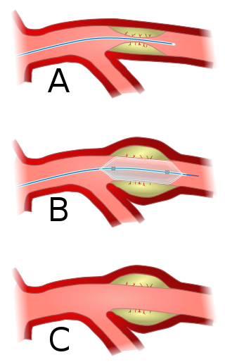
Angioplasty, also known as balloon angioplasty and percutaneous transluminal angioplasty (PTA), is a minimally invasive endovascular procedure used to widen narrowed or obstructed arteries or veins, typically to treat arterial atherosclerosis. A deflated balloon attached to a catheter is passed over a guide-wire into the narrowed vessel and then inflated to a fixed size. The balloon forces expansion of the blood vessel and the surrounding muscular wall, allowing an improved blood flow. A stent may be inserted at the time of ballooning to ensure the vessel remains open, and the balloon is then deflated and withdrawn. Angioplasty has come to include all manner of vascular interventions that are typically performed percutaneously.

<span class="mw-page-title-main">Interventional radiology</span> Medical subspecialty

Interventional radiology (IR) is a medical specialty that performs various minimally-invasive procedures using medical imaging guidance, such as x-ray fluoroscopy, computed tomography, magnetic resonance imaging, or ultrasound. IR performs both diagnostic and therapeutic procedures through very small incisions or body orifices. Diagnostic IR procedures are those intended to help make a diagnosis or guide further medical treatment, and include image-guided biopsy of a tumor or injection of an imaging contrast agent into a hollow structure, such as a blood vessel or a duct. By contrast, therapeutic IR procedures provide direct treatment—they include catheter-based medicine delivery, medical device placement, and angioplasty of narrowed structures.

<span class="mw-page-title-main">Peripheral artery disease</span> Medical condition

Peripheral artery disease (PAD) is an abnormal narrowing of arteries other than those that supply the heart or brain. PAD can happen in any blood vessel, but it is more common in the legs than the arms.

<span class="mw-page-title-main">Vascular surgery</span> Medical specialty, operative procedures for the treatment of vascular disorders

Vascular surgery is a surgical subspecialty in which vascular diseases involving the arteries, veins, or lymphatic vessels, are managed by medical therapy, minimally-invasive catheter procedures and surgical reconstruction. The specialty evolved from general and cardiovascular surgery where it refined the management of just the vessels, no longer treating the heart or other organs. Modern vascular surgery includes open surgery techniques, endovascular techniques and medical management of vascular diseases - unlike the parent specialities. The vascular surgeon is trained in the diagnosis and management of diseases affecting all parts of the vascular system excluding the coronaries and intracranial vasculature. Vascular surgeons also are called to assist other physicians to carry out surgery near vessels, or to salvage vascular injuries that include hemorrhage control, dissection, occlusion or simply for safe exposure of vascular structures.

<span class="mw-page-title-main">Cerebral angiography</span> Angiography that produces images of blood vessels in and around the brain

Cerebral angiography is a form of angiography which provides images of blood vessels in and around the brain, thereby allowing detection of abnormalities such as arteriovenous malformations and aneurysms. It was pioneered in 1927 by the Portuguese neurologist Egas Moniz at the University of Lisbon, who also helped develop thorotrast for use in the procedure.

<span class="mw-page-title-main">Takayasu's arteritis</span> Medical condition

Takayasu's arteritis (TA), also known as aortic arch syndrome, nonspecific aortoarteritis, and pulseless disease, is a form of large vessel granulomatous vasculitis with massive intimal fibrosis and vascular narrowing, most commonly affecting young or middle-aged women of Asian descent, though anyone can be affected. It mainly affects the aorta and its branches, as well as the pulmonary arteries. Females are about 8–9 times more likely to be affected than males.

<span class="mw-page-title-main">Renal artery stenosis</span> Medical condition

Renal artery stenosis (RAS) is the narrowing of one or both of the renal arteries, most often caused by atherosclerosis or fibromuscular dysplasia. This narrowing of the renal artery can impede blood flow to the target kidney, resulting in renovascular hypertension – a secondary type of high blood pressure. Possible complications of renal artery stenosis are chronic kidney disease and coronary artery disease.

Bruit, also called vascular murmur, is the abnormal sound generated by turbulent flow of blood in an artery due to either an area of partial obstruction or a localized high rate of blood flow through an unobstructed artery.

<span class="mw-page-title-main">Carotid artery stenosis</span> Medical condition

Carotid artery stenosis is a narrowing or constriction of any part of the carotid arteries, usually caused by atherosclerosis.

Vertebrobasilar insufficiency (VBI) describes a temporary set of symptoms due to decreased blood flow (ischemia) in the posterior circulation of the brain. The posterior circulation supplies the medulla, pons, midbrain, cerebellum and supplies the posterior cerebellar artery to the thalamus and occipital cortex. As a result, symptoms vary widely depending which brain region is predominantly affected.

<span class="mw-page-title-main">Carotid-cavernous fistula</span> Medical condition

A carotid-cavernous fistula results from an abnormal communication between the arterial and venous systems within the cavernous sinus in the skull. It is a type of arteriovenous fistula. As arterial blood under high pressure enters the cavernous sinus, the normal venous return to the cavernous sinus is impeded and this causes engorgement of the draining veins, manifesting most dramatically as a sudden engorgement and redness of the eye of the same side.

<span class="mw-page-title-main">Renovascular hypertension</span> Medical condition

Renovascular hypertension is a condition in which high blood pressure is caused by the kidneys' hormonal response to narrowing of the arteries supplying the kidneys. When functioning properly this hormonal axis regulates blood pressure. Due to low local blood flow, the kidneys mistakenly increase blood pressure of the entire circulatory system. It is a form of secondary hypertension - a form of hypertension whose cause is identifiable.

<span class="mw-page-title-main">Watershed stroke</span> Medical condition

A watershed stroke is defined as a brain ischemia that is localized to the vulnerable border zones between the tissues supplied by the anterior, posterior and middle cerebral arteries. The actual blood stream blockage/restriction site can be located far away from the infarcts. Watershed locations are those border-zone regions in the brain supplied by the major cerebral arteries where blood supply is decreased. Watershed strokes are a concern because they comprise approximately 10% of all ischemic stroke cases. The watershed zones themselves are particularly susceptible to infarction from global ischemia as the distal nature of the vasculature predisposes these areas to be most sensitive to profound hypoperfusion.

<span class="mw-page-title-main">Vascular disease</span> Medical condition

Vascular disease is a class of diseases of the vessels of the circulatory system in the body, including blood vessels – the arteries and veins, and the lymphatic vessels. Vascular disease is a subgroup of cardiovascular disease. Disorders in this vast network of blood and lymph vessels can cause a range of health problems that can sometimes become severe, and fatal. Coronary heart disease for example, is the leading cause of death for men and women in the United States.

<span class="mw-page-title-main">Vertebral artery dissection</span> Tear of the inner lining of the vertebral artery

Vertebral artery dissection (VAD) is a flap-like tear of the inner lining of the vertebral artery, which is located in the neck and supplies blood to the brain. After the tear, blood enters the arterial wall and forms a blood clot, thickening the artery wall and often impeding blood flow. The symptoms of vertebral artery dissection include head and neck pain and intermittent or permanent stroke symptoms such as difficulty speaking, impaired coordination, and visual loss. It is usually diagnosed with a contrast-enhanced CT or MRI scan.

<span class="mw-page-title-main">Carotid stenting</span>

Carotid artery stenting is an endovascular procedure where a stent is deployed within the lumen of the carotid artery to treat narrowing of the carotid artery and decrease the risk of stroke. It is used to treat narrowing of the carotid artery in high-risk patients, when carotid endarterectomy is considered too risky.

<span class="mw-page-title-main">Computed tomography angiography</span>

Computed tomography angiography is a computed tomography technique used for angiography—the visualization of arteries and veins—throughout the human body. Using contrast injected into the blood vessels, images are created to look for blockages, aneurysms, dissections, and stenosis. CTA can be used to visualize the vessels of the heart, the aorta and other large blood vessels, the lungs, the kidneys, the head and neck, and the arms and legs. CTA can also be used to localise arterial or venous bleed of the gastrointestinal system.

<span class="mw-page-title-main">Cervical artery dissection</span> Medical condition

Cervical artery dissection is dissection of one of the layers that compose the carotid and vertebral artery in the neck (cervix). They include:

Interventional neuroradiology (INR) also known as neurointerventional surgery (NIS), endovascular therapy (EVT), endovascular neurosurgery, and interventional neurology is a medical subspecialty of neurosurgery, neuroradiology, intervention radiology and neurology specializing in minimally invasive image-based technologies and procedures used in diagnosis and treatment of diseases of the head, neck, and spine.

Segmental arterial mediolysis (SAM) is a rare disorder of the arteries characterized by the development of aneurysms, blood clots, narrowing of the arteries (stenoses), and blood collections (hematomas) in the affected distribution.

References

  1. 1 2 3 4 5 6 7 8 9 10 11 12 13 14 Poloskey SL, Olin JW, Mace P, Gornik HL (May 2012). "Fibromuscular dysplasia". Circulation. 125 (18): e636–9. doi: 10.1161/CIRCULATIONAHA.111.090449 . PMID   22566353.
  2. 1 2 3 4 5 Olin J.W.; Froehlich J.; Gu X.; et al. (2012). "The United States registry for fibromuscular dysplasia: results in the first 447 patients". Circulation. 125 (25): 3182–90. doi: 10.1161/CIRCULATIONAHA.112.091223 . PMID   22615343.
  3. 1 2 3 4 5 6 7 8 9 10 Olin JW (April 2007). "Recognizing and managing fibromuscular dysplasia". Cleveland Clinic Journal of Medicine. 74 (4): 273–4, 277–82. doi:10.3949/ccjm.74.4.273. PMID   17438676. S2CID   21928728.
  4. 1 2 3 4 5 6 7 8 Olin JW, Sealove BA (March 2011). "Diagnosis, management, and future developments of fibromuscular dysplasia". Journal of Vascular Surgery. 53 (3): 826–36.e1. doi: 10.1016/j.jvs.2010.10.066 . PMID   21236620.
  5. Hendricks NJ, Matsumoto AH, Angle JF, Baheti A, Sabri SS, Park AW, Stone JR, Patrie JT, Dworkin L, Cooper CJ, Murphy TP, Cutlip DE (October 2014). "Is fibromuscular dysplasia underdiagnosed? A comparison of the prevalence of FMD seen in CORAL trial participants versus a single institution population of renal donor candidates". Vasc Med. 19 (5): 363–7. doi: 10.1177/1358863X14544715 . PMID   25082538.
  6. Lüscher TF, Lie JT, Stanson AW, Houser OW, Hollier LH, Sheps SG (October 1987). "Arterial fibromuscular dysplasia". Mayo Clin. Proc. 62 (10): 931–52. doi: 10.1016/s0025-6196(12)65051-4 . PMID   3309488.
  7. 1 2 3 4 Kirton A.; Crone M.; Benseler S.; Mineyko A.; Armstrong D.; Wade A.; Crous-Tsanaclis A. M. (2013). "Fibromuscular dysplasia and childhood stroke". Brain. 136 (6): 1846–1856. doi: 10.1093/brain/awt111 . PMID   23715093.
  8. 1 2 3 Meyers K. E.; Sharma N. (2007). "Fibromuscular dysplasia in children and adolescents". Cath Lab Digest. 15 (6).
  9. Mace, P., "FMDSA Fibromuscular Dysplasia Society of America." Welcome to FMDSA. N.p., 2011. Web. 22 July 2013.
  10. 1 2 3 4 Gornik HL, Persu A, Adlam D, Aparicio LS, Azizi M, Boulanger M, et al. (April 2019). "First International Consensus on the diagnosis and management of fibromuscular dysplasia". Vasc Med. 24 (2): 164–189. doi: 10.1177/1358863X18821816 . PMID   30648921.
  11. Tullus K (2013). "Renovascular hypertension—is it fibromuscular dysplasia or Takayasu arteritis". Pediatric Nephrology. 28 (2): 191–196. doi:10.1007/s00467-012-2151-7. PMID   22453736. S2CID   737167.
  12. Sos TA, Pickering TG, Sniderman K, Saddekni S, Case DB, Silane MF, Vaughan ED, Laragh JH (August 1983). "Percutaneous transluminal renal angioplasty in renovascular hypertension due to atheroma or fibromuscular dysplasia". The New England Journal of Medicine. 309 (5): 274–9. doi:10.1056/NEJM198308043090504. PMID   6223227.
  13. Belzer FO, Salvatierra O, Palubinskas A, Stoney RJ (October 1975). "Ex vivo renal artery reconstruction". Annals of Surgery. 182 (4): 456–63. doi:10.1097/00000658-197510000-00011. PMC   1344011 . PMID   1180583.
  14. Stawicki SP, Rosenfeld JC, Weger N, Fields EL, Balshi JD (September 2006). "Spontaneous renal artery dissection: three cases and clinical algorithms". J Hum Hypertens. 20 (9): 710–8. doi: 10.1038/sj.jhh.1002045 . PMID   16710291.

Citations