Focused ultrasound-mediated diagnostics

Last updated


Focused-ultrasound-mediated diagnostics or FUS-mediated diagnostics are an area of clinical diagnostic tools that use ultrasound to detect diseases and cancers. Although ultrasound has been used for imaging in various settings, focused-ultrasound refers to the detection of specific cells and biomarkers under flow combining ultrasound with lasers, microbubbles, and imaging techniques. Current diagnostic techniques for detecting tumors and diseases using biopsies often include invasive procedures and require improved accuracy, especially in cases such as glioblastoma and melanoma. The field of FUS-mediated diagnostics targeting cells and biomarkers is being investigated for overcoming these limitations.

Contents

FUS-mediated biopsy uses ultrasound wavelengths as low as those used for imaging to detect biomarkers in the bloodstream, referred to as in-vivo biopsies. Alternatively, studies have used FUS transducer acoustofluidic systems aiming to improve the accuracy of in-vitro cytometry methods for diagnostics of diseases from plasma samples.

In-vivo methods

Blood–brain barrier-disruption

One application of FUS involves the diagnosis of glioma's. Due to the sensitive environment of the brain, open or stereotactic biopsies are not always feasible and noninvasive biopsy methods are sought for detecting glioblastoma (GBM) without the risk of inducing further injury. The use of FUS, and MRI-guided FUS specifically, in combination with microbubbles has been under investigation for enhancing diagnosis methods for those patients. [1]

Blood brain barriers limit biomarker release and intercranial drug delivery. Protective barriers of the brain.jpg
Blood brain barriers limit biomarker release and intercranial drug delivery.

Microbubbles (MB) are gas-filled membranes typically made of polymers or lipids that can induce blood-brain barrier (BBB) opening when combined with ultrasound without majorly affecting surrounding tissues. Studies have shown that when combined with microbubbles or MRI, FUS can be used to locally modulate the blood brain barrier (BBB), the main deterrent to accurate glioblastoma biomarker detection and drug delivery to the brain. [1] When exposed to ultrasound pulses, microbubbles oscillate in size in a process called sonication which makes the brain temporarily permeable to receiving drugs or releasing biomarkers into the blood when exposed to low intensity FUS. [2] [3]

The use of ultrasound to facilitate blood-brain barrier disruption has been tested in animal trials including primates. Research has been focused on using this tool for improving the treatment of glioblastoma, and studies have noted that microbubble-enhanced focused-ultrasound (MB-FUS) systems can be used for drug delivery as well as diagnostics. [1] [4]

BBB disruption by focused ultrasound paired with microbubbles to release biomarkers for glioma detection. BBB disruption by focused ultrasound.png
BBB disruption by focused ultrasound paired with microbubbles to release biomarkers for glioma detection.

Liquid biopsies are one of the noninvasive methods for tumor detection through checking for tumor biomarkers within the blood. However, due to the blood brain barrier (BBB), GBM tumor biomarkers cannot enter the blood at detectable levels. Researchers have aimed to use FUS and microbubbles to enhance glioblastoma detection in liquid biopsies (sonobiopsy). [1] [4] One technique uses MRI-guided FUS sonication of microbubbles to increase BBB permeability and allow GBM tumor biomarkers (EGFRvIII and TERT C228T) to pass the BBB and enter the plasma. This allowed for highly sensitive detection of brain tumors in mouse and pig models through liquid biopsy after BBB opening. Sonobiopsy enhanced EGFRvIII and TERT C228T detection 9-fold and 3-fold, respectively, in mice. In their developed pig GBM model, detection increased to 100% and 71.43% for EGFRvIII and TERT C228T biomarkers respectively. This study reported no significant off-target effects on the brain from sonobiopsy. [4]

Microbubble (MB) oscillations used in these techniques can be controlled by using a secondary feedback transducer or imaging probe on a patient's head in order to prevent MB collapses. Low frequencies correspond to 200 kHz and high frequencies correspond to 650 kHz. [5] Several models such as the INSIGHTEC have been used for clinical contexts. BBB opening and closing can be controlled by decreasing and increasing FUS pulse lengths, respectively. One of the limitations of using FUS-mediated BBB opening is possible negative effects on neuroplasticity in the brain, however, in most studies there has been no effect on parenchymal tissue reported. [5]

Cytometry

Another area of in-vivo diagnostics uses focused ultrasound for non-invasive blood sampling. One study developed a device for "sonocytometry" which detects particle size with high intensity focused ultrasound (HIFU) under flow. A central frequency of 30 MHz can be used for an ultrasonic transducer to measure the diameter of particles in blood flow using the ultrasonic backscatter signal. [6] Ultrasound backscatter uses the variability between acoustic impedance to differentiate between particle versus medium. [7] Such a device is expected to have implications for anemia, leukemia, and other blood-related diseases. [6]

Photoacoustic Imaging can be used in-vivo to detect unusual cells in the bloodstream. PASchematics v2.png
Photoacoustic Imaging can be used in-vivo to detect unusual cells in the bloodstream.

In-vivo systems using photoacoustic (PA) flow cytography have also aimed to allow the detection of unusual melanoma cells in the bloodstream of such patients in mouse models while a coupled laser eradicates the cell on the spot through thermal ablation. Due to the high red to near-infrared absorption spectrum of single and cluster melanoma cells at 1064 nm, they are able to be distinguished using sensitive PA imaging. [8]

in-vivo cytometry methods use FUS paired with lasers to detect and differentiate circulating tumor cells from normal blood components by PA signals and US backscatter with a probe placed over the skin. In vivo cytometry.png
in-vivo cytometry methods use FUS paired with lasers to detect and differentiate circulating tumor cells from normal blood components by PA signals and US backscatter with a probe placed over the skin.

The use of FUS for in-vivo cytometry has been used in humans as well. For example, a group developed an in-vivo cytometry technique, the Cytophone, that uses focused-ultrasound to detect label-free circulating tumor cells (CTCs) in melanoma and healthy patients. This system has passed animal trials and used on humans. [9] The technique uses vapor nano-bubbles that allow for amplification of acoustic signals to detect CTCs in the blood for melanoma screening. Photoacoustic (PA) waves resulting from a 1060 nm laser pulse over the skin hitting CTCs produces peaks in the PA signal compared to a neutral red blood cell reading and negative signals for other normal blood components. The Cytophone technology was able to detect 1 CTC/L of blood and diagnose 27 of 28 patients correctly which is generally regarded as more accurate than existing methods. [9]

These photoacoustic flow cytometry (PAFC) systems coupled with FUS are being investigated aiming to allow non-invasive blood testing in various settings. For example, one application of such device was to determine what tumor manipulation methods might increase CTCs in the bloodstream [10] in-turn providing information that may be used to enhance surgical intervention techniques. Another application used a system that combines photoacoustic and ultrasound to detect tumor angiogenesis at high resolutions in-vivo with mouse models to detect melanoma phantoms. [11]

Acoustofluidics for vasculature imaging

Alternatively, acoustofluidics, or the use of FUS to manipulate particles under flow at lower wavelengths, has been used to separate cells and various particles in diagnostic applications. FUS can further be used for imaging using super-harmonic ultrasound. [12] An example of this is acoustic angiography where FUS is used to reach high resolutions for this technique. [13] Similar studies use targeted microbubbles along with super-harmonic signals to produce high resolution 3D images of microvasculature and molecular imaging. [14] This in-vivo imaging technique allowed diagnosis of fibrosarcoma in a rat model. [14]

Microbubbles and nanobubbles can also have different moieties bound to them to be able to detect, for example, angiogenesis in tumor environments, inflammation, or prostate cancer delineation. [15] Passive targeting involves modulating microbubble composition to allow incorporation into a specific tissue or cell while active targeting involves adding targeting moieties to the MB covalently or using strept(avidin)-biotin click chemistry. They can also be non-targeted and used for routine imaging. [15]

Microbubbles can also pose as theranostics, using ultrasound imaging to locate the tumor via targeted MBs then destroying the MBs to release therapeutic drugs on-site .Microbubbles with molecular markers VEGFR2 and αvβ3 integrin have been used in many preclinical tests for detecting cancers by attaching to tumor vasculature-specific receptors. In cancers including pancreatic cancer, ovarian cancer, and squamous cell carcinoma, targeted microbubbles were successfully used to assess angiogenesis and cytotoxicity by MB accumulation and ultrasound intensity detection. Targeted molecular ultrasound can also be used for non-cancerous applications such as inflammation in atherosclerosis patients where plaque-targeted MBs determined intensity of plaque buildup and severity of atherosclerosis therefrom. [15]

In-vitro methods

In-vitro cytometry methods use FUS paired with lasers to detect biomarkers in a sample flowing through a microfluidic system via PA waves and US backscatter signals. Flow cytometry in vitro.png
In-vitro cytometry methods use FUS paired with lasers to detect biomarkers in a sample flowing through a microfluidic system via PA waves and US backscatter signals.

Applications of FUS also include in-vitro methods. Studies have shown that FUS can be used to overcome limitations of current cytometry methods that often require cyctotoxic flourecent markers and do not usually provide detailed information about cell types. Acoustic waves, often used interchangeably with ultrasound, have been used to irradiate cells and their photoacoustic response in turn measured to differentiate cells without the need for lysis buffers, tagging agents, and further sample preparation methods. [7] One study showed that acoustic flow cytometer (AFC) can use ultra-high frequency ultrasound to detect cells and particles under flow without the need for labelling. By incorporating an ultrasound transducer that detects both ultrasonic backscatter and PA signals, the device is able to distinguish cell types in a polydimethylsiloxane (PDMS)-based microfluidic device with relative accuracy. The red blood cell and white blood cell count using AFC was found to be accurate compared with the conventional florescence-activated cell-sorting (FACS) results. [7]

Similar technologies are being investigated to make improvements to in-vitro testing of diseases. One such device uses acoustofluidics for detecting Alzheimer's disease. [16] Acoustofluidics allowed the detection of Alzheimer-specific biomarkers. This process is in-vitro requiring a patient's plasma sample and the use of ultrasound to detect nano-sized biomarkers. [16] Ultrasound in the form of acoustics has been shown to improve microfluidic techniques by allowing control over the liquid and interaction kinetics through bulk acoustic waves (BAW) or surface acoustic waves (SAW). [17]

Challenges and Limitations

Some of the challenges for acoustofluidic microdevices include manufacturing, since the development must take place in a cleanroom and there is need for expensive materials limiting scaling and industrialization. Additionally, while a device may be portable, concerns include supplementary devices such as amplifiers and generators detering large-scale manufacturing. For example, the manufacturing of acoustic microreactors may hinder their portability potential due to various necessary equipment required with the device. [17]

Health effects are also of concern to researchers and clinicians with the use of focused ultrasound because long duration of FUS at certain wavelengths can induce further damage to surrounding tissues which proposes the need for safety parameters for such devices. [5] [1] Furthermore, side effects such as allergies and other adverse reactions have been noted in some FUS-mediated diagnostic methods which may be worsened by preexisting conditions. In BBB-disruption, specifically, the concern for affected neuroplasticity exists. [1] [5] Determining the exact amplitude of FUS for sonicating is especially important since high amplitudes have been reported to lead to intracranial hemorrhage. [18] [19]

With in-vitro uses of FUS, only size and number of cells can be detected but studies have noted that information about the cell structure or organelles are unavailable. [7] However, improving in-vitro diagnostic systems is still being researched. A common biomarker cancer detection technique relies on ctDNA, but this biomarker is currently suspected to have various limitations. [3] Some limitations related to using ctDNA biomarkers include the lack of specificity for detection of rare cancers and low release rate from certain tumors. In such cases, however, FUS-microbubble sonication systems are being investigated for increase in tumor permeability and detection rate of cancers or allow drug delivery. [20] [3]

Focused-ultrasound mediated diagnostics is an expanding area in research that is often paired with the aim of using FUS to better detect or release biomarkers or to allow for a local drug delivery technique.

Related Research Articles

<span class="mw-page-title-main">Brain tumor</span> Neoplasm in the brain

A brain tumor occurs when abnormal cells form within the brain. There are two main types of tumors: malignant (cancerous) tumors and benign (non-cancerous) tumors. These can be further classified as primary tumors, which start within the brain, and secondary tumors, which most commonly have spread from tumors located outside the brain, known as brain metastasis tumors. All types of brain tumors may produce symptoms that vary depending on the size of the tumor and the part of the brain that is involved. Where symptoms exist, they may include headaches, seizures, problems with vision, vomiting and mental changes. Other symptoms may include difficulty walking, speaking, with sensations, or unconsciousness.

<span class="mw-page-title-main">Blood–brain barrier</span> Semipermeable capillary border that allows selective passage of blood constituents into the brain

The blood–brain barrier (BBB) is a highly selective semipermeable border of endothelial cells that regulates the transfer of solutes and chemicals between the circulatory system and the central nervous system, thus protecting the brain from harmful or unwanted substances in the blood. The blood–brain barrier is formed by endothelial cells of the capillary wall, astrocyte end-feet ensheathing the capillary, and pericytes embedded in the capillary basement membrane. This system allows the passage of some small molecules by passive diffusion, as well as the selective and active transport of various nutrients, ions, organic anions, and macromolecules such as glucose and amino acids that are crucial to neural function.

<span class="mw-page-title-main">Medical ultrasound</span> Diagnostic and therapeutic technique

Medical ultrasound includes diagnostic techniques using ultrasound, as well as therapeutic applications of ultrasound. In diagnosis, it is used to create an image of internal body structures such as tendons, muscles, joints, blood vessels, and internal organs, to measure some characteristics or to generate an informative audible sound. The usage of ultrasound to produce visual images for medicine is called medical ultrasonography or simply sonography, or echography. The practice of examining pregnant women using ultrasound is called obstetric ultrasonography, and was an early development of clinical ultrasonography. The machine used is called an ultrasound machine, a sonograph or an echograph. The visual image formed using this technique is called an ultrasonogram, a sonogram or an echogram.

<span class="mw-page-title-main">Photoacoustic imaging</span> Imaging using the photoacoustic effect

Photoacoustic imaging or optoacoustic imaging is a biomedical imaging modality based on the photoacoustic effect. Non-ionizing laser pulses are delivered into biological tissues and part of the energy will be absorbed and converted into heat, leading to transient thermoelastic expansion and thus wideband ultrasonic emission. The generated ultrasonic waves are detected by ultrasonic transducers and then analyzed to produce images. It is known that optical absorption is closely associated with physiological properties, such as hemoglobin concentration and oxygen saturation. As a result, the magnitude of the ultrasonic emission, which is proportional to the local energy deposition, reveals physiologically specific optical absorption contrast. 2D or 3D images of the targeted areas can then be formed.

<span class="mw-page-title-main">Contrast-enhanced ultrasound</span> Medical imaging technique

Contrast-enhanced ultrasound (CEUS) is the application of ultrasound contrast medium to traditional medical sonography. Ultrasound contrast agents rely on the different ways in which sound waves are reflected from interfaces between substances. This may be the surface of a small air bubble or a more complex structure. Commercially available contrast media are gas-filled microbubbles that are administered intravenously to the systemic circulation. Microbubbles have a high degree of echogenicity. There is a great difference in echogenicity between the gas in the microbubbles and the soft tissue surroundings of the body. Thus, ultrasonic imaging using microbubble contrast agents enhances the ultrasound backscatter, (reflection) of the ultrasound waves, to produce a sonogram with increased contrast due to the high echogenicity difference. Contrast-enhanced ultrasound can be used to image blood perfusion in organs, measure blood flow rate in the heart and other organs, and for other applications.

<span class="mw-page-title-main">Molecular imaging</span> Imaging molecules within living patients

Molecular imaging is a field of medical imaging that focuses on imaging molecules of medical interest within living patients. This is in contrast to conventional methods for obtaining molecular information from preserved tissue samples, such as histology. Molecules of interest may be either ones produced naturally by the body, or synthetic molecules produced in a laboratory and injected into a patient by a doctor. The most common example of molecular imaging used clinically today is to inject a contrast agent into a patient's bloodstream and to use an imaging modality to track its movement in the body. Molecular imaging originated from the field of radiology from a need to better understand fundamental molecular processes inside organisms in a noninvasive manner.

<span class="mw-page-title-main">Circulating tumor cell</span> Cell from a primary tumor carried by blood circulation

A circulating tumor cell (CTC) is a cell that has shed into the vasculature or lymphatics from a primary tumor and is carried around the body in the blood circulation. CTCs can extravasate and become seeds for the subsequent growth of additional tumors (metastases) in distant organs, a mechanism that is responsible for the vast majority of cancer-related deaths. The detection and analysis of CTCs can assist early patient prognoses and determine appropriate tailored treatments. Currently, there is one FDA-approved method for CTC detection, CellSearch, which is used to diagnose breast, colorectal and prostate cancer.

<span class="mw-page-title-main">Sonoporation</span> Technique in molecular biology

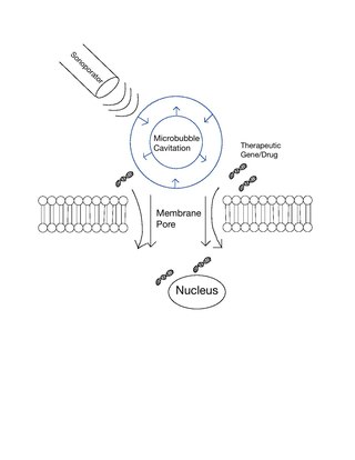
Sonoporation, or cellular sonication, is the use of sound in the ultrasonic range for increasing the permeability of the cell plasma membrane. This technique is usually used in molecular biology and non-viral gene therapy in order to allow uptake of large molecules such as DNA into the cell, in a cell disruption process called transfection or transformation. Sonoporation employs the acoustic cavitation of microbubbles to enhance delivery of these large molecules. The exact mechanism of sonoporation-mediated membrane translocation remains unclear, with a few different hypotheses currently being explored.

Microbubbles are bubbles smaller than one hundredth of a millimetre in diameter, but larger than one micrometre. They have widespread application in industry, medicine, life science, and food technology. The composition of the bubble shell and filling material determine important design features such as buoyancy, crush strength, thermal conductivity, and acoustic properties.

Preclinical imaging is the visualization of living animals for research purposes, such as drug development. Imaging modalities have long been crucial to the researcher in observing changes, either at the organ, tissue, cell, or molecular level, in animals responding to physiological or environmental changes. Imaging modalities that are non-invasive and in vivo have become especially important to study animal models longitudinally. Broadly speaking, these imaging systems can be categorized into primarily morphological/anatomical and primarily molecular imaging techniques. Techniques such as high-frequency micro-ultrasound, magnetic resonance imaging (MRI) and computed tomography (CT) are usually used for anatomical imaging, while optical imaging, positron emission tomography (PET), and single photon emission computed tomography (SPECT) are usually used for molecular visualizations.

<span class="mw-page-title-main">Sonodynamic therapy</span>

Sonodynamic therapy (SDT) is a noninvasive treatment, often used for tumor irradiation, that utilizes a sonosensitizer and the deep penetration of ultrasound to treat lesions of varying depths by reducing target cell number and preventing future tumor growth. Many existing cancer treatment strategies cause systemic toxicity or cannot penetrate tissue deep enough to reach the entire tumor; however, emerging ultrasound stimulated therapies could offer an alternative to these treatments with their increased efficiency, greater penetration depth, and reduced side effects. Sonodynamic therapy could be used to treat cancers and other diseases, such as atherosclerosis, and diminish the risk associated with other treatment strategies since it induces cytotoxic effects only when externally stimulated by ultrasound and only at the cancerous region, as opposed to the systemic administration of chemotherapy drugs.

Multi-spectral optoacoustic tomography (MSOT), also known as functional photoacoustic tomography (fPAT), is an imaging technology that generates high-resolution optical images in scattering media, including biological tissues. MSOT illuminates tissue with light of transient energy, typically light pulses lasting 1-100 nanoseconds. The tissue absorbs the light pulses, and as a result undergoes thermo-elastic expansion, a phenomenon known as the optoacoustic or photoacoustic effect. This expansion gives rise to ultrasound waves (photoechoes) that are detected and formed into an image. Image formation can be done by means of hardware or computed tomography. Unlike other types of optoacoustic imaging, MSOT involves illuminating the sample with multiple wavelengths, allowing it to detect ultrasound waves emitted by different photoabsorbing molecules in the tissue, whether endogenous or exogenous. Computational techniques such as spectral unmixing deconvolute the ultrasound waves emitted by these different absorbers, allowing each emitter to be visualized separately in the target tissue. In this way, MSOT can allow visualization of hemoglobin concentration and tissue oxygenation or hypoxia. Unlike other optical imaging methods, MSOT is unaffected by photon scattering and thus can provide high-resolution optical images deep inside biological tissues.

<span class="mw-page-title-main">Circulating tumor DNA</span> Tumor-derived fragmented DNA in the bloodstream

Circulating tumor DNA (ctDNA) is tumor-derived fragmented DNA in the bloodstream that is not associated with cells. ctDNA should not be confused with cell-free DNA (cfDNA), a broader term which describes DNA that is freely circulating in the bloodstream, but is not necessarily of tumor origin. Because ctDNA may reflect the entire tumor genome, it has gained traction for its potential clinical utility; "liquid biopsies" in the form of blood draws may be taken at various time points to monitor tumor progression throughout the treatment regimen.

A liquid biopsy, also known as fluid biopsy or fluid phase biopsy, is the sampling and analysis of non-solid biological tissue, primarily blood. Like traditional biopsy, this type of technique is mainly used as a diagnostic and monitoring tool for diseases such as cancer, with the added benefit of being largely non-invasive. Liquid biopsies may also be used to validate the efficiency of a cancer treatment drug by taking multiple samples in the span of a few weeks. The technology may also prove beneficial for patients after treatment to monitor relapse.

<span class="mw-page-title-main">Photoacoustic microscopy</span>

Photoacoustic microscopy is an imaging method based on the photoacoustic effect and is a subset of photoacoustic tomography. Photoacoustic microscopy takes advantage of the local temperature rise that occurs as a result of light absorption in tissue. Using a nanosecond pulsed laser beam, tissues undergo thermoelastic expansion, resulting in the release of a wide-band acoustic wave that can be detected using a high-frequency ultrasound transducer. Since ultrasonic scattering in tissue is weaker than optical scattering, photoacoustic microscopy is capable of achieving high-resolution images at greater depths than conventional microscopy methods. Furthermore, photoacoustic microscopy is especially useful in the field of biomedical imaging due to its scalability. By adjusting the optical and acoustic foci, lateral resolution may be optimized for the desired imaging depth.

Tissue image cytometry or tissue cytometry is a method of digital histopathology and combines classical digital pathology and computational pathology into one integrated approach with solutions for all kinds of diseases, tissue and cell types as well as molecular markers and corresponding staining methods to visualize these markers. Tissue cytometry uses virtual slides as they can be generated by multiple, commercially available slide scanners, as well as dedicated image analysis software – preferentially including machine and deep learning algorithms. Tissue cytometry enables cellular analysis within thick tissues, retaining morphological and contextual information, including spatial information on defined cellular subpopulations. In this process, a tissue sample, either formalin-fixed paraffin-embedded (FFPE) or frozen tissue section, also referred to as “cryocut”, is labelled with either immunohistochemistry(IHC) or immunofluorescent markers, scanned with high-throughput slide scanners and the data gathered from virtual slides is processed and analyzed using software that is able to identify individual cells in tissue context automatically and distinguish between nucleus and cytoplasm for each cell. Additional algorithms can identify cellular membranes, subcellular structures and/or multicellular tissue structures.

A specific branch of contrast-enhanced ultrasound, acoustic angiography is a minimally invasive and non-ionizing medical imaging technique used to visualize vasculature. Acoustic angiography was first developed by the Dayton Laboratory at North Carolina State University and provides a safe, portable, and inexpensive alternative to the most common methods of angiography such as Magnetic Resonance Angiography and Computed Tomography Angiography. Although ultrasound does not traditionally exhibit the high resolution of MRI or CT, high-frequency ultrasound (HFU) achieves relatively high resolution by sacrificing some penetration depth. HFU typically uses waves between 20 and 100 MHz and achieves resolution of 16-80μm at depths of 3-12mm. Although HFU has exhibited adequate resolution to monitor things like tumor growth in the skin layers, on its own it lacks the depth and contrast necessary for imaging blood vessels. Acoustic angiography overcomes the weaknesses of HFU by combining contrast-enhanced ultrasound with the use of a dual-element ultrasound transducer to achieve high resolution visualization of blood vessels at relatively deep penetration levels.

Photoacoustic flow cytometry or PAFC is a biomedical imaging modality that utilizes photoacoustic imaging to perform flow cytometry. A flow of cells passes a photoacoustic system producing individual signal response. Each signal is counted to produce a quantitative evaluation of the input sample.

<span class="mw-page-title-main">Focused ultrasound for intracranial drug delivery</span> Medical technique

Focused ultrasound for intracrainial drug delivery is a non-invasive technique that uses high-frequency sound waves to disrupt tight junctions in the blood–brain barrier (BBB), allowing for increased passage of therapeutics into the brain. The BBB normally blocks nearly 98% of drugs from accessing the central nervous system, so FUS has the potential to address a major challenge in intracranial drug delivery by providing targeted and reversible BBB disruption. Using FUS to enhance drug delivery to the brain could significantly improve patient outcomes for a variety of diseases including Alzheimer's disease, Parkinson's disease, and brain cancer.

Epitope Detection in Monocytes (EDIM) is a technology that uses the innate immune system's mechanisms to detect biomarkers or antigens in immune cells. It is a non-invasive form of liquid biopsy, i.e. biopsy from blood, which analyzes activated macrophages (CD14+/CD16+) for disease-specific epitopes, such as tumor cell components.

References

  1. 1 2 3 4 5 6 Lechpammer, Mirna; Rao, Rohan; Shah, Sanjit; Mirheydari, Mona; Bhattacharya, Debanjan; Koehler, Abigail; Toukam, Donatien Kamdem; Haworth, Kevin J.; Pomeranz Krummel, Daniel; Sengupta, Soma (January 2022). "Advances in Immunotherapy for the Treatment of Adult Glioblastoma: Overcoming Chemical and Physical Barriers". Cancers. 14 (7): 1627. doi: 10.3390/cancers14071627 . ISSN   2072-6694. PMC   8997081 . PMID   35406398.
  2. Rincon-Torroella, Jordina; Khela, Harmon; Bettegowda, Anya; Bettegowda, Chetan (2022-01-01). "Biomarkers and focused ultrasound: the future of liquid biopsy for brain tumor patients". Journal of Neuro-Oncology. 156 (1): 33–48. doi:10.1007/s11060-021-03837-0. ISSN   1573-7373. PMC   8714625 . PMID   34613580.
  3. 1 2 3 Campos-Carrillo, Andrea; Weitzel, Jeffrey N.; Sahoo, Prativa; Rockne, Russell; Mokhnatkin, Janet V.; Murtaza, Muhammed; Gray, Stacy W.; Goetz, Laura; Goel, Ajay; Schork, Nicholas; Slavin, Thomas P. (2020-03-01). "Circulating tumor DNA as an early cancer detection tool". Pharmacology & Therapeutics. 207: 107458. doi:10.1016/j.pharmthera.2019.107458. ISSN   0163-7258. PMC   6957244 . PMID   31863816.
  4. 1 2 3 Pacia, Christopher P.; Yuan, Jinyun; Yue, Yimei; Xu, Lu; Nazeri, Arash; Desai, Rupen; Gach, H. Michael; Wang, Xiaowei; Talcott, Michael R.; Chaudhuri, Aadel A.; Dunn, Gavin P.; Leuthardt, Eric C.; Chen, Hong (2022-01-01). "Sonobiopsy for minimally invasive, spatiotemporally-controlled, and sensitive detection of glioblastoma-derived circulating tumor DNA". Theranostics. 12 (1): 362–378. doi:10.7150/thno.65597. ISSN   1838-7640. PMC   8690937 . PMID   34987650. S2CID   244002784.
  5. 1 2 3 4 Conti, Allegra; Kamimura, Hermes A. S.; Novell, Anthony; Duggento, Andrea; Toschi, Nicola (2020). "Magnetic Resonance Methods for Focused Ultrasound-Induced Blood-Brain Barrier Opening". Frontiers in Physics. 8: 393. Bibcode:2020FrP.....8..393C. doi: 10.3389/fphy.2020.547674 . ISSN   2296-424X.
  6. 1 2 Komatsu, Yosuke; Nagaoka, Ryo; Funamoto, Ken-ichi; Hayase, Toshiyuki; Masauzi, Nobuo; Kanai, Hiroshi; Saijo, Yoshifumi (August 2014). ""Sonocytometry" Novel diagnostic method of ultrasonic differentiation of cells in blood flow". 2014 36th Annual International Conference of the IEEE Engineering in Medicine and Biology Society. Vol. 2014. pp. 2761–2764. doi:10.1109/EMBC.2014.6944195. ISBN   978-1-4244-7929-0. PMID   25570563. S2CID   2059865.
  7. 1 2 3 4 Gnyawali, Vaskar; Strohm, Eric M.; Wang, Jun-Zhi; Tsai, Scott S. H.; Kolios, Michael C. (2019-02-07). "Simultaneous acoustic and photoacoustic microfluidic flow cytometry for label-free analysis". Scientific Reports. 9 (1): 1585. Bibcode:2019NatSR...9.1585G. doi:10.1038/s41598-018-37771-5. ISSN   2045-2322. PMC   6367457 . PMID   30733497. S2CID   256995755.
  8. He, Yun; Wang, Lidai; Shi, Junhui; Yao, Junjie; Li, Lei; Zhang, Ruiying; Huang, Chih-Hsien; Zou, Jun; Wang, Lihong V. (2016-12-21). "In vivo label-free photoacoustic flow cytography and on-the-spot laser killing of single circulating melanoma cells". Scientific Reports. 6 (1): 39616. Bibcode:2016NatSR...639616H. doi:10.1038/srep39616. ISSN   2045-2322. PMC   5175175 . PMID   28000788. S2CID   11955888.
  9. 1 2 Galanzha, Ekaterina I.; Menyaev, Yulian A.; Yadem, Aayire C.; Sarimollaoglu, Mustafa; Juratli, Mazen A.; Nedosekin, Dmitry A.; Foster, Stephen R.; Jamshidi-Parsian, Azemat; Siegel, Eric R.; Makhoul, Issam; Hutchins, Laura F.; Suen, James Y.; Zharov, Vladimir P. (2019-06-12). "In vivo liquid biopsy using Cytophone platform for photoacoustic detection of circulating tumor cells in patients with melanoma". Science Translational Medicine. 11 (496): eaat5857. doi:10.1126/scitranslmed.aat5857. ISSN   1946-6234. PMC   9235419 . PMID   31189720.
  10. Juratli, Mazen A.; Sarimollaoglu, Mustafa; Siegel, Eric R.; Nedosekin, Dmitry A.; Galanzha, Ekaterina I.; Suen, James Y.; Zharov, Vladimir P. (August 2014). "Real-time monitoring of circulating tumor cell release during tumor manipulation using in vivo photoacoustic and fluorescent flow cytometry: Circulating Tumor Cell Release During Medical Intervention". Head & Neck. 36 (8): 1207–1215. doi:10.1002/hed.23439. PMC   9212256 . PMID   23913663.
  11. Wang, Yating; Xu, Dong; Yang, Sihua; Xing, Da (2016-02-01). "Toward in vivo biopsy of melanoma based on photoacoustic and ultrasound dual imaging with an integrated detector". Biomedical Optics Express. 7 (2): 279–286. doi:10.1364/BOE.7.000279. ISSN   2156-7085. PMC   4771448 . PMID   26977339.
  12. Fan, Yanping; Wang, Xuan; Ren, Jiaqi; Lin, Francis; Wu, Jiandong (2022-09-01). "Recent advances in acoustofluidic separation technology in biology". Microsystems & Nanoengineering. 8 (1): 94. Bibcode:2022MicNa...8...94F. doi:10.1038/s41378-022-00435-6. ISSN   2055-7434. PMC   9434534 . PMID   36060525.
  13. Gessner, Ryan C.; Frederick, C. Brandon; Foster, F. Stuart; Dayton, Paul A. (2013-07-17). "Acoustic Angiography: A New Imaging Modality for Assessing Microvasculature Architecture". International Journal of Biomedical Imaging. 2013: e936593. doi: 10.1155/2013/936593 . ISSN   1687-4188. PMC   3730364 . PMID   23997762.
  14. 1 2 Shelton, Sarah E.; Lindsey, Brooks D.; Tsuruta, James K.; Foster, F. Stuart; Dayton, Paul A. (2016-03-01). "Molecular Acoustic Angiography: A New Technique for High-resolution Superharmonic Ultrasound Molecular Imaging". Ultrasound in Medicine and Biology. 42 (3): 769–781. doi:10.1016/j.ultrasmedbio.2015.10.015. ISSN   0301-5629. PMC   5653972 . PMID   26678155.
  15. 1 2 3 Kiessling, Fabian; Fokong, Stanley; Koczera, Patrick; Lederle, Wiltrud; Lammers, Twan (2012-03-01). "Ultrasound Microbubbles for Molecular Diagnosis, Therapy, and Theranostics". Journal of Nuclear Medicine. 53 (3): 345–348. doi: 10.2967/jnumed.111.099754 . ISSN   0161-5505. PMID   22393225. S2CID   12846755.
  16. 1 2 Hao, Nanjing; Wang, Zeyu; Liu, Pengzhan; Becker, Ryan; Yang, Shujie; Yang, Kaichun; Pei, Zhichao; Zhang, Peiran; Xia, Jianping; Shen, Liang; Wang, Lin; Welsh-Bohmer, Kathleen A.; Sanders, Laurie H.; Lee, Luke P.; Huang, Tony Jun (2022-01-15). "Acoustofluidic multimodal diagnostic system for Alzheimer's disease". Biosensors and Bioelectronics. 196: 113730. doi:10.1016/j.bios.2021.113730. ISSN   0956-5663. PMC   8643320 . PMID   34736099. S2CID   240241845.
  17. 1 2 Zhao, Xiong; Chen, Zhenzhen; Qiu, Yinan; Hao, Nanjing (2023-02-20). "Acoustic microfluidics for colloidal materials and interface engineering". Materials Advances. 4 (4): 988–994. doi: 10.1039/D2MA00590E . ISSN   2633-5409. S2CID   256051520.
  18. Liu, Hao-Li; Wai, Yau-Yau; Chen, Wen-Shiang; Chen, Jin-Chung; Hsu, Po-Hong; Wu, Xin-Yu; Huang, Wen-Cheng; Yen, Tzu-Chen; Wang, Jiun-Jie (April 2008). "Hemorrhage detection during focused-ultrasound induced blood-brain-barrier opening by using susceptibility-weighted magnetic resonance imaging". Ultrasound in Medicine & Biology. 34 (4): 598–606. doi: 10.1016/j.ultrasmedbio.2008.01.011 . ISSN   0301-5629. PMID   18313204.
  19. McDannold, Nathan; Vykhodtseva, Natalia; Hynynen, Kullervo (April 2007). "Use of Ultrasound Pulses Combined with Definity for Targeted Blood-Brain Barrier Disruption: A Feasibility Study". Ultrasound in Medicine & Biology. 33 (4): 584–590. doi:10.1016/j.ultrasmedbio.2006.10.004. PMC   2066193 . PMID   17337109.
  20. Ng, Serina; Healey, Andrew John; Sontum, Per Christian; Kvåle, Svein; Torp, Sverre H.; Sulheim, Einar; Von Hoff, Daniel; Han, Haiyong (2022-12-01). "Effect of acoustic cluster therapy (ACT) combined with chemotherapy in a patient-derived xenograft mouse model of pancreatic cancer". Journal of Controlled Release. 352: 1134–1143. doi: 10.1016/j.jconrel.2022.11.016 . hdl: 11250/3053212 . ISSN   0168-3659. PMID   36372388. S2CID   253509238.