Functional ultrasound imaging

Last updated
Main applications and features of functional ultrasound (fUS) imaging Main applications and features of functional ultrasound (fUS) imaging.svg
Main applications and features of functional ultrasound (fUS) imaging

Functional ultrasound imaging (fUS) is a medical ultrasound imaging technique of detecting or measuring changes in neural activities or metabolism, for example, the loci of brain activity, typically through measuring blood flow or hemodynamic changes. The method can be seen as an extension of Doppler imaging.

Contents

Background

Main brain functional imaging technique resolutions Main brain functional imaging technique resolutions.svg
Main brain functional imaging technique resolutions

Brain activation can either be directly measured by imaging electrical activity of neurons using voltage sensitive dyes, calcium imaging, electroencephalography, or magnetoencephalography, or indirectly by detecting hemodynamic changes in blood flow in the neurovascular systems through functional magnetic resonance imaging (fMRI), positron emission tomography (PET), Functional near-infrared spectroscopy (fNIRS), or Doppler ultrasonography )... [1]

Optical based methods generally provide the highest spatial and temporal resolutions; however, due to scattering, they are intrinsically limited to the investigation of the cortex. Thus, they are often used on animal models after partially removing or thinning the skull to allow for the light to penetrate into tissue. fMRI and PET, which measure the blood-oxygen level dependent (BOLD) signal, were the only techniques capable of imaging brain activation in depth. BOLD signal increases when neuronal activation exceeds oxygen consumption, where blood flow increases significantly. In fact, in-depth imaging of cerebral hemodynamic responses by fMRI, being noninvasive, paved the way for major discoveries in neurosciences in the early stage, and is applicable on humans. However, fMRI also suffers limitations. First, the cost and size of MR machines can be prohibitive. Also, spatially resolved fMRI is achieved at the expense of a substantial drop in temporal resolution and/or SNR. As a result, the imaging of transient events such as epilepsy is particularly challenging. Finally, fMRI is not appropriate for all clinical applications. For example, fMRI is rarely performed on infants because of specific issues concerning infant sedation. [2]

Like fMRI, Doppler based functional ultrasound approach are based on the neurovascular coupling and are thus limited by the spatiotemporal features of neurovascular coupling as they measure cerebral blood volume (CBV) changes. CBV is a pertinent parameter for functional imaging that is already used by other modalities such as intrinsic optical imaging or CBV-weighted fMRI. The spatiotemporal extent of CBV response was extensively studied. The spatial resolution of sensory-evoked CBV response can go down to cortical column (~100 µm). Temporally, the CBV impulse response function was measured to typically start at ~0.3 s and peak at ~1 s in response to ultrashort stimuli (300µs), which is much slower than the underlying electrical activity. [3]

Conventional Doppler based functional imaging approaches

Hemodynamic changes in the brain are often used as a surrogate indicator of neuronal activity to map the loci of brain activity. Major part of the hemodynamic response occurs in small vessels; however, conventional Doppler ultrasound is not sensitive enough to detect the blood flow in such small vessels. [2]

Functional Transcranial Doppler (fTCD)

Ultrasound Doppler imaging can be used to obtain basic functional measurements of brain activity using blood flow. In functional transcranial Doppler sonography, a low frequency (1-3 MHz) transducer is used through the temporal bone window with a conventional pulse Doppler mode to estimate blood flow at a single focal location. The temporal profile of blood velocity is usually acquired in main large arteries such as the middle cerebral artery (MCA). The peak velocity is compared between rest and task conditions or between right and left sides when studying lateralization. [4]

Power Doppler

Power Doppler is a Doppler sequence that measures the ultrasonic energy backscattered from red blood cells in each pixel of the image. It provides no information on blood velocity but is proportional to blood volume within the pixel. However, conventional power Doppler imaging lacks sensitivity to detect small arterioles/venules and thus is unable to provide local neurofunctional information through neurovascular coupling. [2]

Ultrasensitive Doppler and functional ultrasound imaging (fUS)

Functional ultrasound imaging was pioneered at ESPCI by Mickael Tanter's team [5] following work on ultrafast imaging [6] and ultrafast Doppler. [7]

Ultrasensitive Doppler principle

Ultrasensitive Doppler relies on ultrafast imaging scanners [6] able to acquire images at thousands of frames per second, thus boosting the power Doppler SNR without any contrast agents. Instead of the line by line acquisition of conventional ultrasound devices, ultra-fast ultrasound takes advantage of successive tilted plane wave transmissions that afterward coherently compounded to form images at high frame rates. Coherent Compound Beamforming consists of the recombination of backscattered echoes from different illuminations achieved on the acoustic pressure field with various angles (as opposed to the acoustic intensity for the incoherent case). All images are added coherently to obtain a final compounded image. This very addition is produced without taking the envelope of the beamformed signals or any other nonlinear procedure to ensure a coherent addition. As a result, coherent adding of several echo waves leads to cancellation of out-of-phase waveforms, narrowing the point spread function (PSF), and thus increasing spatial resolution. A theoretical model demonstrates that the gain in sensitivity of the ultrasensitive Doppler method is due to the combination of the high signal-to-noise ratio (SNR) of the gray scale images, due to the synthetic compounding of backscattered echoes and the extensive signal samples averaging due to the high temporal resolution of ultrafast frame rates. [2] The sensitivity was recently further improved using multiple plane wave transmissions [8] and advanced spatiotemporal clutter filters for better discrimination between low blood flow and tissue motion. Ultrasound researchers have been using ultrafast imaging research platforms with parallel acquisition of channels and custom sequences programming to investigate ultrasensitive Doppler/fUS modalities. A custom real-time high-performance GPU beamforming code with a high data transfer rate (several GBytes per second) must then be implemented to perform imaging at high frame rate. Acquisitions can also typically easily provide gigabytes of data depending on acquisition duration.

Ultrasensitive Doppler has a typical 50-200 µm spatial resolution depending on the ultrasound frequency used. [2] It features a temporal resolution in the tens of milliseconds, can image the full depth of the brain and can provide 3D angiography. [9]

functional Ultrasound imaging

This signal boost enables the sensitivity required to map subtle blood variations in small arterioles (down to 1mm/s) related to neuronal activity. By applying an external stimulus such as a sensory, auditory or visual stimulation, it is then possible to construct a map of brain activation from the ultrasensitive Doppler movie.

fUS measures indirectly cerebral blood volume which provides an effect size close to 20% and as such is quite more sensitive than fMRI whose BOLD response is typically only a couple of percents. Correlation maps or statistical parametric maps can be constructed to highlight the activated areas. fUS has been shown to have a spatial resolution on the order 100 micrometers at 15 MHz in ferrets [10] and is sensitive enough to perform single trial detection in awake primates. [11] Other fMRI-like modalities such as functional connectivity can also be implemented.

Commercial scanners with specialized hardware and software [12] should enable fUS to rapidly expand behind ultrasound research labs to the neuroscience community.

4D functional ultrasound imaging

Some researchers conducted 4D functional ultrasound imaging of whole-brain activity in rodents. Currently, two different technological solutions are proposed for the acquisition of 3D and 4D fUS data, each with its own advantages and drawbacks. [13] The first is a tomographic approach based on motorized translation of linear probes. This approach proved to be a successful method for several applications such as 3D retinotopic mapping in the rodent brain [14] [15] and 3D tonotopic mapping of the auditory system in ferrets. [10] The second approach relies on high frequency 2D matrix array transducer technology coupled with a high channel count electronic system for fast 3D imaging. To counterbalance the intrinsically poor sensitivity of matrix elements, they devised a 3D multiplane-wave scheme with 3D spatiotemporal encoding of transmit signals using Hadamard coefficients. For each transmission, the backscattered signals containing mixed echoes from the different plane waves are decoded using the summation of echoes from successive receptions with appropriate Hadamard coefficients. This summation enables the synthetic building of echoes from a virtual individual plane wave transmission with a higher amplitude. Finally, they perform coherent compounding beamforming of decoded echoes to produce 3D ultrasonic images and apply a spatiotemporal clutter filter separating blood flow from tissue motion to compute a power Doppler volume, which is proportional to the cerebral blood volume. [16]

Features

Advantages

• High SNR with large effect size >15% of relative CBV increase compared to ~1% in BOLD fMRI

• High spatial resolution (100 micrometers at 15 MHz for preclinical use),

• Compatibility with other techniques commonly used by physiologists, in particular electrophysiological recordings or optogenetics.

• Can be used in awake animals, headfixed or mobile.

• Inexpensive and more practical (smaller machine, transportable), compared with fMRI.

• Requires no calibration and little setup time. Easy to set up.

• Enabling study of the subcortical structures makes in-depth imaging prospective compared with optical techniques [2]

• Can be used through the transfontanellar window in neonates

• Transcranial in mice

• 3D scans possible using motors or a 2D matrix array

Drawbacks

• Cannot image through the skull (except mice): can be solved by techniques of thinned-skull already developed for chronical optical imaging, [17] the use of TPX window or the use contrast agents to increase blood echogenicity to allow imaging through the skull.

• Capillary blood flow is on the order of 0.5 mm/s, which could be filtered out by HPF and thus could not be detected although advanced spatiotemporal clutter filters have been proposed.

• 2D matrix array technology for 3D fUS imaging is still in research and suffers some sensitivity limitations. 3D scans using motors have typical lower temporal resolution than equivalent 2D scans.

Applications

Functional ultrasound imaging has a wide range of applications in research and in clinical practice.

Preclinical applications

Preclinical applications of fUS imaging Preclinical applications of fUS imaging.svg
Preclinical applications of fUS imaging

fUS can benefit in monitoring cerebral function in the whole brain which is important to understanding how the brain works on a large scale under normal or pathological conditions. The ability to image cerebral blood volume at high spatiotemporal resolution and with high sensitivity using fUS could be of great interest for applications in which fMRI reaches its limits, such as imaging of epileptic-induced changes in blood volume. [18] fUS can be applied for chronic studies in animal models through a thinned-skull [19] or smaller cranial window or directly through the skull in mice.

Brain activity mapping

Tonotopics or retinotopics maps [20] can be constructed by mapping the response of frequency-varying sounds [10] or moving visual targets. [14] [20] [15]

functional connectivity / resting state

When no stimulus is applied, fUS can be used to study functional connectivity during resting state. The method has been demonstrated in rats [21] and awake mice [22] and can be used for pharmacological studies when testing drugs. [23] Seed-based maps, independent component analysis of resting states modes or functional connectivity matrix between atlas-based regions of interests can be constructed with high resolution.

awake fUS imaging

Using dedicated ultralight probes, it is possible to perform freely-moving experiments in rats or mice. [24] [25] The size of the probes and electromagnetic-compatibility of fUS means it can also be used easily on head-fixed setups for mice [15] or in electrophysiology chambers in primate. [11]

Clinical applications

Clinical neuroimaging using ultrasound Clinical neuroimaging using ultrasound.svg
Clinical neuroimaging using ultrasound

Neonates

Thanks to its portability, fUS has also been used in clinics in awake neonates. [26] Functional ultrasound imaging can be applied to neonatal brain imaging in a non-invasive manner through the fontanel window. Ultrasound is usually performed in this case, which means that the current procedures does not have to be changed. High quality angiographic images could help diagnose vascular diseases such as perinatal ischemia or ventricular hemorrhage.

Adults / intraoperative

For adults, this method can be used during neurosurgery to guide the surgeon through the vasculature and to monitor the patient's brain function prior to tumor resection [27] [28]

See also

Related Research Articles

Functional magnetic resonance imaging MRI procedure that measures brain activity by detecting associated changes in blood flow

Functional magnetic resonance imaging or functional MRI (fMRI) measures brain activity by detecting changes associated with blood flow. This technique relies on the fact that cerebral blood flow and neuronal activation are coupled. When an area of the brain is in use, blood flow to that region also increases.

Functional neuroimaging

Functional neuroimaging is the use of neuroimaging technology to measure an aspect of brain function, often with a view to understanding the relationship between activity in certain brain areas and specific mental functions. It is primarily used as a research tool in cognitive neuroscience, cognitive psychology, neuropsychology, and social neuroscience.

The first neuroimaging technique ever is the so-called ‘human circulation balance’ invented by Angelo Mosso in the 1880s and able to non-invasively measure the redistribution of blood during emotional and intellectual activity. Then, in the early 1900s, a technique called pneumoencephalography was set. This process involved draining the cerebrospinal fluid from around the brain and replacing it with air, altering the relative density of the brain and its surroundings, to cause it to show up better on an x-ray, and it was considered to be incredibly unsafe for patients. A form of magnetic resonance imaging (MRI) and computed tomography (CT) were developed in the 1970s and 1980s. The new MRI and CT technologies were considerably less harmful and are explained in greater detail below. Next came SPECT and PET scans, which allowed scientists to map brain function because, unlike MRI and CT, these scans could create more than just static images of the brain's structure. Learning from MRI, PET and SPECT scanning, scientists were able to develop functional MRI (fMRI) with abilities that opened the door to direct observation of cognitive activities.

Near-infrared spectroscopy

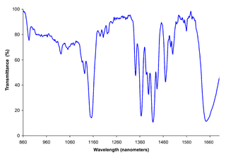
Near-infrared spectroscopy (NIRS) is a spectroscopic method that uses the near-infrared region of the electromagnetic spectrum. Typical applications include medical and physiological diagnostics and research including blood sugar, pulse oximetry, functional neuroimaging, sports medicine, elite sports training, ergonomics, rehabilitation, neonatal research, brain computer interface, urology, and neurology. There are also applications in other areas as well such as pharmaceutical, food and agrochemical quality control, atmospheric chemistry, combustion research and astronomy.

Functional integration is the study of how brain regions work together to process information and effect responses. Though functional integration frequently relies on anatomic knowledge of the connections between brain areas, the emphasis is on how large clusters of neurons – numbering in the thousands or millions – fire together under various stimuli. The large datasets required for such a whole-scale picture of brain function have motivated the development of several novel and general methods for the statistical analysis of interdependence, such as dynamic causal modelling and statistical linear parametric mapping. These datasets are typically gathered in human subjects by non-invasive methods such as EEG/MEG, fMRI, or PET. The results can be of clinical value by helping to identify the regions responsible for psychiatric disorders, as well as to assess how different activities or lifestyles affect the functioning of the brain.

Functional near-infrared spectroscopy

Functional near-infrared spectroscopy (fNIRS) is an optical brain monitoring technique which uses near-infrared spectroscopy for the purpose of functional neuroimaging. Using fNIRS, brain activity is measured by using near-infrared light to estimate cortical hemodynamic activity which occur in response to neural activity. Alongside EEG, fNIRS is one of the most common non-invasive neuroimaging techniques which can be used in portable contexts. The signal is often compared with the BOLD signal measured by fMRI and is capable of measuring changes both in oxy- and deoxyhemoglobin concentration, but can only measure from regions near the cortical surface. fNIRS may also be referred to as Optical Topography (OT) and is sometimes referred to simply as NIRS.

Transcranial Doppler

Transcranial Doppler (TCD) and transcranial color Doppler (TCCD) are types of Doppler ultrasonography that measure the velocity of blood flow through the brain's blood vessels by measuring the echoes of ultrasound waves moving transcranially. These modes of medical imaging conduct a spectral analysis of the acoustic signals they receive and can therefore be classified as methods of active acoustocerebrography. They are used as tests to help diagnose emboli, stenosis, vasospasm from a subarachnoid hemorrhage, and other problems. These relatively quick and inexpensive tests are growing in popularity. The tests are effective for detecting sickle cell disease, ischemic cerebrovascular disease, subarachnoid hemorrhage, arteriovenous malformations, and cerebral circulatory arrest. The tests are possibly useful for perioperative monitoring and meningeal infection. The equipment used for these tests is becoming increasingly portable, making it possible for a clinician to travel to a hospital, to a doctor's office, or to a nursing home for both inpatient and outpatient studies. The tests are often used in conjunction with other tests such as MRI, MRA, carotid duplex ultrasound and CT scans. The tests are also used for research in cognitive neuroscience.

Neuroimaging Set of techniques to measure and visualize aspects of the nervous system

Neuroimaging or brain imaging is the use of various techniques to either directly or indirectly image the structure, function, or pharmacology of the nervous system. It is a relatively new discipline within medicine, neuroscience, and psychology. Physicians who specialize in the performance and interpretation of neuroimaging in the clinical setting are neuroradiologists. Neuroimaging falls into two broad categories:

Event-related optical signal (EROS) is a neuroimaging technique that uses infrared light through optical fibers to measure changes in optical properties of active areas of the cerebral cortex. The fast optical signal (EROS) measures changes in infrared light scattering that occur with neural activity. Whereas techniques such as diffuse optical imaging (DOI) and near-infrared spectroscopy (NIRS) measure optical absorption of hemoglobin, and thus are based on cerebral blood flow, EROS takes advantage of the scattering properties of the neurons themselves, and thus provide a much more direct measure of cellular activity.

FreeSurfer Brain imaging software package

FreeSurfer is a brain imaging software package originally developed by Bruce Fischl, Anders Dale, Martin Sereno, and Doug Greve. Development and maintenance of FreeSurfer is now the primary responsibility of the Laboratory for Computational Neuroimaging at the Athinoula A. Martinos Center for Biomedical Imaging. FreeSurfer contains a set of programs with a common focus of analyzing magnetic resonance imaging (MRI) scans of brain tissue. It is an important tool in functional brain mapping and contains tools to conduct both volume based and surface based analysis. FreeSurfer includes tools for the reconstruction of topologically correct and geometrically accurate models of both the gray/white and pial surfaces, for measuring cortical thickness, surface area and folding, and for computing inter-subject registration based on the pattern of cortical folds.

EEG-fMRI is a multimodal neuroimaging technique whereby EEG and fMRI data are recorded synchronously for the study of electrical brain activity in correlation with haemodynamic changes in brain during the electrical activity, be it normal function or associated with disorders.

Event-related functional magnetic resonance imaging (efMRI) is a technique used in magnetic resonance imaging of medical patients.

Preclinical imaging is the visualization of living animals for research purposes, such as drug development. Imaging modalities have long been crucial to the researcher in observing changes, either at the organ, tissue, cell, or molecular level, in animals responding to physiological or environmental changes. Imaging modalities that are non-invasive and in vivo have become especially important to study animal models longitudinally. Broadly speaking, these imaging systems can be categorized into primarily morphological/anatomical and primarily molecular imaging techniques. Techniques such as high-frequency micro-ultrasound, magnetic resonance imaging (MRI) and computed tomography (CT) are usually used for anatomical imaging, while optical imaging, positron emission tomography (PET), and single photon emission computed tomography (SPECT) are usually used for molecular visualizations.

Anders Martin Dale is a prominent neuroscientist and Professor of Radiology, Neurosciences, Psychiatry, and Cognitive Science at the University of California, San Diego (UCSD), and is one of the world's leading developers of sophisticated computational neuroimaging techniques. He is the founding Director of the Center for Multimodal Imaging Genetics (CMIG) at UCSD.

Magnetic resonance imaging of the brain

Magnetic resonance imaging of the brain uses magnetic resonance imaging (MRI) to produce high quality two-dimensional or three-dimensional images of the brain and brainstem without the use of ionizing radiation (X-rays) or radioactive tracers.

Resting state fMRI

Resting state fMRI is a method of functional magnetic resonance imaging (fMRI) that is used in brain mapping to evaluate regional interactions that occur in a resting or task-negative state, when an explicit task is not being performed. A number of resting-state conditions are identified in the brain, one of which is the default mode network. These resting brain state conditions are observed through changes in blood flow in the brain which creates what is referred to as a blood-oxygen-level dependent (BOLD) signal that can be measured using fMRI.

Functional magnetic resonance spectroscopy of the brain (fMRS) uses magnetic resonance imaging (MRI) to study brain metabolism during brain activation. The data generated by fMRS usually shows spectra of resonances, instead of a brain image, as with MRI. The area under peaks in the spectrum represents relative concentrations of metabolites.

The following outline is provided as an overview of and topical guide to brain mapping:

Dynamic functional connectivity (DFC) refers to the observed phenomenon that functional connectivity changes over a short time. Dynamic functional connectivity is a recent expansion on traditional functional connectivity analysis which typically assumes that functional networks are static in time. DFC is related to a variety of different neurological disorders, and has been suggested to be a more accurate representation of functional brain networks. The primary tool for analyzing DFC is fMRI, but DFC has also been observed with several other mediums. DFC is a recent development within the field of functional neuroimaging whose discovery was motivated by the observation of temporal variability in the rising field of steady state connectivity research.

MRI sequence

An MRI sequence in magnetic resonance imaging (MRI) is a particular setting of pulse sequences and pulsed field gradients, resulting in a particular image appearance.

References

  1. Petersen, C. C. (2007). The functional organization of the barrel cortex. Neuron, 56(2), 339-355.
  2. 1 2 3 4 5 6 Mace, E.; Montaldo, G.; O., B.F.; Cohen, I.; Fink, M.; Tanter, M. (2013). "Functional ultrasound imaging of the brain: theory and basic principles". IEEE Transactions on Ultrasonics, Ferroelectrics and Frequency Control. 60 (3): 492–506. doi:10.1109/tuffc.2013.2592. PMID   23475916. S2CID   27482186.
  3. Deffieux, Thomas, et al. “Functional Ultrasound Neuroimaging: a Review of the Preclinical and Clinical State of the Art.” Current Opinion in Neurobiology, Elsevier Current Trends, 22 Feb. 2018.
  4. Knecht S, Deppe M, Ebner A, Henningsen H, Huber T, Jokeit H, Ringelstein E-B: Noninvasive determination of language lateralization by functional transcranial doppler sonography: a comparison with the Wada test. Stroke 1998, 29:82-86.
  5. Macé E, Montaldo G, Cohen I, Baulac M, Fink M, Tanter M. Functional ultrasound imaging of the brain. Nat Methods. 2011 Jul 3;8(8):662-4. doi:10.1038/nmeth.1641
  6. 1 2 Tanter M, Fink M: Ultrafast imaging in biomedical ultrasound. IEEE Transactions Ultrasonic Ferroelectric Freqquency Control 2014, 61:102-119.
  7. Bercoff J, Montaldo G, Loupas T, Savery D, Mézière F, Fink M, Tanter M. Ultrafast compound Doppler imaging: providing full blood flow characterization. IEEE Trans Ultrason Ferroelectr Freq Control. 2011 Jan;58(1):134-47. doi: 10.1109/TUFFC.2011.1780
  8. Tiran E, Deffieux T, Correia M, Maresca D, Osmanski B-F, Sieu L-A, Bergel A, Cohen I, Pernot M, Tanter M: Multiplane wave imaging increases signal-to-noise ratio in ultrafast ultrasound imaging. Physics in Medicine and Biology 2015, 60:8549-8566.
  9. Demene C, Tiran E, Sieu LA, Bergel A, Gennisson JL, Pernot M, Deffieux T, Cohen I, Tanter M: 4D microvascular imaging based on ultrafast Doppler tomography. Neuroimage 2016.
  10. 1 2 3 Célian Bimbard, Charlie Demene, Constantin Girard, et al., Multi-scale mapping along the auditory hierarchy using high-resolution functional UltraSound in the awake ferret,eLife 2018;7:e35028 doi: 10.7554/eLife.35028
  11. 1 2 Dizeux, A., Gesnik, M., Ahnine, H. et al. Functional ultrasound imaging of the brain reveals propagation of task-related brain activity in behaving primates. Nat Commun 10, 1400 (2019). https://doi.org/10.1038/s41467-019-09349-w
  12. https://iconeus.com/
  13. "The path to 4D fUS" (PDF). Iconeus. Retrieved 25 May 2020.
  14. 1 2 Gesnik M, Blaize K, Deffieux T, Gennisson JL, Sahel JA, Fink M, Picaud S, Tanter M. 3D functional ultrasound imaging of the cerebral visual system in rodents. Neuroimage. 2017 Apr 1;149:267-274. doi:10.1016/j.neuroimage.2017.01.071
  15. 1 2 3 Macé et al, Whole-Brain Functional Ultrasound Imaging Reveals Brain Modules for Visuomotor Integration, Neuron, Volume 100, Issue 5, 2018, Pages 1241-1251.e7,
  16. Rabut, C., Correia, M., Finel, V., Pezet, S., Pernot, M., Deffieux, T., & Tanter, M. (2019). 4D functional ultrasound imaging of whole-brain activity in rodents. Nature methods, 16(10), 994-997.
  17. P. J. Drew, A. Y. Shih, J. D. Driscoll, P. M. Knutsen, P. Blinder, D. Davalos, K. Akassoglou, P. S. Tsai, and D. Kleinfeld, “Chronic optical access through a polished and reinforced thinned skull,” Nature Methods, vol. 7, pp. 981–984, December 2010.
  18. Macé, E., Montaldo, G., Cohen, I. et al. Functional ultrasound imaging of the brain. Nature Methods 8, 662–664 (2011) doi:10.1038/nmeth.1641
  19. Drew, P.J. et al. Nature Methods 7, 981–984 (2010).
  20. 1 2 Kévin Blaize, Fabrice Arcizet, Marc Gesnik, Harry Ahnine, Ulisse Ferrari, Thomas Deffieux, Pierre Pouget, Frédéric Chavane, Mathias Fink, José-Alain Sahel, Mickael Tanter et Serge Picaud,Functional ultrasound imaging of deep visual cortex in awake nonhuman primates, Proceedings of the National Academy of Sciences Jun 2020, 201916787; DOI: 10.1073/pnas.1916787117
  21. Osmanski BF, Pezet S, Ricobaraza A, Lenkei Z, Tanter M. Functional ultrasound imaging of intrinsic connectivity in the living rat brain with high spatiotemporal resolution. Nat Commun. 2014 Oct 3;5:5023. doi: 10.1038/ncomms6023.
  22. Jeremy Ferrier, Elodie Tiran, Thomas Deffieux, Mickael Tanter, Zsolt Lenkei, Functional imaging evidence for task-induced deactivation and disconnection of a major default mode network hub in the mouse brain, Proceedings of the National Academy of Sciences Jun 2020, 201920475; DOI: 10.1073/pnas.1920475117
  23. Claire Rabut, Jeremt Ferrier, Adrien Bertolo, Bruno Osmanski, Xavier Mousset,Sophie Pezet, Thomas Deffieux, Zsolt Lenkei, Mickael Tanter, Pharmaco-fUS: Quantification of pharmacologically-induced dynamic changes in brain perfusion and connectivity by functional ultrasound imaging in awake mice, NeuroImage, 2020, https://doi.org/10.1016/j.neuroimage.2020.117231
  24. Sieu LA, Bergel A, Tiran E, Deffieux T, Pernot M, Gennisson JL, Tanter M,Cohen I. EEG and functional ultrasound imaging in mobile rats. Nat Methods. 2015 Sep;12(9):831-4. doi: 10.1038/nmeth.3506
  25. Tiran E, Ferrier J, Deffieux T, Gennisson JL, Pezet S, Lenkei Z, Tanter M. Transcranial Functional Ultrasound Imaging in Freely Moving Awake Mice and Anesthetized Young Rats without Contrast Agent. Ultrasound Med Biol. 2017 Aug;43(8):1679-1689. doi: 10.1016/j.ultrasmedbio.2017.03.011.
  26. Demene, Charlie; Mairesse, Jerome; Baranger, Jerome; et al. Ultrafast Doppler for neonatal brain imaging NEUROIMAGE Volume: 185 Pages: 851-856
  27. Imbault M, Chauvet D, Gennisson JL, Capelle L, Tanter M. Intraoperative Functional Ultrasound Imaging of Human Brain Activity. Sci Rep. 2017 Aug 4;7(1):7304. doi: 10.1038/s41598-017-06474-8.
  28. Soloukey Sadaf, Vincent Arnaud J. P. E., Satoer Djaina D. et al, Functional Ultrasound (fUS) During Awake Brain Surgery: The Clinical Potential of Intra-Operative Functional and Vascular Brain Mapping, Frontiers in Neuroscience,13,2020,pp. 1384