
Functional magnetic resonance imaging or functional MRI (fMRI) measures brain activity by detecting changes associated with blood flow. This technique relies on the fact that cerebral blood flow and neuronal activation are coupled. When an area of the brain is in use, blood flow to that region also increases.

Brodmann area 9, or BA9, refers to a cytoarchitecturally defined portion of the frontal cortex in the brain of humans and other primates. It contributes to the dorsolateral and medial prefrontal cortex.
The first neuroimaging technique ever is the so-called 'human circulation balance' invented by Angelo Mosso in the 1880s and able to non-invasively measure the redistribution of blood during emotional and intellectual activity. Then, in the early 1900s, a technique called pneumoencephalography was set. This process involved draining the cerebrospinal fluid from around the brain and replacing it with air, altering the relative density of the brain and its surroundings, to cause it to show up better on an x-ray, and it was considered to be incredibly unsafe for patients. A form of magnetic resonance imaging (MRI) and computed tomography (CT) were developed in the 1970s and 1980s. The new MRI and CT technologies were considerably less harmful and are explained in greater detail below. Next came SPECT and PET scans, which allowed scientists to map brain function because, unlike MRI and CT, these scans could create more than just static images of the brain's structure. Learning from MRI, PET and SPECT scanning, scientists were able to develop functional MRI (fMRI) with abilities that opened the door to direct observation of cognitive activities.

The transverse temporal gyri, also called Heschl's gyri or Heschl's convolutions, are gyri found in the area of primary auditory cortex buried within the lateral sulcus of the human brain, occupying Brodmann areas 41 and 42. Transverse temporal gyri are superior to and separated from the planum temporale by Heschl's sulcus. Transverse temporal gyri are found in varying numbers in both the right and left hemispheres of the brain and one study found that this number is not related to the hemisphere or dominance of hemisphere studied in subjects. Transverse temporal gyri can be viewed in the sagittal plane as either an omega shape or a heart shape.
Multisensory integration, also known as multimodal integration, is the study of how information from the different sensory modalities may be integrated by the nervous system. A coherent representation of objects combining modalities enables animals to have meaningful perceptual experiences. Indeed, multisensory integration is central to adaptive behavior because it allows animals to perceive a world of coherent perceptual entities. Multisensory integration also deals with how different sensory modalities interact with one another and alter each other's processing.

In psycholinguistics, language processing refers to the way humans use words to communicate ideas and feelings, and how such communications are processed and understood. Language processing is considered to be a uniquely human ability that is not produced with the same grammatical understanding or systematicity in even human's closest primate relatives.
Sensory processing is the process that organizes and distinguishes sensation from one's own body and the environment, thus making it possible to use the body effectively within the environment. Specifically, it deals with how the brain processes multiple sensory modality inputs, such as proprioception, vision, auditory system, tactile, olfactory, vestibular system, interoception, and taste into usable functional outputs.
Neuromarketing is a commercial marketing communication field that applies neuropsychology to market research, studying consumers' sensorimotor, cognitive, and affective responses to marketing stimuli. The potential benefits to marketers include more efficient and effective marketing campaigns and strategies, fewer product and campaign failures, and ultimately the manipulation of the real needs and wants of people to suit the needs and wants of marketing interests.
Sensory neuroscience is a subfield of neuroscience which explores the anatomy and physiology of neurons that are part of sensory systems such as vision, hearing, and olfaction. Neurons in sensory regions of the brain respond to stimuli by firing one or more nerve impulses following stimulus presentation. How is information about the outside world encoded by the rate, timing, and pattern of action potentials? This so-called neural code is currently poorly understood and sensory neuroscience plays an important role in the attempt to decipher it. Looking at early sensory processing is advantageous since brain regions that are "higher up" contain neurons which encode more abstract representations. However, the hope is that there are unifying principles which govern how the brain encodes and processes information. Studying sensory systems is an important stepping stone in our understanding of brain function in general.

Jonathon Stevens "Jon Driver" was a psychologist and neuroscientist. He was a leading figure in the study of perception, selective attention and multisensory integration in the normal and damaged human brain.
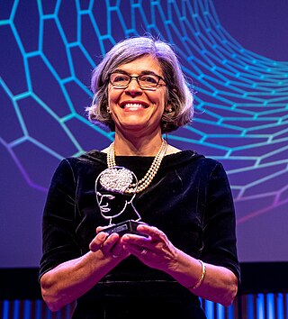
Anna Christina Nobre FBA, MAE, fNASc is a Brazilian and British cognitive neuroscientist working at the University of Oxford in England.

In neuroscience, functional specialization is a theory which suggests that different areas in the brain are specialized for different functions.
The study of memory incorporates research methodologies from neuropsychology, human development and animal testing using a wide range of species. The complex phenomenon of memory is explored by combining evidence from many areas of research. New technologies, experimental methods and animal experimentation have led to an increased understanding of the workings of memory.
Robert Turner is a British neuroscientist, physicist, and social anthropologist. He has been a director and professor at the Max Planck Institute for Human Cognitive and Brain Sciences in Leipzig, Germany, and is an internationally recognized expert in brain physics and magnetic resonance imaging (MRI). Coils inside every MRI scanner owe their shape to his ideas.

Resting state fMRI is a method of functional magnetic resonance imaging (fMRI) that is used in brain mapping to evaluate regional interactions that occur in a resting or task-negative state, when an explicit task is not being performed. A number of resting-state brain networks have been identified, one of which is the default mode network. These brain networks are observed through changes in blood flow in the brain which creates what is referred to as a blood-oxygen-level dependent (BOLD) signal that can be measured using fMRI.
Functional magnetic resonance spectroscopy of the brain (fMRS) uses magnetic resonance imaging (MRI) to study brain metabolism during brain activation. The data generated by fMRS usually shows spectra of resonances, instead of a brain image, as with MRI. The area under peaks in the spectrum represents relative concentrations of metabolites.
The following outline is provided as an overview of and topical guide to brain mapping:
James S. Hyde was an American biophysicist. He held the James S. Hyde chair in Biophysics at the Medical College of Wisconsin (MCW) where he specialized in magnetic resonance instrumentation and methodology development in two distinct areas: electron paramagnetic resonance (EPR) spectroscopy and magnetic resonance imaging (MRI). He is senior author of the widely cited 1995 paper by B.B. Biswal et al. reporting the discovery of resting state functional connectivity (fcMRI) in the human brain. He also served as Director of the National Biomedical EPR Center, a Research Resource supported by the National Institutes of Health. He was author of more than 400 peer-reviewed papers and review articles and held 35 U.S. Patents. He was recognized by Festschrifts in both EPR and fcMRI.
Michelle Hampson is an American neuroscientist who is an Associate Professor of Radiology and Biomedical Imaging at Yale University. She serves as director of real-time functional magnetic resonance imaging.

Sophie Molholm is an American neuroscientist, who is the director of the Cognitive Neurophysiology Laboratory (CNL) and the Human Clinical Phenotyping Core (HCP) at the Albert Einstein College of Medicine in New York. She is professor (tenured) of Paediatrics, Neuroscience and Psychiatry, and Behavioral Sciences, and was endowed as the Muriel and Harold Block Faculty Scholar in Mental Illness at Einstein (2012–2017).