Haidinger's brush

Last updated
Simulated appearance of Haidinger's brush for vertically polarized light. Size and intensity exaggerated for clarity. Orientation varies with that of polarization of light source. Haidinger's brush.jpg
Simulated appearance of Haidinger's brush for vertically polarized light. Size and intensity exaggerated for clarity. Orientation varies with that of polarization of light source.

Haidinger's brush, more commonly known as Haidinger's brushes is an image produced by the eye, an entoptic phenomenon, first described by Austrian physicist Wilhelm Karl von Haidinger in 1844. Haidinger saw it when he looked through various minerals that polarized light. [1] [2]

Contents

Many people are able to perceive polarization of light. [3] Haidinger's brushes may be seen as a yellowish horizontal bar or bow-tie shape (with "fuzzy" ends, hence the name "brush") visible in the center of the visual field against the blue sky viewed while facing away from the sun, or on any bright background. It typically occupies roughly 3–5 degrees of vision, about twice or three times the width of one's thumb held at arm's length. The direction of light polarization is perpendicular to the yellow bar (i.e., vertical if the bar is horizontal). Fainter bluish or purplish areas may be visible between the yellow brushes (see illustration). Haidinger's brush may also be seen by looking at a white area on many LCD flat panel computer screens (due to the polarization effect of the display), in which case it is often diagonal.

Physiological causes

Haidinger's brush is usually attributed to the dichroism of the xanthophyll pigment found in the macula lutea. As described by the Fresnel laws, the behavior and distribution of oblique rays in the cylindrical geometry of the foveal blue cones produce an extrinsic dichroism. The size of the brush is consistent with the size of the macula.

It is thought that the macula's dichroism arises from some of its pigment molecules being arranged circularly; (the small proportion of circularly arranged molecules accounts for the faintness of the phenomenon.) Xanthophyll pigments tend to be parallel to visive nerves that (because the fovea is not flat), are almost orthogonal to the fovea in its central part but nearly parallel in its outer region. As a result, two different areas of the fovea can be sensitive to two different degrees of polarization. [4]

Seeing Haidinger's brush

Simulated appearance of a computer screen viewed through a polarizer, showing typical size and intensity of Haidinger's brush Haidinger's Brush (as demonstrated on Wikipedia's main page).jpg
Simulated appearance of a computer screen viewed through a polarizer, showing typical size and intensity of Haidinger's brush

Many people find it difficult to see Haidinger's brush initially. It is very faint, much more so than generally indicated in illustrations, and, like other stabilized images, tends to appear and disappear.

It is most easily seen when it can be made to move. Because it is always positioned on the macula, there is no way to make it move laterally, but it can be made to rotate, by viewing a white surface through a rotating polarizer, or by slowly tilting one's head to one side.

To see Haidinger's brush, start by using a polarizer, such as a lens from a pair of polarizing sunglasses. Gaze at an evenly lit, textureless surface through the lens and rotate the polarizer.

An option is to use the polarizer built into a computer's LCD screen. Look at a white area on the screen, and slowly tilt the head (a CRT monitor has no polarizer, and will not work for this purpose unless a separate polarizer is used).

It appears with more distinctness against a blue background. With practice, it is possible to see it in the naturally polarized light of a blue sky. Minnaert recommended practicing first with a polarizer, then trying it without. [5] The areas of the sky with the strongest polarization are those 90 degrees away from the sun. Minnaert said that after a minute of gazing at the sky, "a kind of marble effect will appear. This is followed shortly by Haidinger's brush." He commented that not all observers see it in the same way. Some see the yellow pattern as solid and the blue pattern as interrupted, as in the illustrations on this page. Some see the blue as solid and the yellow as interrupted, and some see it alternating between the two states.

Use

The fact that the sensation of Haidinger's brush corresponds with the visual field of the macula means that it can be utilised in training people to look at objects with their macula. People with certain types of strabismus may undergo an adaptation whereupon they look at the object of attention not with their fovea (at the centre of the macula) but with an eccentric region of the retina. This adaptation is known as eccentric fixation. To aid in training a person to look at an object with their fovea rather than their eccentric retinal zone, a training device can be used. One such apparatus utilises a rotating polarised plate backlit with a bright white light. Wearing blue spectacles (to enhance the Haidinger's brush image) and an occluder over the other eye, the user will hopefully notice the Haidinger's brush where their macula correlates with their visual field. The goal of the training is for the user to learn to look at the test object in such a way that the Haidinger's brush overlaps the test object (and the viewer is thus now looking at it with their fovea). The reason for such training is that the healthy fovea is far greater in its resolving power than any other part of the retina. Another diagnostic method that utilises birefringent properties of the retinal tissue is retinal birefringence scanning, that can be used in case of severe amblyopia or when the specialist lacks a cooperation from the patient.

See also

Related Research Articles

<span class="mw-page-title-main">Circular polarization</span> Polarization state

In electrodynamics, circular polarization of an electromagnetic wave is a polarization state in which, at each point, the electromagnetic field of the wave has a constant magnitude and is rotating at a constant rate in a plane perpendicular to the direction of the wave.

<span class="mw-page-title-main">Polarization (waves)</span> Property of waves that can oscillate with more than one orientation

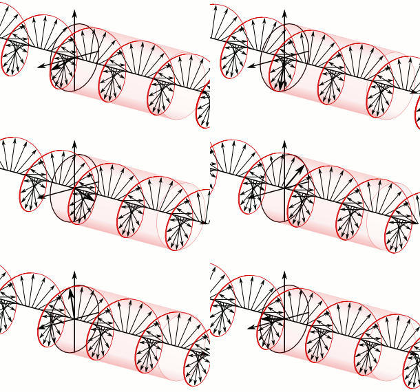
Polarization is a property of transverse waves which specifies the geometrical orientation of the oscillations. In a transverse wave, the direction of the oscillation is perpendicular to the direction of motion of the wave. A simple example of a polarized transverse wave is vibrations traveling along a taut string (see image); for example, in a musical instrument like a guitar string. Depending on how the string is plucked, the vibrations can be in a vertical direction, horizontal direction, or at any angle perpendicular to the string. In contrast, in longitudinal waves, such as sound waves in a liquid or gas, the displacement of the particles in the oscillation is always in the direction of propagation, so these waves do not exhibit polarization. Transverse waves that exhibit polarization include electromagnetic waves such as light and radio waves, gravitational waves, and transverse sound waves in solids.

<span class="mw-page-title-main">Retina</span> Part of the eye

The retina is the innermost, light-sensitive layer of tissue of the eye of most vertebrates and some molluscs. The optics of the eye create a focused two-dimensional image of the visual world on the retina, which then processes that image within the retina and sends nerve impulses along the optic nerve to the visual cortex to create visual perception. The retina serves a function which is in many ways analogous to that of the film or image sensor in a camera.

<span class="mw-page-title-main">Birefringence</span> Property of materials whose refractive index depends on light polarization and direction

Birefringence is the optical property of a material having a refractive index that depends on the polarization and propagation direction of light. These optically anisotropic materials are described as birefringent or birefractive. The birefringence is often quantified as the maximum difference between refractive indices exhibited by the material. Crystals with non-cubic crystal structures are often birefringent, as are plastics under mechanical stress.

<span class="mw-page-title-main">Macula</span> Oval-shaped pigmented area near the center of the retina

The macula (/ˈmakjʊlə/) or macula lutea is an oval-shaped pigmented area in the center of the retina of the human eye and in other animals. The macula in humans has a diameter of around 5.5 mm (0.22 in) and is subdivided into the umbo, foveola, foveal avascular zone, fovea, parafovea, and perifovea areas.

<span class="mw-page-title-main">Peripheral vision</span> Area of ones field of vision outside of the point of fixation

Peripheral vision, or indirect vision, is vision as it occurs outside the point of fixation, i.e. away from the center of gaze or, when viewed at large angles, in the "corner of one's eye". The vast majority of the area in the visual field is included in the notion of peripheral vision. "Far peripheral" vision refers to the area at the edges of the visual field, "mid-peripheral" vision refers to medium eccentricities, and "near-peripheral", sometimes referred to as "para-central" vision, exists adjacent to the center of gaze.

<span class="mw-page-title-main">Xanthophyll</span> Chemical compounds subclass

Xanthophylls are yellow pigments that occur widely in nature and form one of two major divisions of the carotenoid group; the other division is formed by the carotenes. The name is from Greek: xanthos (ξανθός), meaning "yellow", and phyllon (φύλλον), meaning "leaf"), due to their formation of the yellow band seen in early chromatography of leaf pigments.
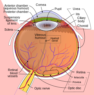
<span class="mw-page-title-main">Fovea centralis</span> Small pit in the retina of the eye responsible for all central vision

The fovea centralis is a small, central pit composed of closely packed cones in the eye. It is located in the center of the macula lutea of the retina.

<span class="mw-page-title-main">3D display</span> Display device

A 3D display is a display device capable of conveying depth to the viewer. Many 3D displays are stereoscopic displays, which produce a basic 3D effect by means of stereopsis, but can cause eye strain and visual fatigue. Newer 3D displays such as holographic and light field displays produce a more realistic 3D effect by combining stereopsis and accurate focal length for the displayed content. Newer 3D displays in this manner cause less visual fatigue than classical stereoscopic displays.

Entoptic phenomena are visual effects whose source is within the human eye itself.

<span class="mw-page-title-main">Polarimetry</span> Measurement and interpretation of the polarization of transverse waves

Polarimetry is the measurement and interpretation of the polarization of transverse waves, most notably electromagnetic waves, such as radio or light waves. Typically polarimetry is done on electromagnetic waves that have traveled through or have been reflected, refracted or diffracted by some material in order to characterize that object.

<span class="mw-page-title-main">Central retinal artery</span>

The central retinal artery branches off the ophthalmic artery, running inferior to the optic nerve within its dural sheath to the eyeball.

<span class="mw-page-title-main">Retinal pigment epithelium</span>

The pigmented layer of retina or retinal pigment epithelium (RPE) is the pigmented cell layer just outside the neurosensory retina that nourishes retinal visual cells, and is firmly attached to the underlying choroid and overlying retinal visual cells.

Scanning laser polarimetry is the use of polarised light to measure the thickness of the retinal nerve fiber layer (RNFL) as part of a glaucoma workup. The GDx-VCC is one example.

<span class="mw-page-title-main">Bird vision</span> Senses for birds

Vision is the most important sense for birds, since good eyesight is essential for safe flight. Birds have a number of adaptations which give visual acuity superior to that of other vertebrate groups; a pigeon has been described as "two eyes with wings". Birds are theropod dinosaurs, and the avian eye resembles that of other reptiles, with ciliary muscles that can change the shape of the lens rapidly and to a greater extent than in the mammals. Birds have the largest eyes relative to their size in the animal kingdom, and movement is consequently limited within the eye's bony socket. In addition to the two eyelids usually found in vertebrates, bird's eyes are protected by a third transparent movable membrane. The eye's internal anatomy is similar to that of other vertebrates, but has a structure, the pecten oculi, unique to birds.

<span class="mw-page-title-main">Mammalian eye</span>

Mammals normally have a pair of eyes. Although mammalian vision is not so excellent as bird vision, it is at least dichromatic for most of mammalian species, with certain families possessing a trichromatic color perception.

<span class="mw-page-title-main">Sunstone (medieval)</span> Ancient navigational aid

The sunstone is a type of mineral attested in several 13th–14th-century written sources in Iceland, one of which describes its use to locate the Sun in a completely overcast sky. Sunstones are also mentioned in the inventories of several churches and one monastery in 14th–15th-century Iceland and Germany.

<span class="mw-page-title-main">Optical phenomenon</span> Observable events that result from the interaction of light and matter

Optical phenomena are any observable events that result from the interaction of light and matter.

Microperimetry, sometimes called fundus-controlled perimetry, is a type of visual field test which uses one of several technologies to create a "retinal sensitivity map" of the quantity of light perceived in specific parts of the retina in people who have lost the ability to fixate on an object or light source. The main difference with traditional perimetry instruments is that, microperimetry includes a system to image the retina and an eye tracker to compensate eye movements during visual field testing.

References

  1. Haidinger, Wilhelm (1844). "Über das directe Erkennen des polarisirten Lichts und der Lage der Polarisationsebene" [On direct observation of polarized light and the orientation of the plane of polarization]. Annalen der Physik. 139 (9): 29–39. Bibcode:1844AnP...139...29H. doi:10.1002/andp.18441390903.
  2. O'Shea, R. P.; Temple, S. E.; Misson, G. P.; Wade, N. J.; Bach, M. (2020). "Historical context, scientific context, and translation of Haidinger's (1844) discovery of naked-eye visibility of the polarization of light". arXiv: 2010.15252 [physics.hist-ph].
  3. O'Shea, R. P.; Misson, G. P.; Temple, S. E. (2021). "Seeing polarization of light with the naked eye". Current Biology. 31 (4): R178–R179. Bibcode:2021CBio...31.R178O. doi: 10.1016/j.cub.2020.12.037 . PMID   33621501. S2CID   231991919.
  4. Le Floch, A; Ropars G; Enoch J; Lakshminarayanan V (2010). "The polarization sense in human vision". Vision Res. 50 (20): 2048–2054. doi: 10.1016/j.visres.2010.07.007 . PMID   20638403. Open Access logo PLoS transparent.svg
  5. Minnaert, M. G. J. (1940). Light and colour in the open air (H. M. Kremer-Priest, Trans.). London: G. Bell and Sons.

Further reading