Floater

Last updated
Floater
Floaters.png
Simulated image of separated, unclumped floaters against a blue sky
Specialty Ophthalmology, optometry
Differential diagnosis Migraine aura [1]

Floaters or eye floaters are sometimes visible deposits (e.g., the shadows of tiny structures of protein or other cell debris projected onto the retina) within the eye's vitreous humour ("the vitreous"), which is normally transparent, or between the vitreous and retina. [2] [3] They can become particularly noticeable when looking at a blank surface or an open monochromatic space, such as blue sky. Each floater can be measured by its size, shape, consistency, refractive index, and motility. [2] They are also called muscae volitantes (Latin for 'flying flies'), or mouches volantes (from the same phrase in French). [4] The vitreous usually starts out transparent, but imperfections may gradually develop as one ages. The common type of floater, present in most people's eyes, is due to these degenerative changes of the vitreous. The perception of floaters, which may be annoying or problematic to some people, is known as myodesopsia, [5] or, less commonly, as myodaeopsia, myiodeopsia, or myiodesopsia. It is not often treated, except in severe cases, where vitrectomy (surgery), laser vitreolysis, and medication may be effective.

Contents

Floaters are visible either because of the shadows that imperfections cast on the retina, [6] or because of the refraction of light that passes through them, and can appear alone or together with several others as a clump in one's visual field. They may appear as spots, threads, or fragments of "cobwebs", which float slowly before the observer's eyes, and move especially in the direction the eyes move. [3] As these objects exist within the eye itself, they are not optical illusions but are entoptic phenomena (caused by the eye itself). They are not to be confused with visual snow, which is similar to the static on a television screen, although these two conditions may co-exist as part of a number of visual disturbances which include starbursts, trails, and afterimages.

Signs and symptoms

External videos
Corps-flottant.jpg
Nuvola apps kaboodle.svg What are those floaty things in your eye? - Michael Mauser, 4:04, TED-Ed

Floaters are from objects in pockets of liquid within the vitreous humour, the thick fluid or gel that fills the eye, [7] or between the vitreous and the retina. The vitreous humour, or vitreous body, is a jelly-like, transparent substance that fills the majority of the eye. It lies within the vitreous chamber behind the lens, and is one of the four optical components of the eye. [8] Thus, floaters follow the rapid motions of the eye, while drifting slowly within the pocket of liquid. [9] When they are first noticed, the natural reaction is to attempt to look directly at them. However, attempting to shift one's gaze toward them can be difficult because floaters follow the motion of the eye, remaining to the side of the direction of gaze. Floaters are, in fact, visible only because they do not remain perfectly fixed within the eye. Although the blood vessels of the eye also obstruct light, they are invisible under normal circumstances because they are fixed in location relative to the retina, and the brain "tunes out" stabilized images through neural adaptation. [3]

Floaters are particularly noticeable when looking at a blank surface or an open monochromatic space, such as blue sky. Despite the name "floaters", many of these specks have a tendency to sink toward the bottom of the eyeball, in whichever way the eyeball is oriented; the supine position (looking up or lying back) tends to concentrate them near the fovea, which is the center of gaze, while the textureless and evenly lit sky forms an ideal background against which to view them. [7] The brightness of the daytime sky also causes the eyes' pupils to contract, reducing the aperture, which makes floaters less blurry and easier to see.

Floaters present at birth usually remain lifelong, while those that appear later may disappear within weeks or months. [10] They are not uncommon, and do not cause serious problems for most people. [9] A survey of optometrists in 2002 suggested that an average of 14 patients per month per optometrist presented with symptoms of floaters in the UK. [11] However, floaters are more than a nuisance and a distraction to those with severe cases, especially if the spots seem constantly to drift through the field of vision. The shapes are shadows projected onto the retina by tiny structures of protein or other cell debris discarded over the years and trapped in the vitreous humour or between the vitreous and retina. Floaters can even be seen when the eyes are closed on especially bright days, when sufficient light penetrates the eyelids to cast the shadows.[ citation needed ] It is not, however, only elderly persons who are troubled by floaters; they can also become a problem to younger people, especially if they are myopic. [12] They are also common after cataract or clear lens extraction operations or after trauma.[ citation needed ]

Floaters are able to catch and refract light in ways that somewhat blur vision temporarily until the floater moves to a different area. Often they trick persons who are troubled by floaters into thinking they see something out of the corner of their eye that really is not there. Most people come to terms with the problem, after a time, and learn to ignore their floaters. For persons with severe floaters it is nearly impossible to ignore completely the large masses that constantly stay within almost direct view.[ citation needed ]

In the case of young people, particularly those under 35, symptomatic floaters are likely suspended within a posterior region of the eye known as the pre-macular bursa. Such floaters appear well-defined and usually bear the appearance of a 'crystal worm' or cobweb. Due to their proximity to the retina, the floaters have a significant effect on the visual field for patients. In addition, such floaters are often in the central visual axis as it moves with the intravitreal currents of the eye. Research on floaters of the pre-macular bursa is minimal, and safe treatment for patients with this disturbance that does not warrant major vitrectomy has yet to be discovered. Moreover, the cause and prognosis for such floaters also remains to be found. Some doctors suggest such floaters may resolve over time, should the floaters move away from the retina.[ citation needed ]

Causes

There are various causes for the appearance of floaters, of which the most common are described here.

Floaters can occur when eyes age; in rare cases, floaters may be a sign of retinal detachment or a retinal tear. [13] [9]

Vitreous syneresis

Vitreous syneresis (liquefaction) and contraction with age can cause vitreous floaters. Additionally, trauma or injury to the globe can cause them. [14] [9]

Vitreous detachments and retinal detachments

Weiss ring: a large, ring shaped floater that is sometimes seen if the vitreous body releases from the back of the eye Vitreous Floaters.jpg
Weiss ring: a large, ring shaped floater that is sometimes seen if the vitreous body releases from the back of the eye

In time, the liquefied vitreous body loses support and its framework contracts. This leads to posterior vitreous detachment, in which the vitreous membrane is released from the sensory retina. [9] During this detachment, the shrinking vitreous can stimulate the retina mechanically, [9] causing the patient to see random flashes across the visual field, sometimes referred to as "flashers", a symptom more formally referred to as photopsia. The ultimate release of the vitreous around the optic nerve head sometimes makes a large floater appear, usually in the shape of a ring ("Weiss ring"). [15] As a complication, part of the retina might be torn off by the departing vitreous membrane, in a process known as retinal detachment. This will often leak blood into the vitreous, which is seen by the patient as a sudden appearance of numerous small dots, moving across the whole field of vision. Retinal detachment requires immediate medical attention, [9] as it can easily cause blindness. Consequently, both the appearance of flashes and the sudden onset of numerous small floaters should be rapidly investigated by an eye care provider. [16]

Posterior vitreous detachment is more common in people who:

Regression of the hyaloid artery

The hyaloid artery, an artery running through the vitreous humour during the fetal stage of development, regresses in the third trimester of pregnancy. Its disintegration can sometimes leave cell matter. [18]

Other common causes

Patients with retinal tears may experience floaters if red blood cells are released from leaky blood vessels, and those with uveitis or vitritis, as in toxoplasmosis, may experience multiple floaters and decreased vision due to the accumulation of white blood cells in the vitreous humour. [19]

Other causes for floaters include cystoid macular edema and asteroid hyalosis. The latter is an anomaly of the vitreous humour, whereby calcium clumps attach themselves to the collagen network. The bodies that are formed in this way move slightly with eye movement, but then return to their fixed position.[ citation needed ]

Diagnosis

Floaters are often readily observed by an ophthalmologist or an optometrist with the use of an ophthalmoscope or slit lamp. However, if the floater is near the retina, it may not be visible to the observer even if it appears large to the patient.

Increasing background illumination or using a pinhole to effectively decrease pupil diameter may allow a person to obtain a better view of his or her own floaters. The head may be tilted in such a way that one of the floaters drifts towards the central axis of the eye. In the sharpened image the fibrous elements are more conspicuous. [20]

The presence of retinal tears with new onset of floaters was surprisingly high (14%; 95% confidence interval, 12–16%) as reported in a meta-analysis published as part of the Rational Clinical Examination Series in the Journal of the American Medical Association. [21] Patients with new onset flashes and/or floaters, especially when associated with visual loss or restriction in the visual field, should seek more urgent ophthalmologic evaluation.

Treatment

While surgeries do exist to correct for severe cases of floaters, there are no medications (including eye drops) that can correct for this vitreous deterioration. Floaters are often caused by the normal aging process and will usually become less bothersome as a person learns to ignore them. Looking up/down and left/right will cause the floaters to leave the direct field of vision as the vitreous humour swirls around due to the sudden movement. [22] If floaters significantly increase in numbers and/or severely affect vision, then one of the treatments below may be necessary.

As of 2017, insufficient evidence is available to compare the safety and efficacy of surgical vitrectomy with laser vitreolysis for the treatment of floaters. A 2017 Cochrane Review did not find any relevant studies that compared the two treatments. [23]

Aggressive marketing campaigns have promoted the use of laser vitreolysis for the treatment of floaters. [24] [25] No strong evidence currently exists for the treatment of floaters with laser vitreolysis. The strongest available evidence comparing these two treatment modalities are retrospective case series. [26]

Surgery

Vitrectomy may be successful in treating more severe cases. [27] [28] The technique usually involves making three openings through the part of the sclera known as the pars plana. Of these small gauge instruments, one is an infusion port to resupply a saline solution and maintain the pressure of the eye, the second is a fiber optic light source, and the third is a vitrector. The vitrector has a reciprocating cutting tip attached to a suction device. This design reduces traction on the retina via the vitreous material. A variant sutureless, self-sealing technique is sometimes used.

Like most invasive surgical procedures, however, vitrectomy carries a risk of complications, [29] including: retinal detachment, anterior vitreous detachment and macular edema – which can threaten vision or worsen existing floaters (in the case of retinal detachment).

Laser vitreolysis

Laser vitreolysis is a possible treatment option for the removal of vitreous strands and opacities (floaters). In this procedure an ophthalmic laser (usually a yttrium aluminium garnet (YAG) laser) applies a series of nanosecond pulses of low-energy laser light to evaporate the vitreous opacities and to sever the vitreous strands. When performed with a YAG laser designed specifically for vitreolysis, reported side effects and complications associated with vitreolysis are rare. However, YAG lasers have traditionally been designed for use in the anterior portion of the eye, i.e. posterior capsulotomy and iridotomy treatments. As a result, they often provide a limited view of the vitreous, which can make it difficult to identify the targeted floaters and membranes. They also carry a high risk of damage to surrounding ocular tissue. Accordingly, vitreolysis is not widely practised, being performed by very few specialists. One of them, John Karickhoff, has performed the procedure more than 1,400 times and claims a 90 percent success rate. [30] However, the MedicineNet web site states that "there is no evidence that this [laser treatment] is effective. The use of a laser also poses significant risks to the vision in what is otherwise a healthy eye." [31]

Medication

Enzymatic vitreolysis has been trialed to treat vitreomacular adhesion (VMA) and anomalous posterior vitreous detachment. Although the mechanism of action may have an effect on clinically significant floaters, as of March 2015 there are no clinical trials being undertaken to determine whether this may be a therapeutic alternative to either conservative management or vitrectomy. [32]

Atropine

Dropping low doses of atropine onto the eye dilates the pupil, thus reducing shadow formation on the retina by floaters. [33] [34]

Research

The VDM project aims to find an effective, low-risk treatment for floaters. [35] So far, there have been studies using colloidal gold or indocyanine green (ICG) injected into the eye followed by a low-energy laser to target problematic floaters, and this has shown to be successful on vitreous opacities obtained during vitrectomy and in rabbits. [36] [37] [38] [39] [40]

Epidemiology

A vitreous detachment typically affects patients older than the age of 50 and increases in prevalence by age 80. Individuals who are myopic or nearsighted have an increased risk of vitreous floaters. Additionally, eyes with an inflammatory disease after direct trauma to the globe or have recently undergone eye surgery have an increased chance of developing a vitreous floater. Men and women appear to be affected equally. [14]

In other animals

It has been theorized that non-human animals are capable of seeing floaters, as most mammals have anatomically similar eye structures to humans. However, floaters in animals are capable of damaging their vision. Animals with synchysis have an increased risk for retinal detachment and may also require surgery. [41]

See also

Related Research Articles

<span class="mw-page-title-main">Diabetic retinopathy</span> Medical condition

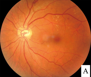
Diabetic retinopathy, is a medical condition in which damage occurs to the retina due to diabetes. It is a leading cause of blindness in developed countries.

<span class="mw-page-title-main">Vitrectomy</span> Surgery to remove vitreous humor from the eye

Vitrectomy is a surgery to remove some or all of the vitreous humor from the eye.

<span class="mw-page-title-main">Retinoschisis</span> Eye disease involving splitting of the retina

Retinoschisis is an eye disease characterized by the abnormal splitting of the retina's neurosensory layers, usually in the outer plexiform layer. Retinoschisis can be divided into degenerative forms which are very common and almost exclusively involve the peripheral retina and hereditary forms which are rare and involve the central retina and sometimes the peripheral retina. The degenerative forms are asymptomatic and involve the peripheral retina only and do not affect the visual acuity. Some rarer forms result in a loss of vision in the corresponding visual field.

The National Eye Institute (NEI) is part of the U.S. National Institutes of Health (NIH), an agency of the U.S. Department of Health and Human Services. The mission of NEI is "to eliminate vision loss and improve quality of life through vision research." NEI consists of two major branches for research: an extramural branch that funds studies outside NIH and an intramural branch that funds research on the NIH campus in Bethesda, Maryland. Most of the NEI budget funds extramural research.

<span class="mw-page-title-main">Eye surgery</span> Surgery performed on the eye or its adnexa

Eye surgery, also known as ophthalmic surgery or ocular surgery, is surgery performed on the eye or its adnexa. Eye surgery is part of ophthalmology and is performed by an ophthalmologist or eye surgeon. The eye is a fragile organ, and requires due care before, during, and after a surgical procedure to minimize or prevent further damage. An eye surgeon is responsible for selecting the appropriate surgical procedure for the patient, and for taking the necessary safety precautions. Mentions of eye surgery can be found in several ancient texts dating back as early as 1800 BC, with cataract treatment starting in the fifth century BC. It continues to be a widely practiced class of surgery, with various techniques having been developed for treating eye problems.

<span class="mw-page-title-main">Phacoemulsification</span> Method of cataract surgery

Phacoemulsification is a cataract surgery method in which the internal lens of the eye which has developed a cataract is emulsified with the tip of an ultrasonic handpiece and aspirated from the eye. Aspirated fluids are replaced with irrigation of balanced salt solution to maintain the volume of the anterior chamber during the procedure. This procedure minimises the incision size and reduces the recovery time and risk of surgery induced astigmatism.

<span class="mw-page-title-main">Retinal detachment</span> Medical condition of the eye

Retinal detachment is a disorder of the eye in which the retina peels away from its underlying layer of support tissue. Initial detachment may be localized, but without rapid treatment the entire retina may detach, leading to vision loss and blindness. It is a surgical emergency.

<span class="mw-page-title-main">Posterior vitreous detachment</span> Eye condition where vitreous membrane detaches from the retina

A posterior vitreous detachment (PVD) is a condition of the eye in which the vitreous membrane separates from the retina. It refers to the separation of the posterior hyaloid membrane from the retina anywhere posterior to the vitreous base.

A scleral buckle is one of several ophthalmologic procedures that can be used to repair a retinal detachment. Retinal detachments are usually caused by retinal tears, and a scleral buckle can be used to close the retinal break, both for acute and chronic retinal detachments.

<span class="mw-page-title-main">Photopsia</span> Presence of perceived flashes of light in ones field of vision

Photopsia is the presence of perceived flashes of light in the field of vision.

Eales disease is a type of obliterative vasculopathy, also known as angiopathia retinae juvenilis, periphlebitis retinae or primary perivasculitis of the retina. It was first described by the British ophthalmologist Henry Eales (1852–1913) in 1880 and is a rare ocular disease characterized by inflammation and possible blockage of retinal blood vessels, abnormal growth of new blood vessels (neovascularization), and recurrent retinal and vitreal hemorrhages.

<span class="mw-page-title-main">Intermediate uveitis</span> Medical condition

Intermediate uveitis is a form of uveitis localized to the vitreous and peripheral retina. Primary sites of inflammation include the vitreous of which other such entities as pars planitis, posterior cyclitis, and hyalitis are encompassed. Intermediate uveitis may either be an isolated eye disease or associated with the development of a systemic disease such as multiple sclerosis or sarcoidosis. As such, intermediate uveitis may be the first expression of a systemic condition. Infectious causes of intermediate uveitis include Epstein–Barr virus infection, Lyme disease, HTLV-1 virus infection, cat scratch disease, and hepatitis C.

<span class="mw-page-title-main">Epiretinal membrane</span> Eye disease

Epiretinal membrane or macular pucker is a disease of the eye in response to changes in the vitreous humor or more rarely, diabetes. Sometimes, as a result of immune system response to protect the retina, cells converge in the macular area as the vitreous ages and pulls away in posterior vitreous detachment (PVD).

<span class="mw-page-title-main">Optic pit</span> Medical condition

Optic pit, optic nerve pit, or optic disc pit (ODP) is rare a congenital excavation (or regional depression) of the optic disc (also optic nerve head), resulting from a malformation during development of the eye. The incidence of ODP is 1 in 10,000 people with no predilection for either gender. There is currently no known risk factors for their development. Optic pits are important because they are associated with posterior vitreous detachments (PVD) and even serous retinal detachments.

<span class="mw-page-title-main">Macular hole</span> Medical condition

A macular hole is a small break in the macula, located in the center of the eye's light-sensitive tissue called the retina.

<span class="mw-page-title-main">Intraocular hemorrhage</span> Medical condition

Intraocular hemorrhage is bleeding inside the eye. Bleeding can occur from any structure of the eye where there is vasculature or blood flow, including the anterior chamber, vitreous cavity, retina, choroid, suprachoroidal space, or optic disc.

<span class="mw-page-title-main">Vitreous hemorrhage</span> Medical condition

Vitreous hemorrhage is the extravasation, or leakage, of blood into the areas in and around the vitreous humor of the eye. The vitreous humor is the clear gel that fills the space between the lens and the retina of the eye. A variety of conditions can result in blood leaking into the vitreous humor, which can cause impaired vision, floaters, and photopsia.

<span class="mw-page-title-main">Vitreomacular adhesion</span> Human medical condition

Vitreomacular adhesion (VMA) is a human medical condition where the vitreous gel of the human eye adheres to the retina in an abnormally strong manner. As the eye ages, it is common for the vitreous to separate from the retina. But if this separation is not complete, i.e. there is still an adhesion, this can create pulling forces on the retina that may result in subsequent loss or distortion of vision. The adhesion in of itself is not dangerous, but the resulting pathological vitreomacular traction (VMT) can cause severe ocular damage.

Robert Machemer was a German-American ophthalmologist, ophthalmic surgeon, and inventor. He is sometimes called the "father of modern retinal surgery."

Sickle cell retinopathy can be defined as retinal changes due to blood vessel damage in the eye of a person with a background of sickle cell disease. It can likely progress to loss of vision in late stages due to vitreous hemorrhage or retinal detachment. Sickle cell disease is a structural red blood cell disorder leading to consequences in multiple systems. It is characterized by chronic red blood cell destruction, vascular injury, and tissue ischemia causing damage to the brain, eyes, heart, lungs, kidneys, spleen, and musculoskeletal system.

References

  1. Johnson, D.; Hollands, H. (2011-11-28). "Acute-onset floaters and flashes". Canadian Medical Association Journal. 184 (4). Canadian Medical Association: 431. doi:10.1503/cmaj.110686. ISSN   0820-3946. PMC   3291672 . PMID   22125334.
  2. 1 2 Cline D; Hofstetter HW; Griffin JR. Dictionary of Visual Science. 4th ed. Butterworth-Heinemann, Boston 1997. ISBN   0-7506-9895-0
  3. 1 2 3 "Facts About Floaters". National Eye Institute . October 2009. Archived from the original on March 21, 2022. Retrieved September 8, 2018.
  4. Archived at Ghostarchive and the Wayback Machine : "Adult Eye Health: Mayo Clinic Radio (5 minutes in to 8 minute talk)". YouTube .
  5. From Greek μυιώδης "fly-like" (Myiodes was also the name of a fly-deterring deity) and ὄψις "sight."
  6. American Academy of Ophthalmology. "Floaters and Flashes: A Closer Look" (pamphlet) San Francisco: AAO, 2006. ISBN   1-56055-371-5
  7. 1 2 "Eye floaters and spots; Floaters or spots in the eye". National Eye Institute. Archived from the original on October 23, 2007. Retrieved 2008-02-01.
  8. Saladin, Kenneth (2012). Anatomy & Physiology: A Unity of Form and Function. New York: McGraw-Hill. p. 614. ISBN   978-0-07-337825-1.
  9. 1 2 3 4 5 6 7 Eye Floaters and Flashes, Animation (Youtube video). Alila Medical Media. October 13, 2020.
  10. "Floaters may remain indefinitely". Archived from the original on September 24, 2015.
  11. Craig Goldsmith; Tristan McMullan; Ted Burton (2007). "Floaterectomy Versus Conventional Pars Plana Vitrectomy For Vitreous Floaters". Digital Journal of Ophthalmology. Archived from the original on 2021-04-18. Retrieved 2008-04-11.{{cite journal}}: Cite journal requires |journal= (help)
  12. "Flashes and Floaters". kindermanneye.com. Retrieved 2022-08-02.
  13. "Floaters". NHS choices. NHS GOV.UK. Retrieved September 17, 2017.
  14. 1 2 Reece Bergstrom; Craig N. Czyz. (2020). "Vitreous Floaters". Statpearls. PMID   29262091. Creative Commons by small.svg  This article incorporates text available under the CC BY 4.0 license.
  15. "Flashes & Floaters". The Eye Digest. Retrieved 2008-02-24.
  16. "Flashes and Floaters (Posterior Vitreous Detachment)". St. Luke's Cataract & Laser Institute. Archived from the original on 2010-05-02. Retrieved 2008-02-01.
  17. American Academy of Ophthalmology – patient education pamphlet
  18. Petersen, Hans Peter (1968). "PERSISTENCE OF THE BERGMEISTER PAPILLA WITH GLIAL OVERGROWTH: Various Diagnostic Problems". Acta Ophthalmologica. 46 (3): 430–440. doi:10.1111/j.1755-3768.1968.tb02826.x. ISSN   1755-375X.
  19. Alan G. Kabat; Joseph W. Sowka (April 2009). "A clinician's guide to flashes and floaters" (PDF). optometry.co.uk. Archived from the original (PDF) on 2012-07-08. Retrieved 2008-04-10.
  20. Judith Lee, and Gretchyn Bailey; Dr. Vance Thompson. "Eye floaters and spots". All about vision. Retrieved 2008-02-01.
  21. Hollands H, Johnson D, Brox AC, Almeida D, Simel DL, Sharma S. Acute-onset floaters and flashes: is this patient at risk for retinal detachment? JAMA. 2009 November 25;302(20):2243–9.
  22. "Flashes and Floaters" (PDF). National Library of Medicine . 19 May 2010. Archived from the original (PDF) on July 28, 2013. Retrieved 9 December 2013.
  23. Kokavec J, Wu Z, Sherwin JC, Ang AJ, Ang GS (2017). "Nd:YAG laser vitreolysis versus pars plana vitrectomy for vitreous floaters". Cochrane Database Syst Rev. 2017 (6): CD011676. doi:10.1002/14651858.CD011676.pub2. PMC   6481890 . PMID   28570745.
  24. "Vitreolysis: Overview". Ellex.com. Archived from the original on 2016-05-01. Retrieved 2016-04-11.
  25. "Floaters". Ellex.com. Archived from the original on 2016-05-02. Retrieved 2016-04-11.
  26. Delaney YM, Oyinloye A, Benjamin L (January 2002). "Nd:YAG vitreolysis and pars plana vitrectomy: surgical treatment for vitreous floaters". Eye (Lond). 16 (1): 21–6. doi: 10.1038/sj.eye.6700026 . PMID   11913884. S2CID   25021874.
  27. Roth M, Trittibach P, Koerner F, Sarra G (Sep 2005). "[Pars plana vitrectomy for idiopathic vitreous floaters.]". Klin Monatsbl Augenheilkd. 222 (9): 728–32. doi:10.1055/s-2005-858497. PMID   16175483. S2CID   260205524.
  28. "Pars plana vitrectomy (PPV) & floater only vitrectomy". Archived from the original on 2007-01-05. Retrieved 2008-02-01.
  29. "Facts About Floaters". nih.gov. Archived from the original on 2013-12-07.
  30. "Surgery To Rid Eye "Floaters" Scrutinized". CBSNews HealthWatch. Retrieved December 5, 2011.
  31. "Eye Floaters". MedicineNet . Retrieved December 5, 2011.
  32. "Pharmacologic vitreolysis". One Clear Vision. Archived from the original on 2015-03-28. Retrieved 2015-03-22.
  33. Kaymak, H.; Frickle, A.; Hagan, P.; Auffarth, G.; Breyer, D.; Klabe, K.; Fulga, R. (2017-10-10). "Training and Innovation". ESCRS | EUROPEAN SOCIETY OF CATARACTS AND REFRACTIVE SURGEONS. Archived from the original on 2022-03-21. Retrieved 2022-03-21.
  34. Kaymak, Hakan; Fricke, Andreas; Mauritz, Yvonne; Löwinger, Anne; Klabe, Karsten; Breyer, Detlev; Lagenbucher, Achim; Seitz, Berthold; Schaeffel, Frank (November 2018). "Short-term effects of low-concentration atropine eye drops on pupil size and accommodation in young adult subjects". Graefe's Archive for Clinical and Experimental Ophthalmology = Albrecht von Graefes Archiv für Klinische und Experimentelle Ophthalmologie. 256 (11): 2211–2217. doi:10.1007/s00417-018-4112-8. ISSN   1435-702X. PMC   6208716 . PMID   30145612.
  35. "Home". vdmresearch.org.
  36. Sauvage, Félix; Fraire, Juan C.; Remaut, Katrien; Sebag, J.; Peynshaert, Karen; Harrington, Michael; Van de Velde, Frans J.; Xiong, Ranhua; Tassignon, Marie-José; Brans, Toon; Braeckmans, Kevin; De Smedt, Stefaan C. (23 July 2019). "Photoablation of Human Vitreous Opacities by Light-Induced Vapor Nanobubbles". ACS Nano. 13 (7): 8401–8416. doi:10.1021/acsnano.9b04050. hdl: 1854/LU-8627551 . PMID   31287662. S2CID   195870794.
  37. "Laser-heated nanoparticles bust eye "floaters"".
  38. Sauvage, Félix (21 June 2021). "Gold nanoparticles for the treatment of eye floaters by light-induced vapor nanobubbles". Investigative Ophthalmology & Visual Science. 62 (8): 3307.
  39. Sauvage, Félix; Nguyen, Van Phuc; Li, Yanxiu; Harizaj, Aranit; Sebag, J.; Roels, Dimitri; Van Havere, Viktor; Peynshaert, Karen; Xiong, Ranhua; Fraire, Juan C.; Tassignon, Marie-José; Remaut, Katrien; Paulus, Yannis M.; Braeckmans, Kevin; De Smedt, Stefaan C. (17 March 2022). "Laser-induced nanobubbles safely ablate vitreous opacities in vivo". Nature Nanotechnology. 17 (5): 552–559. Bibcode:2022NatNa..17..552S. doi:10.1038/s41565-022-01086-4. hdl: 1854/LU-8755668 . PMID   35302088. S2CID   247501386.
  40. "Safer eye floater treatments come with a burst of nanobubbles".
  41. "Eye Can See Clearly Now: Vitreal Floaters in Pets". 8 May 2021.