Sclera

Last updated
Sclera
Sclera.PNG
The sclera, as separated from the cornea by the corneal limbus.
Details
Part of Eye
System Visual system
Artery Anterior ciliary arteries, long posterior ciliary arteries, short posterior ciliary arteries
Identifiers
Latin sclera
MeSH D012590
TA98 A15.2.02.002
TA2 6750
FMA 58269
Anatomical terminology

The sclera, [note 1] also known as the white of the eye or, in older literature, as the tunica albuginea oculi, is the opaque, fibrous, protective outer layer of the eye containing mainly collagen and some crucial elastic fiber. [2]

Contents

In the development of the embryo, the sclera is derived from the neural crest. [3] In children, it is thinner and shows some of the underlying pigment, appearing slightly blue. In the elderly, fatty deposits on the sclera can make it appear slightly yellow. People with dark skin can have naturally darkened sclerae, the result of melanin pigmentation. [4]

In humans, and some other vertebrates, the whole sclera is white or pale, contrasting with the coloured iris. The cooperative eye hypothesis suggests that the pale sclera evolved as a method of nonverbal communication that makes it easier for one individual to identify where another individual is looking. Other mammals with white or pale sclera include chimpanzees, many orangutans, some gorillas, and bonobos. [5]

Structure

Closeup of goat eye.jpg
Goat eye with relatively dark sclera and horizontal pupil
A cow's eye.jpg
Cow eye with dark sclera
Western Lowland Gorilla2010501A (cropped).jpg
Rare whites in a western lowland gorilla

The sclera forms the posterior five-sixths of the connective tissue coat of the human eyeball. It is continuous with the dura mater and the cornea, and maintains the shape of the eyeball, offering resistance to internal and external forces, and provides an attachment for the extraocular muscle insertions. The sclera is perforated by many nerves and vessels passing through the posterior scleral foramen, the hole that is formed by the optic nerve. At the optic disc, the outer two-thirds of the sclera continues with the dura mater (outer coat of the brain) via the dural sheath of the optic nerve. The inner third joins with some choroidal tissue to form a plate (lamina cribrosa) across the optic nerve with perforations through which the optic fibers (fasciculi) pass. The thickness of the sclera varies from 1 mm at the posterior pole to 0.3 mm just behind the insertions of the four rectus muscles. The sclera's blood vessels are mainly on the surface. Along with the vessels of the conjunctiva (which is a thin layer covering the sclera), those in the episclera render the inflamed eye bright red. [6]

In many vertebrates, the sclera is reinforced with plates of cartilage or bone, together forming a circular structure called the sclerotic ring . In primitive fish, this ring consists of four plates, but the number is lower in many living ray-finned fishes, and much higher in lobe-finned fishes, crocodilians, various reptiles, and birds. The ring has disappeared in many groups, including living amphibians, some reptiles and fish, and all mammals. [7]

The eyes of all non-human primates have been thought to be dark with small, barely visible sclera, but recent research has suggested that white sclera are not uncommon in chimpanzees, and are also present in other mammals. [8]

Histology

Histologically speaking, it is characterized as dense connective tissue made primarily of type 1 collagen fibers. [9] The collagen of the sclera is continuous with the cornea. From outer to innermost, the four layers of the sclera are:

The sclera is opaque due to the irregularity of the Type I [10] collagen fibers, as opposed to the near-uniform thickness and parallel arrangement of the corneal collagen. Moreover, the cornea bears more mucopolysaccharide (a carbohydrate that has among its repeating units a nitrogenous sugar, hexosamine) to embed the fibrils.

The cornea, unlike the sclera, has six layers. The middle (third in number; provided the first layer being the anterior and outermost and the sixth layer being the posterior and the inner most), thickest layer is also called the stroma. The sclera, like the cornea, contains a basal endothelium, above which there is the lamina fusca, containing a high count of pigment cells. [6]

Sometimes, very small gray-blue spots can appear on the sclera, a harmless condition called scleral melanocytosis.

Function

Human eyes are somewhat distinctive in the animal kingdom in that the sclera is very plainly visible whenever the eye is open. This is not just due to the white color of the human sclera, which many other species share, but also to the fact that the human iris is relatively small and comprises a significantly smaller portion of the exposed eye surface compared to other animals. It is theorized that this adaptation evolved because of humans’ social nature as the eye became a useful communication tool in addition to a sensory organ. It is believed that the exposed sclera of the human eye makes it easier for one individual to identify where another individual is looking, increasing the efficacy of this particular form of nonverbal communication, called cooperative eye hypothesis. [11] Animal researchers have also found that, in the course of their domestication, dogs have also developed the ability to pick up visual cues from the eyes of humans. Dogs do not seem to use this form of communication with one another and only look for visual information from the eyes of humans. [12]

Another hypothesis states that the distinctiveness of the human's sclera is due to genetic drift and sexual selection. A visibly white sclera is perceived as a sign of good health and youthfullness, [13] this could have been a criteria of selection when looking for a mate. In this case, humans' ability to communicate with their eyes (glancing, cluing at others) would only be a consequence of a very visible sclera. [14]

Trauma

The bony area that makes up the human eye socket provides exceptional protection to the sclera. However, if the sclera is ruptured by a blunt force or is penetrated by a sharp object, the recovery of full former vision is usually rare. If pressure is applied slowly, the eye is actually very elastic. However, most ruptures involve objects moving at some velocity. The cushion of orbital fat protects the sclera from head-on blunt forces, but damage from oblique forces striking the eye from the side is not prevented by this cushion. Hemorrhaging and a dramatic drop in intraocular pressure are common, along with a reduction in visual perception to only broad hand movements and the presence or absence of light. However, a low-velocity injury which does not puncture and penetrate the sclera requires only superficial treatment and the removal of the object. Sufficiently small objects which become embedded and which are subsequently left untreated may eventually become surrounded by a benign cyst, causing no other damage or discomfort. [15]

Thermal trauma

The sclera is rarely damaged by brief exposure to heat: the eyelids provide exceptional protection, and the fact that the sclera is covered in layers of moist tissue means that these tissues are able to cause much of the offending heat to become dissipated as steam before the sclera itself is damaged. Even relatively low-temperature molten metals when splashed against an open eye have been shown to cause very little damage to the sclera, even while creating detailed casts of the surrounding eyelashes. Prolonged exposure, however—on the order of 30 seconds—at temperatures above 45 °C (113 °F) will begin to cause scarring, and above 55 °C (131 °F) will cause extreme changes in the sclera and surrounding tissue. Such long exposures even in industrial settings are virtually nonexistent. [15]

Chemical injury

The sclera is highly resistant to injury from brief exposure to toxic chemicals. The reflexive production of tears at the onset of chemical exposure tends to quickly wash away such irritants, preventing further harm. Acids with a pH below 2.5 are the source of greatest acidic burn risk, with sulfuric acid, the kind present in car batteries and therefore commonly available, being among the most dangerous in this regard. However, acid burns, even severe ones, seldom result in loss of the eye. [15]

Alkali burns, on the other hand, such as those resulting from exposure to ammonium hydroxide or ammonium chloride or other chemicals with a pH above 11.5, will cause cellular tissue in the sclera to saponify and should be considered medical emergencies requiring immediate treatment. [15]

Abnormal coloring

Redness of the sclera is typically caused by eye irritation causing blood vessels to expand, such as in conjunctivitis ("pink eye"). Episcleritis is a generally benign condition of the episclera causing eye redness. Scleritis is a serious inflammatory disease of the sclera causing redness of the sclera often progressing to purple.

Yellowing or a light green color of the sclera is a visual symptom of jaundice.

In cases of osteogenesis imperfecta, the sclera may appear to have a blue tint, more pronounced than the slight blue tint seen in children. The blue tint is caused by the showing of the underlying uveal tract (choroid and retinal pigment epithelium).

In those with Ehlers-Danlos syndrome, the sclera may be tinted blue due to the lack of proper connective tissue. [16]

In very rare but severe cases of kidney failure and liver failure, the sclera may turn black.[ citation needed ]

See also

Notes

  1. The word sclera ( /ˈsklɛərə/ or /ˈsklɪərə/ ; both are common), plural sclerae ( /ˈsklɛəri/ or /ˈsklɪəri/ ) or scleras, is from the Greek skleros, meaning hard. [1]

Related Research Articles

<span class="mw-page-title-main">Iris (anatomy)</span> Colored part of an eye

The iris is a thin, annular structure in the eye in most mammals and birds, responsible for controlling the diameter and size of the pupil, and thus the amount of light reaching the retina. In optical terms, the pupil is the eye's aperture, while the iris is the diaphragm. Eye color is defined by the iris.

<span class="mw-page-title-main">Cornea</span> Transparent front layer of the eye

The cornea is the transparent front part of the eye that covers the iris, pupil, and anterior chamber. Along with the anterior chamber and lens, the cornea refracts light, accounting for approximately two-thirds of the eye's total optical power. In humans, the refractive power of the cornea is approximately 43 dioptres. The cornea can be reshaped by surgical procedures such as LASIK.

<span class="mw-page-title-main">Choroid</span> Vascular layer of the eye, containing connective tissue, and lying between the retina and the sclera

The choroid, also known as the choroidea or choroid coat, is a part of the uvea, the vascular layer of the eye. It contains connective tissues, and lies between the retina and the sclera. The human choroid is thickest at the far extreme rear of the eye, while in the outlying areas it narrows to 0.1 mm. The choroid provides oxygen and nourishment to the outer layers of the retina. Along with the ciliary body and iris, the choroid forms the uveal tract.

<span class="mw-page-title-main">Eye surgery</span> Surgery performed on the eye or its adnexa

Eye surgery, also known as ophthalmic surgery or ocular surgery, is surgery performed on the eye or its adnexa. Eye surgery is part of ophthalmology and is performed by an ophthalmologist or eye surgeon. The eye is a fragile organ, and requires due care before, during, and after a surgical procedure to minimize or prevent further damage. An eye surgeon is responsible for selecting the appropriate surgical procedure for the patient, and for taking the necessary safety precautions. Mentions of eye surgery can be found in several ancient texts dating back as early as 1800 BC, with cataract treatment starting in the fifth century BC. It continues to be a widely practiced class of surgery, with various techniques having been developed for treating eye problems.

<span class="mw-page-title-main">Superior oblique muscle</span> Part of the eye

The superior oblique muscle or obliquus oculi superior is a fusiform muscle originating in the upper, medial side of the orbit which abducts, depresses and internally rotates the eye. It is the only extraocular muscle innervated by the trochlear nerve.

<span class="mw-page-title-main">Human eye</span> Sensory organ of vision

The human eye is an organ of the sensory nervous system that reacts to visible light and allows the use of visual information for various purposes including seeing things, keeping balance, and maintaining circadian rhythm.

<span class="mw-page-title-main">Extraocular muscles</span> Seven extrinsic muscles of the eye

The extraocular muscles, or extrinsic ocular muscles, are the seven extrinsic muscles of the eye in humans and other animals. Six of the extraocular muscles, the four recti muscles, and the superior and inferior oblique muscles, control movement of the eye. The other muscle, the levator palpebrae superioris, controls eyelid elevation. The actions of the six muscles responsible for eye movement depend on the position of the eye at the time of muscle contraction.

<span class="mw-page-title-main">Descemet's membrane</span> Membrane in the cornea of the eye

Descemet's membrane is the basement membrane that lies between the corneal proper substance, also called stroma, and the endothelial layer of the cornea. It is composed of different kinds of collagen than the stroma. The endothelial layer is located at the posterior of the cornea. Descemet's membrane, as the basement membrane for the endothelial layer, is secreted by the single layer of squamous epithelial cells that compose the endothelial layer of the cornea.

<span class="mw-page-title-main">Tenon's capsule</span> Membrane surrounding the eye forming a socket in which it moves

Tenon's capsule, also known as the Tenon capsule, fascial sheath of the eyeball or the fascia bulbi, is a thin membrane which envelops the eyeball from the optic nerve to the corneal limbus, separating it from the orbital fat and forming a socket in which it moves.

<span class="mw-page-title-main">Pinguecula</span> Medical condition

A pinguecula is a common type of conjunctival stromal degeneration in the eye. It appears as an elevated yellow-white plaque in the bulbar conjunctiva near the limbus. Calcification may also seen occasionally.

<span class="mw-page-title-main">Ocular prosthesis</span> Type of craniofacial prosthesis

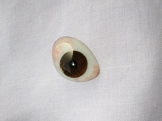
An ocular prosthesis, artificial eye or glass eye is a type of craniofacial prosthesis that replaces an absent natural eye following an enucleation, evisceration, or orbital exenteration. The prosthesis fits over an orbital implant and under the eyelids. Though often referred to as a glass eye, the ocular prosthesis roughly takes the shape of a convex shell and is made of medical grade plastic acrylic. A few ocular prostheses today are made of cryolite glass. A variant of the ocular prosthesis is a very thin hard shell known as a scleral shell which can be worn over a damaged or eviscerated eye. Makers of ocular prosthetics are known as ocularists. An ocular prosthesis does not provide vision; this would be a visual prosthesis. Someone with an ocular prosthesis is altogether blind on the affected side and has monocular vision.

<span class="mw-page-title-main">Equine vision</span>

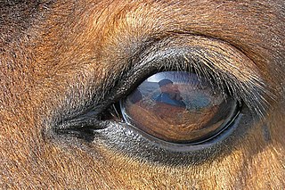
The equine eye is one of the largest of any land mammal. Its visual abilities are directly related to the animal's behavior; for example, it is active during both day and night, and it is a prey animal. Both the strengths and weaknesses of the horse's visual abilities should be taken into consideration when training the animal, as an understanding of the horse's eye can help to discover why the animal behaves the way it does in various situations.

The nerve fibers forming the optic nerve exit the eye posteriorly through a hole in the sclera that is occupied by a mesh-like structure called the lamina cribrosa. It is formed by a multilayered network of collagen fibers that extend from the scleral canal wall. The nerve fibers that comprise the optic nerve run through pores formed by these collagen beams. In humans, a central retinal artery is located slightly off-center in the nasal direction.

A staphyloma is an abnormal protrusion of the uveal tissue through a weak point in the eyeball. The protrusion is generally black in colour, due to the inner layers of the eye. It occurs due to weakening of outer layer of eye by an inflammatory or degenerative condition. It may be of five types, depending on the location on the eyeball.

<span class="mw-page-title-main">Mammalian eye</span>

Mammals normally have a pair of eyes. Although mammalian vision is not so excellent as bird vision, it is at least dichromatic for most of mammalian species, with certain families possessing a trichromatic color perception.

Histology is the study of the minute structure, composition, and function of tissues. Mature human vocal cords are composed of layered structures which are quite different at the histological level.

<span class="mw-page-title-main">Globe (human eye)</span> Region of the eyeball apart from its appendages

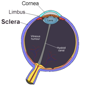
The globe of the eye, or bulbus oculi, is the frontmost sensory organ of the human ocular system, going from the cornea at the front, to the anterior part of the optic nerve at the back. More simply, the eyeball itself, as well as the ganglion cells in the retina that eventually transmit visual signals through the optic nerve. A hollow structure, the bulbus oculi is composed of a wall enclosing a cavity filled with fluid with three coats: the sclera, choroid, and the retina. Normally, the bulbus oculi is bulb-like structure. However, the bulbus oculi is not completely spherical. Its anterior surface, transparent and more curved, is known as the cornea of the bulbus oculi.

<span class="mw-page-title-main">Accessory visual structures</span> External parts of the eye including eyebrow, eyelid, and lacrimal apparatus

The accessory visual structures are the protecting and supporting structures (adnexa) of the eye, including the eyebrow, eyelids, and lacrimal apparatus. The eyebrows, eyelids, eyelashes, lacrimal gland and drainage apparatus all play a crucial role with regards to globe protection, lubrication, and minimizing the risk of ocular infection. The adnexal structures also help to keep the cornea moist and clean.

<span class="mw-page-title-main">Scleral reinforcement surgery</span>

Scleral reinforcement is a surgical procedure used to reduce or stop further macular damage caused by high myopia, which can be degenerative.

<span class="mw-page-title-main">Eagle eye</span>

The eagle eye is among the sharpest in the animal kingdom, with an eyesight estimated at 4 to 8 times stronger than that of the average human. Although an eagle may only weigh 10 pounds (4.5 kg), its eyes are roughly the same size as those of a human. Eagle weight varies: a small eagle could weigh 700 grams (1.5 lb), while a larger one could weigh 6.5 kilograms (14 lb); an eagle of about 10 kilograms (22 lb) weight could have eyes as big as that of a human who weighs 200 pounds (91 kg). Although the size of the eagle eye is about the same as that of a human being, the back side shape of the eagle eye is flatter. Their eyes are stated to be larger in size than their brain, by weight. Color vision with resolution and clarity are the most prominent features of eagles' eyes, hence sharp-sighted people are sometimes referred to as "eagle-eyed". Eagles can identify five distinctly colored squirrels and locate their prey even if hidden.

References

  1. Mosby's Medical, Nursing, and Allied Health Dictionary (4th ed.). St. Louis: Mosby. 1994. p. 1402. ISBN   978-0815161134.
  2. Cassin, Barbara; Solomon, Sheila A.B. (1990). Dictionary of Eye Terminology (2nd ed.). Gainesville, Fla.: Triad Pub. Co. ISBN   978-0937404331.
  3. Hermann D. Schubert. Anatomy of the Orbit "New York Eye and Ear Infirmary of Mount Sinai - New York City - NYEE" (PDF). Archived from the original (PDF) on 2008-10-08. Retrieved 2008-05-19.
  4. Mukamal, Reena (30 July 2020). "Why Are the Whites of My Eyes Discolored?". American Academy of Ophthalmology. Retrieved 11 December 2020.
  5. Clark, Isabelle R.; Lee, Kevin C.; Poux, Tucker; Langergraber, Kevin E.; Mitani, John C.; Watts, David; Reed, James; Sandel, Aaron A. (2023-03-01). "White sclera is present in chimpanzees and other mammals". Journal of Human Evolution. 176: 103322. doi: 10.1016/j.jhevol.2022.103322 . ISSN   0047-2484. PMC   9998187 . PMID   36706647. S2CID   256314941.
  6. 1 2 "eye, human."Encyclopædia Britannica from Encyclopædia Britannica 2006 Ultimate Reference Suite DVD 2009
  7. Romer, Alfred Sherwood; Parsons, Thomas S. (1977). The Vertebrate Body. Philadelphia, PA: Holt-Saunders International. p. 461. ISBN   978-0-03-910284-5.
  8. Clark, Isabella R.; Lee, Kevin C.; Poux, Tucker; Langergraber, Kevin E.; Mitani, John C.; Watts, David; Reed, James; Sandel, Aaron A. (March 2023). "White sclera is present in chimpanzees and other mammals". Journal of Human Evolution. 176. doi:10.1016/j.jhevol.2022.103322. PMC   9998187 . PMID   36706647.
  9. Pradeep, Tejus; Mehra, Divy; Le, Patrick H. (2023), "Histology, Eye", StatPearls, Treasure Island (FL): StatPearls Publishing, PMID   31335063 , retrieved 2023-11-11
  10. Keeley, FW; Morin, JD; Vesely, S (November 1984). "Characterization of collagen from normal human sclera". Experimental Eye Research. 39 (5): 533–42. doi:10.1016/0014-4835(84)90053-8. PMID   6519194.
  11. Michael Tomasello, Brian Hare, Hagen Lehmann, Josep Call. "Reliance on head versus eyes in the gaze following of great apes and human infants: the cooperative eye hypothesis" http://www.chrisknight.co.uk/wp-content/uploads/2008/06/eyes-cooperation.pdf
  12. Director and Producer: Dan Child, Executive Producer: Andrew Kohen (2010-01-06). "The Secret Life of the Dog". Horizon. BBC. BBC2.
  13. Russell, R., Sweda, J. R., Porcheron, A., & Mauger, E. (2014). Sclera color changes with age and is a cue for perceiving age, health, and beauty. Psychology and Aging, 29(3), 626–635. https://doi.org/10.1037/a0036142
  14. Caspar, K.R., Biggemann, M., Geissmann, T. et al. Ocular pigmentation in humans, great apes, and gibbons is not suggestive of communicative functions. Sci Rep 11, 12994 (2021). https://doi.org/10.1038/s41598-021-92348-z
  15. 1 2 3 4 Peter G Watson (11 April 2012). "Chapter 9". The Sclera and Systemic Disorders. JP Medical Ltd. p. 293. ISBN   978-1-907816-07-9.
  16. Asanad, S., Bayomi, M., Brown, D., Buzzard, J., Lai, E., Ling, C., Miglani, T., Mohammed, T., Tsai, J., Uddin, O., & Singman, E. (2022). Ehlers-Danlos syndromes and their manifestations in the visual system. Frontiers in Medicine, 9. https://doi.org/10.3389/fmed.2022.996458