In anatomy, the orbit is the cavity or socket/hole of the skull in which the eye and its appendages are situated. "Orbit" can refer to the bony socket, or it can also be used to imply the contents. In the adult human, the volume of the orbit is 30 millilitres, of which the eye occupies 6.5 ml. The orbital contents comprise the eye, the orbital and retrobulbar fascia, extraocular muscles, cranial nerves II, III, IV, V, and VI, blood vessels, fat, the lacrimal gland with its sac and duct, the eyelids, medial and lateral palpebral ligaments, cheek ligaments, the suspensory ligament, septum, ciliary ganglion and short ciliary nerves.

An eyelid is a thin fold of skin that covers and protects an eye. The levator palpebrae superioris muscle retracts the eyelid, exposing the cornea to the outside, giving vision. This can be either voluntarily or involuntarily. "Palpebral" means relating to the eyelids. Its key function is to regularly spread the tears and other secretions on the eye surface to keep it moist, since the cornea must be continuously moist. They keep the eyes from drying out when asleep. Moreover, the blink reflex protects the eye from foreign bodies. A set of specialized hairs known as lashes grow from the upper and lower eyelid margins to further protect the eye from dust and debris.

A facelift, technically known as a rhytidectomy, is a type of cosmetic surgery procedure intended to give a more youthful facial appearance. There are multiple surgical techniques and exercise routines. Surgery usually involves the removal of excess facial skin, with or without the tightening of underlying tissues, and the redraping of the skin on the patient's face and neck. Exercise routines tone underlying facial muscles without surgery. Surgical facelifts are effectively combined with eyelid surgery (blepharoplasty) and other facial procedures and are typically performed under general anesthesia or deep twilight sleep.

The ophthalmic artery (OA) is an artery of the head. It is the first branch of the internal carotid artery distal to the cavernous sinus. Branches of the ophthalmic artery supply all the structures in the orbit around the eye, as well as some structures in the nose, face, and meninges. Occlusion of the ophthalmic artery or its branches can produce sight-threatening conditions.

The corrugator supercilii muscle is a small, narrow, pyramidal muscle of the face. It arises from the medial end of the superciliary arch; it inserts into the deep surface of the skin of the eyebrow.

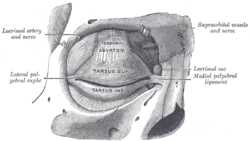
The orbicularis oculi is a muscle in the face that closes the eyelids. It arises from the nasal part of the frontal bone, from the frontal process of the maxilla in front of the lacrimal groove, and from the anterior surface and borders of a short fibrous band, the medial palpebral ligament.

The tarsi or tarsal plates are two comparatively thick, elongated plates of dense connective tissue, about 10 mm (0.39 in) in length for the upper eyelid and 5 mm for the lower eyelid; one is found in each eyelid, and contributes to its form and support. They are located directly above the lid margins. The tarsus has a lower and upper part making up the palpebrae.

The fascia lata is the deep fascia of the thigh. It encloses the thigh muscles and forms the outer limit of the fascial compartments of thigh, which are internally separated by the medial intermuscular septum and the lateral intermuscular septum. The fascia lata is thickened at its lateral side where it forms the iliotibial tract, a structure that runs to the tibia and serves as a site of muscle attachment.

The lacrimal canaliculi are the small channels in each eyelid that drain lacrimal fluid, from the lacrimal puncta to the lacrimal sac. This forms part of the lacrimal apparatus that drains lacrimal fluid from the surface of the eye to the nasal cavity.

The lacrimal sac or lachrymal sac is the upper dilated end of the nasolacrimal duct, and is lodged in a deep groove formed by the lacrimal bone and frontal process of the maxilla. It connects the lacrimal canaliculi, which drain tears from the eye's surface, and the nasolacrimal duct, which conveys this fluid into the nasal cavity. Lacrimal sac occlusion leads to dacryocystitis.

The posterior lacrimal crest is a vertical bony ridge on the orbital surface of the lacrimal bone. It divides the bone into two parts. It gives origin to the lacrimal part of the orbicularis oculi muscle.
The anterior lacrimal crest is a bony projection on the frontal process of the maxilla. It creates the lateral margin of the lacrimal sac fossa and is continuous with the orbital margin. The medial palpebral ligament is attached to anterior lacrimal crest. It is an important structure to avoid damaging during rhinoplasty.

The frontal process of the maxilla is a strong plate, which projects upward, medialward, and backward from the maxilla, forming part of the lateral boundary of the nose.

The angular artery is an artery of the face. It is the terminal part of the facial artery. It ascends to the medial angle of the eye's orbit. It is accompanied by the angular vein. It ends by anastomosing with the dorsal nasal branch of the ophthalmic artery. It supplies the lacrimal sac, the orbicularis oculi muscle, and the outer side of the nose.
The medial palpebral arteries are arteries of the head that contribute arterial blood supply to the eyelids. They are derived from the ophthalmic artery; a single medial palpebral artery issues from the ophthalmic artery before splitting into a superior and an inferior medial palpebral artery, each supplying one eyelid.

In anatomy, the orbital septum is a membranous sheet that acts as the anterior (frontal) boundary of the orbit. It extends from the orbital rims to the eyelids. It forms the fibrous portion of the eyelids.

The zygomatic branches of the facial nerve (malar branches) are nerves of the face. They run across the zygomatic bone to the lateral angle of the orbit. Here, they supply the orbicularis oculi muscle, and join with filaments from the lacrimal nerve and the zygomaticofacial branch of the maxillary nerve (CN V2).
Oculoplastics, or oculoplastic surgery, includes a wide variety of surgical procedures that deal with the orbit, eyelids, tear ducts, and the face. It also deals with the reconstruction of the eye and associated structures.
The lateral palpebral raphe is a ligamentous band near the eye. Its existence is contentious, and many sources describe it as the continuation of nearby muscles. It is formed from the lateral ends of the orbicularis oculi muscle. It connects the orbicularis oculi muscle, the frontosphenoidal process of the zygomatic bone, and the tarsi of the eyelids.

The following outline is provided as an overview of and topical guide to human anatomy: