
Glaucoma is a group of eye diseases that result in damage to the optic nerve and cause vision loss. The most common type is open-angle glaucoma, in which the drainage angle for fluid within the eye remains open, with less common types including closed-angle glaucoma and normal-tension glaucoma. Open-angle glaucoma develops slowly over time without pain. Peripheral vision may begin to decrease, followed by central vision, resulting in blindness if not treated. Closed-angle glaucoma can present gradually or suddenly. The sudden presentation may involve severe eye pain, blurred vision, mid-dilated pupil, redness of the eye, and nausea. Vision loss from glaucoma, once it has occurred, is permanent. Eyes affected by glaucoma are referred to as being glaucomatous.

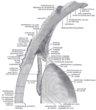
In humans and most mammals and birds, the iris is a thin, annular structure in the eye, responsible for controlling the diameter and size of the pupil, and thus the amount of light reaching the retina. Eye color is defined by the iris. In optical terms, the pupil is the eye's aperture, while the iris is the diaphragm.

The cornea is the transparent front part of the eye that covers the iris, pupil, and anterior chamber. Along with the anterior chamber and lens, the cornea refracts light, accounting for approximately two-thirds of the eye's total optical power. In humans, the refractive power of the cornea is approximately 43 dioptres. The cornea can be reshaped by surgical procedures such as LASIK.

The aqueous humour is a transparent water-like fluid similar to blood plasma, but containing low protein concentrations. It is secreted from the ciliary body, a structure supporting the lens of the eyeball. It fills both the anterior and the posterior chambers of the eye, and is not to be confused with the vitreous humour, which is located in the space between the lens and the retina, also known as the posterior cavity or vitreous chamber. Blood cannot normally enter the eyeball.

The ciliary body is a part of the eye that includes the ciliary muscle, which controls the shape of the lens, and the ciliary epithelium, which produces the aqueous humor. The aqueous humor is produced in the non-pigmented portion of the ciliary body. The ciliary body is part of the uvea, the layer of tissue that delivers oxygen and nutrients to the eye tissues. The ciliary body joins the ora serrata of the choroid to the root of the iris.

The ciliary muscle is an intrinsic muscle of the eye formed as a ring of smooth muscle in the eye's middle layer, uvea. It controls accommodation for viewing objects at varying distances and regulates the flow of aqueous humor into Schlemm's canal. It also changes the shape of the lens within the eye but not the size of the pupil which is carried out by the sphincter pupillae muscle and dilator pupillae.
Cyclospasm is the contraction of the ciliary muscle in the eye, in the accommodation of focus for near vision. Cyclospasm may also exert tensions on the trabecular meshwork, opening the pores and facilitating outflow of the aqueous humour into the canal of Schlemm. The increase in aqueous humour outflow is desirable for patients with glaucoma.
The anterior chamber (AC) is the aqueous humor-filled space inside the eye between the iris and the cornea's innermost surface, the endothelium. Hyphema, anterior uveitis and glaucoma are three main pathologies in this area. In hyphema, blood fills the anterior chamber as a result of a hemorrhage, most commonly after a blunt eye injury. Anterior uveitis is an inflammatory process affecting the iris and ciliary body, with resulting inflammatory signs in the anterior chamber. In glaucoma, blockage of the trabecular meshwork prevents the normal outflow of aqueous humour, resulting in increased intraocular pressure, progressive damage to the optic nerve head, and eventually blindness.
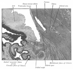
Schlemm's canal is a circular lymphatic-like vessel in the eye. It collects aqueous humor from the anterior chamber and delivers it into the episcleral blood vessels. Canaloplasty may be used to widen it.

Trabeculectomy is a surgical procedure used in the treatment of glaucoma to relieve intraocular pressure by removing part of the eye's trabecular meshwork and adjacent structures. It is the most common glaucoma surgery performed and allows drainage of aqueous humor from within the eye to underneath the conjunctiva where it is absorbed. This outpatient procedure was most commonly performed under monitored anesthesia care using a retrobulbar block or peribulbar block or a combination of topical and subtenon anesthesia. Due to the higher risks associated with bulbar blocks, topical analgesia with mild sedation is becoming more common. Rarely general anesthesia will be used, in patients with an inability to cooperate during surgery.
Glaucoma is a group of diseases affecting the optic nerve that results in vision loss and is frequently characterized by raised intraocular pressure (IOP). There are many glaucoma surgeries, and variations or combinations of those surgeries, that facilitate the escape of excess aqueous humor from the eye to lower intraocular pressure, and a few that lower IOP by decreasing the production of aqueous humor.

The scleral spur in the visual system is a protrusion of the sclera into the anterior chamber. The spur is an annular structure composed of collagen in the human eye.

Myocilin, trabecular meshwork inducible glucocorticoid response (TIGR), also known as MYOC, is a protein which in humans is encoded by the MYOC gene. Mutations in MYOC are a major cause of glaucoma.
Pseudoexfoliation syndrome, often abbreviated as PEX and sometimes as PES or PXS, is an aging-related systemic disease manifesting itself primarily in the eyes which is characterized by the accumulation of microscopic granular amyloid-like protein fibers. Its cause is unknown, although there is speculation that there may be a genetic basis. It is more prevalent in women than men, and in persons past the age of seventy. Its prevalence in different human populations varies; for example, it is prevalent in Scandinavia. The buildup of protein clumps can block normal drainage of the eye fluid called the aqueous humor and can cause, in turn, a buildup of pressure leading to glaucoma and loss of vision. As worldwide populations become older because of shifts in demography, PEX may become a matter of greater concern.
The Trabectome is a surgical device that can be used for ab interno trabeculotomy, a minimally invasive glaucoma surgery for the surgical management of adult, juvenile and infantile glaucoma. The trabecular meshwork is a major site of resistance to aqueous humor outflow. As angle surgeries such as Trabectome follow the physiologic outflow pathway, the risk of complications is significantly lower than filtering surgeries. Hypotony with damage to the macula, can occur with pressures below 5 mmHg for instance after traditional trabeculectomy, because of the episcleral venous pressure limit. The Trabectome handpiece is inserted into the anterior chamber, its tip positioned into Schlemm's canal, and advanced to the left and to the right. Different from cautery, the tip generates plasma to molecularize the trabecular meshwork and remove it drag-free and with minimal thermal effect. Active irrigation of the trabectome surgery system helps to keep the anterior chamber formed during the procedure and precludes the need for ophthalmic viscoelastic devices. Viscoelastic devices tend to trap produced debris or gas bubbles and diminish visualization. The Trabectome decreases the intra-ocular pressure typically to a mid-teen range and reduces the patient's requirement to take glaucoma eye drops and glaucoma medications. The theoretically lowest pressure that can be achieved is equal to 8 mmHg in the episcleral veins. This procedure is performed through a small incision and can be done on an outpatient basis.
Micro-invasive glaucoma surgery (MIGS) is the latest advance in surgical treatment for glaucoma, which aims to reduce intraocular pressure by either increasing outflow of aqueous humor or reducing its production. MIGS comprises a group of surgical procedures which share common features. MIGS procedures involve a minimally invasive approach, often with small cuts or micro-incisions through the cornea that causes the least amount of trauma to surrounding scleral and conjunctival tissues. The techniques minimize tissue scarring, allowing for the possibility of traditional glaucoma procedures such as trabeculectomy or glaucoma valve implantation to be performed in the future if needed.
The anterior chamber angle is a part of the eye located between the cornea and iris which contains the trabecular meshwork. The size of this angle is an important determinant of the rate aqueous humour flows out of the eye, and thus, the intraocular pressure. The anterior chamber angle is the structure which determines the anterior chamber depth. An extremely narrow anterior chamber angle is a feature of angle closure glaucoma.

Secondary glaucoma is a collection of progressive optic nerve disorders associated with a rise in intraocular pressure (IOP) which results in the loss of vision. In clinical settings, it is defined as the occurrence of IOP above 21 mmHg requiring the prescription of IOP-managing drugs. It can be broadly divided into two subtypes: secondary open-angle glaucoma and secondary angle-closure glaucoma, depending on the closure of the angle between the cornea and the iris. Principal causes of secondary glaucoma include optic nerve trauma or damage, eye disease, surgery, neovascularization, tumours and use of steroid and sulfa drugs. Risk factors for secondary glaucoma include uveitis, cataract surgery and also intraocular tumours. Common treatments are designed according to the type and the underlying causative condition, in addition to the consequent rise in IOP. These include drug therapy, the use of miotics, surgery or laser therapy.
Schwartz–Matsuo syndrome is a human eye disease characterised by rhegmatogenous retinal detachment, elevated intraocular pressure (IOP) and open angle of anterior chamber.

Uveitic glaucoma is most commonly a progression stage of noninfectious anterior uveitis or iritis.


