
Glaucoma is a group of eye diseases that result in damage to the optic nerve and cause vision loss. The most common type is open-angle glaucoma, in which the drainage angle for fluid within the eye remains open, with less common types including closed-angle glaucoma and normal-tension glaucoma. Open-angle glaucoma develops slowly over time and there is no pain. Peripheral vision may begin to decrease, followed by central vision, resulting in blindness if not treated. Closed-angle glaucoma can present gradually or suddenly. The sudden presentation may involve severe eye pain, blurred vision, mid-dilated pupil, redness of the eye, and nausea. Vision loss from glaucoma, once it has occurred, is permanent. Eyes affected by glaucoma are referred to as being glaucomatous.

In neuroanatomy, the optic nerve, also known as the second cranial nerve, cranial nerve II, or simply CN II, is a paired cranial nerve that transmits visual information from the retina to the brain. In humans, the optic nerve is derived from optic stalks during the seventh week of development and is composed of retinal ganglion cell axons and glial cells; it extends from the optic disc to the optic chiasma and continues as the optic tract to the lateral geniculate nucleus, pretectal nuclei, and superior colliculus.
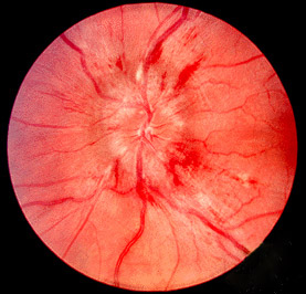
Papilledema or papilloedema is optic disc swelling that is caused by increased intracranial pressure due to any cause. The swelling is usually bilateral and can occur over a period of hours to weeks. Unilateral presentation is extremely rare.
The visual field is the "spatial array of visual sensations available to observation in introspectionist psychological experiments". Or simply, visual field can be defined as the entire area that can be seen when an eye is fixed straight at a point.
Optic nerve hypoplasia (ONH) is a medical condition arising from the underdevelopment of the optic nerve(s). This condition is the most common congenital optic nerve anomaly. The optic disc appears abnormally small, because not all the optic nerve axons have developed properly. It is often associated with endocrinopathies, developmental delay, and brain malformations. The optic nerve, which is responsible for transmitting visual signals from the retina to the brain, has approximately 1.2 million nerve fibers in the average person. In those diagnosed with ONH, however, there are noticeably fewer nerves.

The optic disc or optic nerve head is the point of exit for ganglion cell axons leaving the eye. Because there are no rods or cones overlying the optic disc, it corresponds to a small blind spot in each eye.

A coloboma is a hole in one of the structures of the eye, such as the iris, retina, choroid, or optic disc. The hole is present from birth and can be caused when a gap called the choroid fissure, which is present during early stages of prenatal development, fails to close up completely before a child is born. Ocular coloboma is relatively uncommon, affecting less than one in every 10,000 births.

Pigment dispersion syndrome (PDS) is an eye disorder that can lead to a form of glaucoma known as pigmentary glaucoma. It takes place when pigment cells slough off from the back of the iris and float around in the aqueous humor. Over time, these pigment cells can accumulate in the anterior chamber in such a way that they begin to clog the trabecular meshwork, which can in turn prevent the aqueous humour from draining and therefore increases the pressure inside the eye. A common finding in PDS are central, vertical corneal endothelial pigment deposits, known as Krukenberg spindle. With PDS, the intraocular pressure tends to spike at times and then can return to normal. Exercise has been shown to contribute to spikes in pressure as well. When the pressure is great enough to cause damage to the optic nerve, this is called pigmentary glaucoma. As with all types of glaucoma, when damage happens to the optic nerve fibers, the vision loss that occurs is irreversible and painless.

The retinal nerve fiber layer (RNFL) or nerve fiber layer, stratum opticum, is formed by the expansion of the fibers of the optic nerve; it is thickest near the optic disc, gradually diminishing toward the ora serrata.
Optic neuropathy is damage to the optic nerve from any cause. The optic nerve is a bundle of millions of fibers in the retina that sends visual signals to the brain. [1].

Papillorenal syndrome is an autosomal dominant genetic disorder marked by underdevelopment (hypoplasia) of the kidney and colobomas of the optic nerve.

Optic disc drusen (ODD) are globules of mucoproteins and mucopolysaccharides that progressively calcify in the optic disc. They are thought to be the remnants of the axonal transport system of degenerated retinal ganglion cells. ODD have also been referred to as congenitally elevated or anomalous discs, pseudopapilledema, pseudoneuritis, buried disc drusen, and disc hyaline bodies.

Optic pit, optic nerve pit, or optic disc pit (ODP) is rare a congenital excavation of the optic disc, resulting from a malformation during development of the eye. The incidence of ODP is 1 in 10,000 people with no predilection for either gender. There is currently no known risk factors for their development. Optic pits are important because they are associated with posterior vitreous detachments (PVD) and even serous retinal detachments.
Scanning laser polarimetry is the use of polarised light to measure the thickness of the retinal nerve fiber layer as part of a glaucoma workup. The GDx-VCC is one example.
Sohan Singh Hayreh was an ophthalmologist, clinical scientist, and professor emeritus of ophthalmology at the University of Iowa. As one of the pioneers in the field of fluorescein angiography, he was generally acknowledged to be a leading authority in vascular diseases of the eye and the optic nerve. For over 60 years, Hayreh was actively involved in basic, experimental, and clinical research in ophthalmology, publishing over 400 original peer-reviewed articles in various international ophthalmic journals, six classical monographs and books in his field of research, and more than 50 chapters in ophthalmic books. He made many seminal observations dealing with the ocular circulation in health and disease, the optic disc and the optic nerve, retinal and choroidal vascular disorders, glaucomatous optic neuropathy, fundus changes in malignant arterial hypertension, ocular neovascularization, rheumatologic disorders of the eye, and nocturnal arterial hypotension. He was an elected fellow of the National Academy of Medical Sciences.
Normal tension glaucoma (NTG) is an eye disease, a neuropathy of the optic nerve, that shows all the characteristics of primary open angle glaucoma except one: the elevated intraocular pressure (IOP) - the classic hallmark of glaucoma - is missing. Normal tension glaucoma is in many cases closely associated with general issues of blood circulation and of organ perfusion like arterial hypotension, metabolic syndrome, and Flammer syndrome.
Rohit Varma is an Indian-American ophthalmologist and professor of ophthalmology and preventive medicine. In 2014, he was named director of the USC Eye Institute and chairman of the Department of Ophthalmology for Keck School of Medicine of USC. In March 2016, Varma was named the interim dean of the Keck School of Medicine, and in November was named dean. In October 2017, USC announced that he stepped down as dean. In October 2018, Varma became the founding director of the Southern California Eyecare and Vision Research Institute.

James C. Tsai is a physician and scientist who serves as president of the New York Eye and Ear Infirmary of Mount Sinai. He also serves as the Delafield-Rodgers Professor of Ophthalmology at the Icahn School of Medicine at Mount Sinai and Chair of the Department of Ophthalmology at the Mount Sinai Health System.
Helen Victoria Danesh-Meyer is a New Zealand ophthalmology academic, and as of 2018 is a full professor at the University of Auckland.
Megalopapilla is a non-progressive human eye condition in which the optic nerve head has an enlarged diameter, exceeding 2.1 mm with no other morphological abnormalities.

