Parafovea or the parafoveal belt is a region in the retina that circumscribes the fovea and is part of the macula lutea. [1] It is circumscribed by the perifovea.
Contents

Parafovea or the parafoveal belt is a region in the retina that circumscribes the fovea and is part of the macula lutea. [1] It is circumscribed by the perifovea.
In reading, information within 1° (approximately 6–8 characters) of the point of fixation is processed in foveal vision, while information up to 6° of visual angle benefits from parafoveal preview. [2] Studies have shown that people can tell the difference in the letters of a word in the fovea and near-parafovea (the part of the parafovea closest to the fovea), but not in the outer edges of the parafovea. [3] In languages that read from left to right, the word immediately to the right of the fixated word is known as the parafoveal word. [3] Information present in the parafovea can interact with information present in the fovea. [4] The benefit the parafoveal preview has is also mediated by how common the word in the parafovea is, with less common words providing less of a reduction in fixation duration when they reach foveal fixation. [5] As the clarity of information in the parafovea is not as great as in the fovea, the SWIFT model of eye movements in reading, while allowing for parallel processing, accounts for this difference by assigning the parafoveal less processing power the further away it is from the foveal fixation. [2]
Information in the parafovea can influence the processing of a scene. In categorization tasks of natural scenes information from the parafovea can be used to determine the gist of a scene well enough for a categorization judgement, though with reduced sensitivity and speed in comparison to foveal vision. [6] An effect of parafoveal preview has also been found for emotional scenes presented in the parafovea, with people more likely shift their fixation point on emotional stimuli than neutral stimuli, when both options are presented parafoveally. [7]


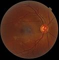

The retina is the innermost, light-sensitive layer of tissue of the eye of most vertebrates and some molluscs. The optics of the eye create a focused two-dimensional image of the visual world on the retina, which then processes that image within the retina and sends nerve impulses along the optic nerve to the visual cortex to create visual perception. The retina serves a function which is in many ways analogous to that of the film or image sensor in a camera.

Haidinger's brush, more commonly known as Haidinger's brushes is an image produced by the eye, an entoptic phenomenon, first described by Austrian physicist Wilhelm Karl von Haidinger in 1844. Haidinger saw it when he looked through various minerals that polarized light.

A saccade is a quick, simultaneous movement of both eyes between two or more phases of fixation in the same direction. In contrast, in smooth pursuit movements, the eyes move smoothly instead of in jumps. The phenomenon can be associated with a shift in frequency of an emitted signal or a movement of a body part or device. Controlled cortically by the frontal eye fields (FEF), or subcortically by the superior colliculus, saccades serve as a mechanism for fixation, rapid eye movement, and the fast phase of optokinetic nystagmus. The word appears to have been coined in the 1880s by French ophthalmologist Émile Javal, who used a mirror on one side of a page to observe eye movement in silent reading, and found that it involves a succession of discontinuous individual movements.

The macula (/ˈmakjʊlə/) or macula lutea is an oval-shaped pigmented area in the center of the retina of the human eye and in other animals. The macula in humans has a diameter of around 5.5 mm (0.22 in) and is subdivided into the umbo, foveola, foveal avascular zone, fovea, parafovea, and perifovea areas.

Peripheral vision, or indirect vision, is vision as it occurs outside the point of fixation, i.e. away from the center of gaze or, when viewed at large angles, in the "corner of one's eye". The vast majority of the area in the visual field is included in the notion of peripheral vision. "Far peripheral" vision refers to the area at the edges of the visual field, "mid-peripheral" vision refers to medium eccentricities, and "near-peripheral", sometimes referred to as "para-central" vision, exists adjacent to the center of gaze.

Visual acuity (VA) commonly refers to the clarity of vision, but technically rates a person's ability to recognize small details with precision. Visual acuity depends on optical and neural factors. Optical factors of the eye influence the sharpness of an image on its retina. Neural factors include the health and functioning of the retina, of the neural pathways to the brain, and of the interpretative faculty of the brain.

The fovea centralis is a small, central pit composed of closely packed cones in the eye. It is located in the center of the macula lutea of the retina.
The visual field is "that portion of space in which objects are visible at the same moment during steady fixation of the gaze in one direction"; in ophthalmology and neurology the emphasis is on the structure inside the visual field and it is then considered “the field of functional capacity obtained and recorded by means of perimetry”.
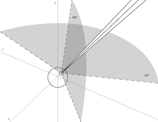
Vision span or perceptual span is a controversial concept referring to the angular span, within which the human eye has sharp enough vision to perform an action accurately. The visual field of the human eye spans approximately 120 degrees of arc. However, most of that arc is peripheral vision. The human eye has much greater resolution in the macula, where there is a higher density of cone cells. The macula has a diameter of about 16 degrees of the retina. The field of view that is observed with sufficient resolution to read text typically spans about 6 degrees of arc, which is wide enough to allow a clear view of about five words in a row when printed text at ordinary size is held about 50 centimeters from the eyes. Regarding face processing, the field of view with a sufficient amount of information in order to recognise faces accurately spans about 7° which represents about 45% of a face. The brain creates the illusion of having a greater visual span by automatically and unconsciously moving the center of vision into any area of interest in the field of view.

Eye movement includes the voluntary or involuntary movement of the eyes. Eye movements are used by a number of organisms to fixate, inspect and track visual objects of interests. A special type of eye movement, rapid eye movement, occurs during REM sleep.

The central retinal artery branches off the ophthalmic artery, running inferior to the optic nerve within its dural sheath to the eyeball.
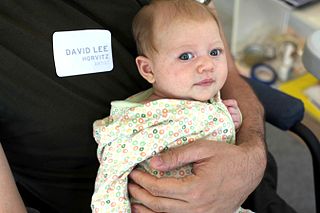
Infant vision concerns the development of visual ability in human infants from birth through the first years of life. The aspects of human vision which develop following birth include visual acuity, tracking, color perception, depth perception, and object recognition.
Eye movement in reading involves the visual processing of written text. This was described by the French ophthalmologist Louis Émile Javal in the late 19th century. He reported that eyes do not move continuously along a line of text, but make short, rapid movements (saccades) intermingled with short stops (fixations). Javal's observations were characterised by a reliance on naked-eye observation of eye movement in the absence of technology. From the late 19th to the mid-20th century, investigators used early tracking technologies to assist their observation, in a research climate that emphasised the measurement of human behaviour and skill for educational ends. Most basic knowledge about eye movement was obtained during this period. Since the mid-20th century, there have been three major changes: the development of non-invasive eye-movement tracking equipment; the introduction of computer technology to enhance the power of this equipment to pick up, record, and process the huge volume of data that eye movement generates; and the emergence of cognitive psychology as a theoretical and methodological framework within which reading processes are examined. Sereno & Rayner (2003) believed that the best current approach to discover immediate signs of word recognition is through recordings of eye movement and event-related potential.
Within computer technology, the gaze-contingency paradigm is a general term for techniques allowing a computer screen display to change in function depending on where the viewer is looking. Gaze-contingent techniques are part of the eye movement field of study in psychology.
Macular hypoplasia is a rare medical condition involving the underdevelopment of the macula, a small area on the retina responsible for seeing in detail and sensing light. Macular hypoplasia is often associated with albinism.

Foveated imaging is a digital image processing technique in which the image resolution, or amount of detail, varies across the image according to one or more "fixation points". A fixation point indicates the highest resolution region of the image and corresponds to the center of the eye's retina, the fovea.

Visual perception is the ability to interpret the surrounding environment through photopic vision, color vision, scotopic vision, and mesopic vision, using light in the visible spectrum reflected by objects in the environment. This is different from visual acuity, which refers to how clearly a person sees. A person can have problems with visual perceptual processing even if they have 20/20 vision.

The foveola is located within a region called the macula, a yellowish, cone photoreceptor filled portion of the human retina. Approximately 0.35 mm in diameter, the foveola lies in the center of the fovea and contains only cone cells and a cone-shaped zone of Müller cells. In this region the cone receptors are found to be longer, slimmer, and more densely packed than anywhere else in the retina, thus allowing that region to have the potential to have the highest visual acuity in the eye.
Perifovea is a region in the retina that circumscribes the parafovea and fovea and is a part of the macula lutea. The perifovea is a belt that covers a 10° radius around the fovea and is 1.5 mm wide. The perifovea ends when the Henle's fiber layer disappears and the ganglion cells are one-layered.
Microperimetry, sometimes called fundus-controlled perimetry, is a type of visual field test which uses one of several technologies to create a "retinal sensitivity map" of the quantity of light perceived in specific parts of the retina in people who have lost the ability to fixate on an object or light source. The main difference with traditional perimetry instruments is that, microperimetry includes a system to image the retina and an eye tracker to compensate eye movements during visual field testing.
| Fibrous tunic (outer) |
|  | |||||
|---|---|---|---|---|---|---|---|
| Uvea / vascular tunic (middle) |
| ||||||
| Retina (inner) |
| ||||||
| Anatomical regions of the eye |
| ||||||
| Other | |||||||