Optic neuritis

Last updated
Optic neuritis
Optic Neuritis.png
Optic Neuritis following a febrile infection in a young woman
Specialty Ophthalmology, optometry, neurology
Symptoms loss of vision, loss of colour vision, pain worsening on eye movements
Complications multiple sclerosis, MOG-disease, NMO
Usual onsetsubacute
Duration1-3 months
TypesMS-ON, MOG-ON, AQP4-ON, CRMP5-ON, SION, RION, CRION, post-infectious ON, post-vaccination ON, ON as complication of systemic diseases or meidication
Causesautoimmune, infection, vaccination, medication
Risk factors genetic
Diagnostic method Diagnostic criteria
Prognosis Prognosis depends on the subtype of ON
Frequencycan be relapsing

Optic neuritis describes any condition that causes inflammation of the optic nerve; it may be associated with demyelinating diseases, or infectious or inflammatory processes. [1]

Contents

It is also known as optic papillitis (when the head of the optic nerve is involved), neuroretinitis (when there is a combined involvement of the optic disc and surrounding retina in the macular area) and retrobulbar neuritis (when the posterior part of the nerve is involved). Prelaminar optic neuritis describes involvement of the non-myelinated axons in the retina. [1]

Classification, diagnosis and symptoms

Example of how optic neuritis affected one eye of a person with multiple sclerosis OpticNeuritisExample.jpg
Example of how optic neuritis affected one eye of a person with multiple sclerosis

Classification and diagnosis

The World Health Organization's ICD-11 classification includes optic neuritis. [2] However a 2022 review found that there is no consensus regarding the classification of optic neuritis, and precise diagnostic criteria are not available. [3]

Symptoms

Major symptoms are

Many patients with optic neuritis may lose some of their color vision in the affected eye (especially red), with colors appearing subtly washed out compared to the other eye. Patients may also experience difficulties judging movement in depth, which can be particular troublesome during driving or sport (Pulfrich effect). Likewise, transient worsening of vision with increase of body temperature (Uhthoff's phenomenon) and glare disability are a frequent complaint.

However not everyone who has optic neuritis has problems with their vision. [6]

Early symptoms

Early symptoms that require investigation include symptoms from multiple sclerosis (twitching, lack of coordination, slurred speech, frequent episodes of partial vision loss or blurred vision), episodes of "disturbed/blackened" rather than blurry indicate moderate stage and require immediate medical attention to prevent further loss of vision. Other early symptoms are reduced night vision, photophobia and red eyes.

Variation in symptoms with age

Several case studies in children have demonstrated the absence of pain in more than half of cases (approximately 60%) in their pediatric study population, with the most common symptom reported simply as "blurriness". [7] [8] Other remarkable differences between the presentation of adult optic neuritis as compared to pediatric cases include more often unilateral optic neuritis in adults, while children much predominantly present with bilateral involvement.

Observation

On medical examination the head of the optic nerve can easily be visualized by a slit lamp with a high positive lens or by using direct ophthalmoscopy; however, frequently there is no abnormal appearance of the nerve head in optic neuritis (in cases of retrobulbar optic neuritis), though it may be swollen in some patients (anterior papillitis or more extensive optic neuritis). In many cases, only one eye is affected, and patients may not be aware of the loss of color vision until they are asked to close or cover the healthy eye.

Imaging

Imaging of the optic nerve with MRI shows increased signal on the affected side. There is contrast enhancment of the symptomatic optic nerve and sheaths acutely or intrinsic signal increase (looking brighter) within ≥ 3 months. [1]

Magnetic Resonnance Imaging (MRI) during an episode of optic neuritis. Case-3-mri.png
Magnetic Resonnance Imaging (MRI) during an episode of optic neuritis.

Advanced imaging using optical coherence tomography (OCT) is very sensitive reveal damage to the optic nerve. The OCT shows corresponding optic disc swelling acutely or an inter-eye difference in the thickness of the neurons and their nerves connecting the eye with the brain in above 4-5% within ≥ 3 months after onset. [1]

Asymmetry in thickness of RNFL

Asymmetry between the eyes in thickness of RNFL has been proposed as a strong indicator of optic neuritis. [9] [10] [11]

Cause

The optic nerve comprises axons that emerge from the retina of the eye and carry visual information to the primary visual nuclei, most of which is relayed to the occipital cortex of the brain to be processed into vision. Inflammation of the optic nerve causes loss of vision, usually because of the swelling and destruction of the myelin sheath covering the optic nerve.

A 2019 review divided optic neuritis into

The most common cause is multiple sclerosis (MS) or ischemic optic neuropathy due to thrombosis or embolism of the vessel that supplies the optic nerve. [13] [14] Up to 50% of patients with MS will develop an episode of optic neuritis, and 20–30% of the time optic neuritis is the presenting sign of MS.[ citation needed ] The presence of demyelinating white matter lesions on brain MRI at the time of presentation of optic neuritis is the strongest predictor for developing clinically definite MS. Almost half of the patients with optic neuritis have white matter lesions consistent with multiple sclerosis.

Some other common causes of optic neuritis include infection (e.g. a tooth abscess in the upper jaw, syphilis, Lyme disease, herpes zoster), autoimmune disorders (e.g. lupus, neurosarcoidosis, neuromyelitis optica), methanol poisoning, vitamin B12 deficiency, beriberi, dysautonomia (i.e. autonomic nervous system dysfunction), and diabetes, or an injury to the eye. [15] In neuromyelitis optica higher AQP4 autoantibody levels are associated with the occurrence of optic neuritis. [16]

Less common causes are: papilledema, brain tumor or abscess in the occipital region, cerebral trauma or hemorrhage, meningitis, arachnoidal adhesions, sinus thrombosis, liver dysfunction, or late stage kidney disease.

Approximate Cause Data (Other TOP Ranking diseases include: Cancer, Heart Disease, Respiratory, Stroke, Diabetes, Alzheimers, Kidney Disease, Parkinsons, Suicide) [17]
Cause and Rank based on DeathsAnnual Num Cases TOTAL (US) (2011)Annual Cases leading to Optic NeuritisPercentPrognosis and Treatment
Multiple sclerosis (Rank 33)400,042146,23245%Most common cause. Almost all patients will experience some form of vision dysfunction. Partial vision loss can occur through the duration of the disease; total vision loss occurs in severe cases and late stages. It may lead to complete or partial loss of vision in one or both eyes. Partial, transient vision loss (lasting less than one hour) can be an indication of early onset multiple sclerosis. [18]
Blood clot (Rank 29) (Optic ONLY)17,00016,7775%Reversible if early and before reduced blood flow causes permanent damage.
Nerve pinch (0)NOT REPORTED-4%Usually heals itself, treatment not needed.
Injury to optic nerve (including poisoning, i.e. methanol) (0)23,82720,121<1%Depends on severity, usually treatable.
Liver dysfunction (Rank 19); if untreated can lead to Failure (Rank 8)141,21111,9827%Poor outcomes and progresses and can lead to total vision loss.
Reduced kidney function (treatable with diet change) (Rank 67 – If untreated, can progress to late stage with much greater mortality rates)509,89816,2819%Good outcomes if early, and can usually be treated with diet changes, progresses and can lead to total vision loss.
Late stage kidney failure (Rank 7)33,2121,1122%Poor outcomes – usually permanent nerve damage at this stage.
Papilledema (brain tumor or abscess) (Rank 10)45,8889,2313%Depends on severity.
Meningitis (Rank 61)2,521189<1%Depends on severity.
Other infections (not from abscess)5,561-<1%Good outcomes, treatable with antibiotics or other microbial drugs.
Diabetes (early stage treatable, late Stage has worse prognosis) (Rank 6)49,56221,11215% Type 1 carries poor prognosis, type 2 can be treated and vision returned.
Unknown--2%


Other diseases associated with optic neuritis include: [19]

Demyelinating recurrent optic neuritis and non-demyelinating (CRION)

The repetition of an idiopathic optic neuritis is considered a distinct clinical condition, and when it shows demyelination, it has been found to be associated to anti-MOG and AQP4-negative neuromyelitis optica. [20]

When an inflammatory recurrent optic neuritis is not demyelinating, it is called chronic relapsing inflammatory optic neuropathy (CRION). [21]

When it is anti-MOG related, it is demyelinating and it is considered inside the anti-MOG associated inflammatory demyelinating diseases.

Some reports point to the possibility to establish a difference via optical coherence tomography. [22]

Outlook and Treatment

Many patients see full recovery but some see some lasting effects. [5] [23] [24]

Steroids

High dose steroids may be given intravenously or orally. [5]

In most MS-associated optic neuritis, visual function spontaneously improves over 2–3 months, and there is evidence that corticosteroid treatment does not affect the long term outcome. However, for optic neuritis that is not MS-associated (or atypical optic neuritis) the evidence is less clear and therefore the threshold for treatment with intravenous corticosteroids is lower. [1] Intravenous corticosteroids also reduce the risk of developing MS in the following two years in patients with MRI lesions; but this effect disappears by the third year of follow up. [25]

Paradoxically, oral administration of corticosteroids in this situation may lead to more recurrent attacks than in non-treated patients (though oral steroids are generally prescribed after the intravenous course, to wean the patient off the medication). This effect of corticosteroids seems to be limited to optic neuritis and has not been observed in other diseases treated with corticosteroids. [26]

A Cochrane systematic review studied the effect of corticosteroids for treating people with acute optic neuritis. [27] Specific corticosteroids studied included intravenous and oral methylprednisone, and oral prednisone. The authors conclude that current evidence does not show a benefit of either intravenous or oral corticosteroids for rate of recovery of vision (in terms of visual acuity, contrast sensitivity, or visual fields). [27] There are a number of reasons why this might be the case. [25] [28]

Immunosuppressants

Immunosuppressants may also be used in treatment. [5]

Pain relief

Pain relief may also be used. [5]

Epidemiology

Optic neuritis typically affects young adults ranging 18–45 years of age, with a mean age of 30–35 years. There is a strong female predominance. The annual incidence is approximately 5/100,000, with a prevalence estimated to be 115/100,000 (0.12%). [29]

Society and culture

In Charles Dickens' Bleak House , the main character, Esther Summerville, has a transient episode of visual loss, the symptoms of which are also seen in people who have optic neuritis. [30] Legal historian William Searle Holdsworth suggested that the events in Bleak House took place in 1827.

In an episode of Dr. Quinn, Medicine Woman ("Season of Miracles", season five), Reverend Timothy Johnson is struck blind by optic neuritis on Christmas Day 1872. He remains blind for the duration of the series.

See also

Related Research Articles

<span class="mw-page-title-main">Acute disseminated encephalomyelitis</span> Autoimmune disease

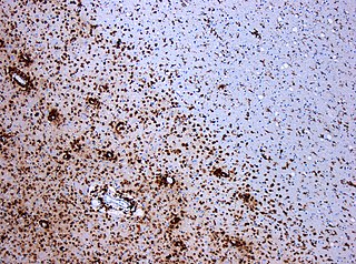
Acute disseminated encephalomyelitis (ADEM), or acute demyelinating encephalomyelitis, is a rare autoimmune disease marked by a sudden, widespread attack of inflammation in the brain and spinal cord. As well as causing the brain and spinal cord to become inflamed, ADEM also attacks the nerves of the central nervous system and damages their myelin insulation, which, as a result, destroys the white matter. The cause is often a trigger such as from viral infection or vaccinations.

<span class="mw-page-title-main">Transverse myelitis</span> Medical condition of the spinal cord

Transverse myelitis (TM) is a rare neurological condition wherein the spinal cord is inflamed. The adjective transverse implies that the spinal inflammation (myelitis) extends horizontally throughout the cross section of the spinal cord; the terms partial transverse myelitis and partial myelitis are sometimes used to specify inflammation that affects only part of the width of the spinal cord. TM is characterized by weakness and numbness of the limbs, deficits in sensation and motor skills, dysfunctional urethral and anal sphincter activities, and dysfunction of the autonomic nervous system that can lead to episodes of high blood pressure. Signs and symptoms vary according to the affected level of the spinal cord. The underlying cause of TM is unknown. The spinal cord inflammation seen in TM has been associated with various infections, immune system disorders, or damage to nerve fibers, by loss of myelin. As opposed to leukomyelitis which affects only the white matter, it affects the entire cross-section of the spinal cord. Decreased electrical conductivity in the nervous system can result.

<span class="mw-page-title-main">Multiple sclerosis</span> Disease that damages the myelin sheaths around nerves

Multiple sclerosis (MS) is an autoimmune disease in which the insulating covers of nerve cells in the brain and spinal cord are damaged. This damage disrupts the ability of parts of the nervous system to transmit signals, resulting in a range of signs and symptoms, including physical, mental, and sometimes psychiatric problems. Specific symptoms can include double vision, vision loss, eye pain, muscle weakness, and loss of sensation or coordination. MS takes several forms, with new symptoms either occurring in isolated attacks or building up over time. In the relapsing forms of MS, between attacks, symptoms may disappear completely, although some permanent neurological problems often remain, especially as the disease advances. In the progressive forms of MS, bodily function slowly deteriorates and disability worsens once symptoms manifest and will steadily continue to do so if the disease is left untreated.

<span class="mw-page-title-main">Demyelinating disease</span> Any neurological disease in which the myelin sheath of neurons is damaged

A demyelinating disease refers to any disease affecting the nervous system where the myelin sheath surrounding neurons is damaged. This damage disrupts the transmission of signals through the affected nerves, resulting in a decrease in their conduction ability. Consequently, this reduction in conduction can lead to deficiencies in sensation, movement, cognition, or other functions depending on the nerves affected.

<span class="mw-page-title-main">Polyneuropathy</span> Medical condition

Polyneuropathy is damage or disease affecting peripheral nerves in roughly the same areas on both sides of the body, featuring weakness, numbness, and burning pain. It usually begins in the hands and feet and may progress to the arms and legs and sometimes to other parts of the body where it may affect the autonomic nervous system. It may be acute or chronic. A number of different disorders may cause polyneuropathy, including diabetes and some types of Guillain–Barré syndrome.

Neuromyelitis optica spectrum disorders (NMOSD) are a spectrum of autoimmune diseases characterized by acute inflammation of the optic nerve and the spinal cord (myelitis). Episodes of ON and myelitis can be simultaneous or successive. A relapsing disease course is common, especially in untreated patients.

Anterior ischemic optic neuropathy (AION) is a medical condition involving loss of vision caused by damage to the optic nerve as a result of insufficient blood supply (ischemia). This form of ischemic optic neuropathy is generally categorized as two types: arteritic AION, in which the loss of vision is the result of an inflammatory disease of arteries in the head called temporal arteritis, and non-arteritic AION, which is due to non-inflammatory disease of small blood vessels.

<span class="mw-page-title-main">Myelin oligodendrocyte glycoprotein</span>

Myelin oligodendrocyte glycoprotein (MOG) is a glycoprotein believed to be important in the myelination of nerves in the central nervous system (CNS). In humans this protein is encoded by the MOG gene. It is speculated to serve as a necessary "adhesion molecule" to provide structural integrity to the myelin sheath and is known to develop late on the oligodendrocyte.

<span class="mw-page-title-main">Neuritis</span> Inflammation of a nerve or generally any part of the nervous system

Neuritis, from the Greek νεῦρον), is inflammation of a nerve or the general inflammation of the peripheral nervous system. Inflammation, and frequently concomitant demyelination, cause impaired transmission of neural signals and leads to aberrant nerve function. Neuritis is often conflated with neuropathy, a broad term describing any disease process which affects the peripheral nervous system. However, neuropathies may be due to either inflammatory or non-inflammatory causes, and the term encompasses any form of damage, degeneration, or dysfunction, while neuritis refers specifically to the inflammatory process.

Uhthoff's phenomenon is the worsening of neurologic symptoms in multiple sclerosis (MS) and other demyelinating diseases when the body is overheated. This may occur due to hot weather, exercise, fever, saunas, hot tubs, hot baths, and hot food and drink. Increased temperature slows nerve conduction, but the exact mechanism remains unknown. With an increased body temperature, nerve impulses are either blocked or slowed in a damaged nerve. Once the body temperature is normalized, signs and symptoms typically reverse.

<span class="mw-page-title-main">Chronic inflammatory demyelinating polyneuropathy</span> Medical condition

Chronic inflammatory demyelinating polyneuropathy (CIDP) is an acquired autoimmune disease of the peripheral nervous system characterized by progressive weakness and impaired sensory function in the legs and arms. The disorder is sometimes called chronic relapsing polyneuropathy (CRP) or chronic inflammatory demyelinating polyradiculoneuropathy. CIDP is closely related to Guillain–Barré syndrome and it is considered the chronic counterpart of that acute disease. Its symptoms are also similar to progressive inflammatory neuropathy. It is one of several types of neuropathy.

Optic neuropathy is damage to the optic nerve from any cause. The optic nerve is a bundle of millions of fibers in the retina that sends visual signals to the brain.

Inflammatory demyelinating diseases (IDDs), sometimes called Idiopathic (IIDDs) due to the unknown etiology of some of them, are a heterogenous group of demyelinating diseases - conditions that cause damage to myelin, the protective sheath of nerve fibers - that occur against the background of an acute or chronic inflammatory process. IDDs share characteristics with and are often grouped together under Multiple Sclerosis. They are sometimes considered different diseases from Multiple Sclerosis, but considered by others to form a spectrum differing only in terms of chronicity, severity, and clinical course.

<span class="mw-page-title-main">Marburg acute multiple sclerosis</span> Medical condition

Marburg acute multiple sclerosis, also known as Marburg multiple sclerosis or acute fulminant multiple sclerosis, is considered one of the multiple sclerosis borderline diseases, which is a collection of diseases classified by some as MS variants and by others as different diseases. Other diseases in this group are neuromyelitis optica (NMO), Balo concentric sclerosis, and Schilder's disease. The graver course is one form of malignant multiple sclerosis, with patients reaching a significant level of disability in less than five years from their first symptoms, often in a matter of months.

<span class="mw-page-title-main">Signs and symptoms of multiple sclerosis</span> Neurological signs and symptoms

The signs and symptoms of multiple sclerosis (MS) encompass a wide range of neurological and physical manifestations, including vision problems, muscle weakness, coordination difficulties, and cognitive impairment, varying significantly in severity and progression among individuals.

<span class="mw-page-title-main">Tumefactive multiple sclerosis</span> Medical condition

Tumefactive multiple sclerosis is a condition in which the central nervous system of a person has multiple demyelinating lesions with atypical characteristics for those of standard multiple sclerosis (MS). It is called tumefactive as the lesions are "tumor-like" and they mimic tumors clinically, radiologically and sometimes pathologically.

Autoimmune optic neuropathy (AON), sometimes called autoimmune optic neuritis, may be a forme fruste of systemic lupus erythematosus (SLE) associated optic neuropathy. AON is more than the presence of any optic neuritis in a patient with an autoimmune process, as it describes a relatively specific clinical syndrome. AON is characterized by chronically progressive or recurrent vision loss associated with serological evidence of autoimmunity. Specifically, this term has been suggested for cases of optic neuritis with serological evidence of vasculitis by positive ANA, despite the lack of meeting criteria for SLE. The clinical manifestations include progressive vision loss that tends to be steroid-responsive and steroid dependent.

Chronic relapsing inflammatory optic neuropathy (CRION) is a form of recurrent optic neuritis that is steroid responsive and dependent. Patients typically present with pain associated with visual loss. CRION is a clinical diagnosis of exclusion, and other demyelinating, autoimmune, and systemic causes should be ruled out. An accurate antibody test which became available commercially in 2017 has allowed most patients previously diagnosed with CRION to be re-identified as having MOG antibody disease, which is not a diagnosis of exclusion. Early recognition is crucial given risks for severe visual loss and because it is treatable with immunosuppressive treatment such as steroids or B-cell depleting therapy. Relapse that occurs after reducing or stopping steroids is a characteristic feature.

MOG antibody disease (MOGAD) or MOG antibody-associated encephalomyelitis (MOG-EM) is an inflammatory demyelinating disease of the central nervous system. Serum anti-myelin oligodendrocyte glycoprotein antibodies are present in up to half of patients with an acquired demyelinating syndrome and have been described in association with a range of phenotypic presentations, including acute disseminated encephalomyelitis, optic neuritis, transverse myelitis, and neuromyelitis optica.

Anti-AQP4 diseases, are a group of diseases characterized by auto-antibodies against aquaporin 4.

References

  1. 1 2 3 4 5 Petzold A, Fraser CL, Abegg M, Alroughani R, Alshowaeir D, Alvarenga R, et al. (December 2022). "Diagnosis and classification of optic neuritis". The Lancet. Neurology. 21 (12): 1120–1134. doi:10.1016/S1474-4422(22)00200-9. PMID   36179757. S2CID   252564095.
  2. 1 2 "ICD-11 for Mortality and Morbidity Statistics". icd.who.int.
  3. "Diagnosis and classification of optic neuritis - The Lancet Neurology".
  4. "Optic neuritis". Mayo Clinic.
  5. 1 2 3 4 5 "Optic neuritis". RNIB.
  6. "Optic Neuritis". www.hopkinsmedicine.org. March 22, 2024.
  7. Lucchinetti CF, Kiers L, O'Duffy A, Gomez MR, Cross S, Leavitt JA, et al. (November 1997). "Risk factors for developing multiple sclerosis after childhood optic neuritis". Neurology. 49 (5): 1413–1418. doi:10.1212/WNL.49.5.1413. PMID   9371931. S2CID   33205877.
  8. Lana-Peixoto MA, Andrade GC (June 2001). "The clinical profile of childhood optic neuritis". Arquivos de Neuro-Psiquiatria. 59 (2–B): 311–317. doi: 10.1590/S0004-282X2001000300001 . PMID   11460171.
  9. "An intereye difference of 5–6 μm in RNFL thickness is a robust structural threshold for identifying the presence of a unilateral optic nerve lesion in MS." Nolan RC, Galetta SL, Frohman TC, Frohman EM, Calabresi PA, Castrillo-Viguera C, et al. (December 2018). "Optimal Intereye Difference Thresholds in Retinal Nerve Fiber Layer Thickness for Predicting a Unilateral Optic Nerve Lesion in Multiple Sclerosis". Journal of Neuro-Ophthalmology. 38 (4): 451–458. doi:10.1097/WNO.0000000000000629. PMC   8845082 . PMID   29384802.
  10. Jiang H, Delgado S, Wang J (February 2021). "Advances in ophthalmic structural and functional measures in multiple sclerosis: do the potential ocular biomarkers meet the unmet needs?". Current Opinion in Neurology. 34 (1): 97–107. doi:10.1097/WCO.0000000000000897. PMC   7856092 . PMID   33278142.
  11. Nij Bijvank J, Uitdehaag BM, Petzold A (February 2022). "Retinal inter-eye difference and atrophy progression in multiple sclerosis diagnostics". Journal of Neurology, Neurosurgery, and Psychiatry. 93 (2): 216–219. doi:10.1136/jnnp-2021-327468. PMC   8785044 . PMID   34764152.
  12. "Critical review: Typical and atypical optic neuritis - Survey of Ophthalmology".
  13. Rizzo JF, Lessell S (December 1991). "Optic neuritis and ischemic optic neuropathy. Overlapping clinical profiles". Archives of Ophthalmology. 109 (12): 1668–1672. doi:10.1001/archopht.1991.01080120052024. PMID   1841572.
  14. Biousse V, Newman NJ (June 2015). "Ischemic Optic Neuropathies". The New England Journal of Medicine. 372 (25): 2428–2436. doi:10.1056/NEJMra1413352. PMID   26083207.
  15. Riordan-Eva P (November 2004). "Clinical assessment of optic nerve disorders". Eye. 18 (11): 1161–1168. doi: 10.1038/sj.eye.6701575 . PMID   15534601.
  16. Isobe N, Yonekawa T, Matsushita T, et al. Clinical relevance of serum aquaporin-4 antibody levels in neuromyelitis optica. Neurochem Res. 2013;38(5):997-1001. doi:10.1007/s11064-013-1009-0
  17. Nicolaie MA, van Houwelingen HC, Putter H (December 2015). "Vertical modelling: Analysis of competing risks data with missing causes of failure". Statistical Methods in Medical Research. 24 (6): 891–908. doi:10.1177/0962280211432067. PMID   22179822. S2CID   43277083.
  18. Marcovitch H, ed. (2018). Black's Medical Dictionary (43rd ed.). A&C Black via Credo Reference.
  19. Khurana AK, Khurana BP (2019). Comprehensive Ophthalmology (7th ed.). New Delhi: Jaypee Brothers Medical Publishers. ISBN   978-93-5270-686-0.
  20. Chalmoukou K, Stathopoulos P, Alexopoulos H, Akrivou S, Dalakas M (2015). "Recurrent Optic Neuritis (rON) is characterised by Anti-MOG Antibodies: A follow-up study". Neurology. 84 (14): 274. doi:10.1212/WNL.84.14_supplement.P5.274.
  21. Kidd D, Burton B, Plant GT, Graham EM (February 2003). "Chronic relapsing inflammatory optic neuropathy (CRION)". Brain. 126 (Pt 2): 276–284. doi: 10.1093/brain/awg045 . PMID   12538397.
  22. Narayan et al. Unique characteristics of optical coherence tomography (OCT) results and visual acuity testing in myelin oligodendrocyte glycoprotein (MOG) antibody positive pediatric patients, November 2018, MS and related disorders, Volume 28, February 2019, Pages 86-90, doi: https://doi.org/10.1016/j.msard.2018.11.026
  23. https://www.mssociety.org.uk/about-ms/signs-and-symptoms/eyes-and-sight/optic-neuritis#:~:text=After%20optic%20neuritis%2C%20your%20vision,when%20you%20get%20hot%2C%20too.
  24. https://mft.nhs.uk/app/uploads/sites/2/2019/10/REH-173.pdf
  25. 1 2 Beck RW, Cleary PA, Trobe JD, Kaufman DI, Kupersmith MJ, Paty DW, et al. (December 1993). "The effect of corticosteroids for acute optic neuritis on the subsequent development of multiple sclerosis. The Optic Neuritis Study Group". The New England Journal of Medicine. 329 (24): 1764–1769. doi: 10.1056/NEJM199312093292403 . PMID   8232485.
  26. Beck RW, Cleary PA, Anderson MM, Keltner JL, Shults WT, Kaufman DI, et al. (February 1992). "A randomized, controlled trial of corticosteroids in the treatment of acute optic neuritis. The Optic Neuritis Study Group". The New England Journal of Medicine. 326 (9): 581–588. doi: 10.1056/NEJM199202273260901 . PMID   1734247.
  27. 1 2 Gal RL, Vedula SS, Beck R (August 2015). "Corticosteroids for treating optic neuritis". The Cochrane Database of Systematic Reviews. 2015 (8): CD001430. doi:10.1002/14651858.CD001430.pub4. PMC   4730547 . PMID   26273799.
  28. Petzold A, Braithwaite T, van Oosten BW, Balk L, Martinez-Lapiscina EH, Wheeler R, et al. (January 2020). "Case for a new corticosteroid treatment trial in optic neuritis: review of updated evidence". Journal of Neurology, Neurosurgery, and Psychiatry. 91 (1): 9–14. doi: 10.1136/jnnp-2019-321653 . PMC   6952848 . PMID   31740484.
  29. Rodriguez M, Siva A, Cross SA, O'Brien PC, Kurland LT (February 1995). "Optic neuritis: a population-based study in Olmsted County, Minnesota". Neurology. 45 (2): 244–250. doi:10.1212/WNL.45.2.244. PMID   7854520. S2CID   25800388.
  30. Petzold A (2013). "Optic Neuritis: Another Dickensian Diagnosis". Neuro-Ophthalmology. 37 (6): 247–250. doi:10.3109/01658107.2013.830313. PMC   5291069 . PMID   28167994.