Slit lamp

Last updated
Eye examination with the aid of a slit lamp. Spaltlampe-2.jpg
Eye examination with the aid of a slit lamp.
Side view of a slit lamp machine. Retina Group slit lamp (side view).jpg
Side view of a slit lamp machine.
Cataract in human eye: magnified view seen on examination with the slit lamp Cataract in human eye.png
Cataract in human eye: magnified view seen on examination with the slit lamp

In ophthalmology and optometry, a slit lamp is an instrument consisting of a high-intensity light source that can be focused to shine a thin sheet of light into the eye. It is used in conjunction with a biomicroscope. The lamp facilitates an examination of the anterior segment and posterior segment of the human eye, which includes the eyelid, sclera, conjunctiva, iris, natural crystalline lens, and cornea. The binocular slit-lamp examination provides a stereoscopic magnified view of the eye structures in detail, enabling anatomical diagnoses to be made for a variety of eye conditions. A second, hand-held lens is used to examine the retina.

Contents

History

Two conflicting trends emerged in the development of the slit lamp. One trend originated from clinical research and aimed to apply the increasingly complex and advanced technology of the time. [1] The second trend originated from ophthalmologic practice and aimed at technical perfection and a restriction to useful methods. The first man credited with developments in this field was Hermann von Helmholtz (1850) when he invented the ophthalmoscope. [2]

In ophthalmology and optometry, the instrument is called a "slit lamp", although it is more correctly called a "slit lamp instrument". [3] Today's instrument is a combination of two separate developments, the corneal microscope and the slit lamp itself. The first concept of a slit lamp dates back to 1911 credited to Allvar Gullstrand and his "large reflection-free ophthalmoscope." [3] The instrument was manufactured by Zeiss and consisted of a special illuminator connected to a small stand base through a vertical adjustable column. The base was able to move freely on a glass plate. The illuminator employed a Nernst glower which was later converted into a slit through a simple optical system. [4] However, the instrument never received much attention and the term "slit lamp" did not appear in any literature again until 1914.

It was not until 1919 that several improvements were made to the Gullstrand slit lamp made by Vogt Henker. First, a mechanical connection was made between lamp and ophthalmoscopic lens. This illumination unit was mounted to the table column with a double articulated arm. The binocular microscope was supported on a small stand and could be moved freely across the tabletop. Later, a cross slide stage was used for this purpose. Vogt introduced Koehler illumination, and the reddish Nernst glower was replaced with the brighter and whiter incandescent lamp. [4] Special mention should be paid to the experiments that followed Henker's improvements in 1919. On his improvements the Nitra lamp was replaced with a carbon arc lamp with a liquid filter. At this time the great importance of color temperature and the luminance of the light source for slit lamp examinations were recognized and the basis created for examinations in red-free light. [4]

In the year 1926, the slit lamp instrument was redesigned. The vertical arrangement of the projector made it easy to handle. For the first time, the axis through the patient's eye was fixed along a common swiveling axis, although the instrument still lacked a coordinate cross-slide stage for instrument adjustment. The importance of focal illumination had not yet been fully recognized. [5]

In 1927, stereo cameras were developed and added to the slit lamp to further its use and application. In 1930, Rudolf Theil further developed the slit lamp, encouraged by Hans Goldmann. [6] Horizontal and vertical co-ordinate adjustments were performed with three control elements on the cross-slide stage. The common swivel axis for microscope and illumination system was connected to the cross-slide stage, which allowed it to be brought to any part of the eye to be examined. [7] A further improvement was made in 1938. A control lever or joystick was used for the first time to allow for horizontal movement.

Following World War II the slit lamp was improved again. On this particular improvement the slit projector could be swiveled continuously across the front of the microscope. This was improved again in 1950, when a company named Littmann redesigned the slit lamp. They adopted the joystick control from the Goldmann instrument and the illumination path present in the Comberg instrument. Additionally, Littmann added the stereo telescope system with a common objective magnification changer. [8]

In 1965, the Model 100/16 Slit Lamp was produced based on the slit lamp by Littmann. This was soon followed by the Model 125/16 Slit Lamp in 1972. The only difference between the two models was their operating distances of 100 mm to 125 mm. With the introduction of the photo slit lamp further advancements were possible. In 1976, the development of the Model 110 Slit Lamp and the 210/211 Photo Slit Lamps were an innovation by which each were constructed from standard modules allowing for a wide range of different configurations. [9] At the same time, halogen lamps replaced the older illumination systems to make them brighter and essentially daylight quality. From 1994 onwards, new slit lamps were introduced which took advantage of new technologies. The last major development was in 1996 in which included new slit lamp optics. [9] See also "From Lateral Illumination to Slit Lamp - An Outline of Medical History". [10]

General procedure

While a patient is seated in the examination chair, they rest their chin and forehead on a support area to steady the head. Using the biomicroscope, the ophthalmologist or optometrist then proceeds to examine the patient's eye. A fine strip of paper, stained with fluorescein, a fluorescent dye, may be touched to the side of the eye; this stains the tear film on the surface of the eye to aid examination. The dye is naturally rinsed out of the eye by tears.

A subsequent test may involve placing drops in the eye in order to dilate the pupils. The drops take about 15 to 20 minutes to work, after which the examination is repeated, allowing the back of the eye to be examined. Patients will experience some light sensitivity for a few hours after this exam, and the dilating drops may also cause increased pressure in the eye, leading to nausea and pain. Patients who experience serious symptoms are advised to seek medical attention immediately.

Adults need no special preparation for the test; however children may need some preparation, depending on age, previous experiences, and level of trust.

Illuminations

Various methods of slitlamp illumination are required to obtain full advantage of slit-lamp biomicroscope. There are mainly six type of illuminating options:[ citation needed ]

  1. Diffuse illumination,
  2. Direct focal illumination,
  3. Specular reflection,
  4. Transillumination or retroillumination,
  5. Indirect lateral illumination or Indirect proximal illumination and
  6. Sclerotic scatter.

Oscillatory Illumination is sometimes considered an illumination technique. [11] Observation with an optical section or direct focal illumination is the most frequently applied method of examination with the slit lamp. With this method, the axes of illuminating and viewing path intersect in the area of the anterior eye media to be examined, for example, the individual corneal layers. [12]

Diffuse illumination

Diffuse Illumination of anterior segment Diffuse Illumination.jpg
Diffuse Illumination of anterior segment

If media, especially that of the cornea, are opaque, optical section images are often impossible depending on severity. In these cases, diffuse illumination may be used to advantage. For this, the slit is opened very wide and a diffuse, attenuated survey illumination is produced by inserting a ground glass screen or diffuser in the illuminating path. [13] "Wide beam" illumination is the only type that has the light source set wide open. Its main purpose is to illuminate as much of the eye and its adnexa at once for general observation. [14]

Direct focal illumination

Lesions are seen in superficial layers of cornea by direct focal illumination Focal illumination.jpg
Lesions are seen in superficial layers of cornea by direct focal illumination

Observation with an optical section or direct focal illumination is the most frequently applied method. It is achieved by directing a full-height, hairline to medium width, medium-bright beam obliquely into the eye and focusing it on the cornea so that a quadrilateral block of light illuminates the transparent medias of eye. Viewing arm and illuminating arm are kept parfocal. This type of illumination is useful for depth localization. Direct focal illumination is used for grading cells and flare in anterior chamber by shortening height of beam to 2–1 mm. [15]

Specular reflection

Specular reflection, or reflected illumination is just like patches of reflection seen on sunlit lake water surface. To achieve specular reflection, the examiner directs a medium to narrow beam of light (it must be thicker than an optical section) toward the eye from the temporal side. The angle of illumination should be wide (50°-60°) relative to the examiners axis of observation (which should be slightly nasal to the patients visual axis). A bright zone of specular reflection will be evident on the temporal, midperipheral corneal epithelium. It is used to see endothelial outline of cornea. [16]

Transillumination or retroillumination

Retro-illumination of anterior subcapsular cataract Retro-illumination.jpg
Retro-illumination of anterior subcapsular cataract

In certain cases, illumination by optical section does not yield sufficient information or is impossible. This is the case, for example, when larger, extensive zones or spaces of the ocular media are opaque. Then the scattered light that is not very bright normally is absorbed. A similar situation arises when areas behind the crystalline lens are to be observed. In this case the observation beam must pass a number of interfaces that may reflect and attenuate the light. [17]

Indirect illumination

Indirect lateral illumination of corneal ulcer Indirect lateral illumination.jpg
Indirect lateral illumination of corneal ulcer

With this method, light enters the eye through a narrow to medium slit (2 to 4 mm) to one side of the area to be examined. The axes of illuminating and viewing path do not intersect at the point of image focus, to achieve this; the illuminating prism is decentered by rotating it about its vertical axis off the normal position. In this way, reflected, indirect light illuminates the area of the anterior chamber or cornea to be examined. The observed corneal area then lies between the incident light section through the cornea and the irradiated area of the iris. Observation is thus against a comparatively dark background. [17]

Sclerotic scatter or scattering sclero-corneal illumination

Sclerotic scatter illumination showing KP on cornea Sclerotic Scatter.jpg
Sclerotic scatter illumination showing KP on cornea

With this type of illumination, a wide light beam is directed onto the limbal region of the cornea at an extremely low angle of incidence and with a laterally de-centered illuminating prism. Adjustment must allow the light beam to transmit through the corneal parenchymal layers according to the principle of total reflection allowing the interface with the cornea to be brightly illuminated. The magnification should be selected so that the entire cornea can be seen at a glance. [18]

Special techniques

Fundus observation and gonioscopy with the slit lamp

Fundoscopy by using +90 diopter lens with the slit lamp Slit lamp fundoscopy.jpg
Fundoscopy by using +90 diopter lens with the slit lamp

Fundus observation is generally performed via ophthalmoscopy, where the observer (fundus camera or observing eye) is focused to infinity, which brings the subject's fundus into focus due to the refractive power of the subject's optical media. In contrast, the microscope in slit lamp biomicroscopy is focused to the anterior segments of the eye, such that direct observation of the fundus is impossible due to the subject's refractive power. However, with the use of auxiliary optics, the fundus can be brought within the focusing range of the microscope. These optics usually take the form of a lens placed on or near the subject's cornea, which range in optical properties and practical application. [19]

Watzke–Allen test is a test used in diagnosis of a full thickness macular hole and also to assess retinal function after surgical closure of the hole, with the help of slit lamp. [20] [21]

Light filters

Most slit-lamps have five light filters options:

  1. Unfiltered,
  2. Heat absorption- for increased patient comfort
  3. Grey filter,
  4. Red free- for better visualisation of nerve fibre layer and haemorrhages and blood vessels.
  5. Cobalt blue- after staining with fluorescein dye, for seeing corneal ulcers, contact lens fitting, Seidel's test

Cobalt blue light

Slit lamps produce light of the wavelength 450 to 500 nm, known as "cobalt blue". This light is specifically useful for looking for problems in the eye once it has been stained with fluorescein. [22]

Zeiss Type slit lamp Slit lamp in use in Koforidua hospital, ghana.jpg
Zeiss Type slit lamp
Haag Streit Type slit lamp Slit lamp in a clinic at Suhum Ghana.jpg
Haag Streit Type slit lamp

Types

There are two distinct slit lamp types based on the location of their illumination system:

Zeiss type

In the Zeiss type slit lamp, the illumination is located below the microscope. This type of slit lamp is named after the manufacturing company Carl Zeiss.

Haag Streit type

In the Haag Streit type slit lamp, the illumination is located above the microscope. This type of slit lamp is named after the manufacturing company Haag Streit. [23] [24]

Interpretation

The slit lamp exam may detect many diseases of the eye, including:

A sign that may be seen in slit lamp examination is a "flare", which is when the slit-lamp beam is visible in the anterior chamber. This occurs when there is breakdown of the blood-aqueous barrier with resultant exudation of protein. [25]

Related Research Articles

<span class="mw-page-title-main">Keratoconus</span> Medical condition involving the eye

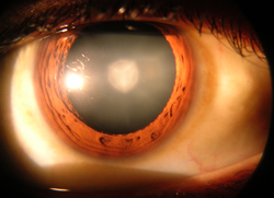
Keratoconus (KC) is a disorder of the eye that results in progressive thinning of the cornea. This may result in blurry vision, double vision, nearsightedness, irregular astigmatism, and light sensitivity leading to poor quality-of-life. Usually both eyes are affected. In more severe cases a scarring or a circle may be seen within the cornea.

<span class="mw-page-title-main">Scanning laser ophthalmoscopy</span>

Scanning laser ophthalmoscopy (SLO) is a method of examination of the eye. It uses the technique of confocal laser scanning microscopy for diagnostic imaging of the retina or cornea of the human eye.

<span class="mw-page-title-main">Eye examination</span> Series of tests assessing vision and pertaining to the eyes

An eye examination is a series of tests performed to assess vision and ability to focus on and discern objects. It also includes other tests and examinations pertaining to the eyes. Eye examinations are primarily performed by an optometrist, ophthalmologist, or an orthoptist. Health care professionals often recommend that all people should have periodic and thorough eye examinations as part of routine primary care, especially since many eye diseases are asymptomatic.

<span class="mw-page-title-main">Fuchs' dystrophy</span> Medical condition

Fuchs dystrophy, also referred to as Fuchs endothelial corneal dystrophy (FECD) and Fuchs endothelial dystrophy (FED), is a slowly progressing corneal dystrophy that usually affects both eyes and is slightly more common in women than in men. Although early signs of Fuchs dystrophy are sometimes seen in people in their 30s and 40s, the disease rarely affects vision until people reach their 50s and 60s.

Eye banks recover, prepare and deliver donated eyes for cornea transplants and research. The first successful cornea transplant was performed in 1905 and the first eye bank was founded in 1944. Currently, in the United States, eye banks provide tissue for over 80,000 cornea transplants each year to treat conditions such as keratoconus and corneal scarring. In some cases, the white of the eye (sclera) is used to surgically repair recipient eyes. Unlike other organs and tissues, corneas are in adequate supply for transplants in the United States, and excess tissue is exported internationally, where there are shortages in many countries, due to greater demand and a less-developed eye banking infrastructure.

<span class="mw-page-title-main">Gonioscopy</span> Measurement of the angle between an eyes iris and cornea

In ophthalmology, gonioscopy is a routine procedure that measures the angle between the iris and the cornea, using a goniolens together with a slit lamp or operating microscope. Its use is important in diagnosing and monitoring various eye conditions associated with glaucoma.

<span class="mw-page-title-main">Corneal topography</span> Medical imaging technique

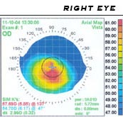
Corneal topography, also known as photokeratoscopy or videokeratography, is a non-invasive medical imaging technique for mapping the anterior curvature of the cornea, the outer structure of the eye. Since the cornea is normally responsible for some 70% of the eye's refractive power, its topography is of critical importance in determining the quality of vision and corneal health.

<span class="mw-page-title-main">Corneal ulcer</span> Medical condition of the eye

Corneal ulcer, also called keratitis, is an inflammatory or, more seriously, infective condition of the cornea involving disruption of its epithelial layer with involvement of the corneal stroma. It is a common condition in humans particularly in the tropics and in farming. In developing countries, children afflicted by vitamin A deficiency are at high risk for corneal ulcer and may become blind in both eyes persisting throughout life. In ophthalmology, a corneal ulcer usually refers to having an infection, while the term corneal abrasion refers more to a scratch injury.

Scanning laser polarimetry is the use of polarised light to measure the thickness of the retinal nerve fiber layer (RNFL) as part of a glaucoma workup. The GDx-VCC is one example.

<span class="mw-page-title-main">Fundus photography</span> Medical imaging of the eyes

Fundus photography involves photographing the rear of an eye, also known as the fundus. Specialized fundus cameras consisting of an intricate microscope attached to a flash enabled camera are used in fundus photography. The main structures that can be visualized on a fundus photo are the central and peripheral retina, optic disc and macula. Fundus photography can be performed with colored filters, or with specialized dyes including fluorescein and indocyanine green.

<span class="mw-page-title-main">Intraocular hemorrhage</span> Medical condition

Intraocular hemorrhage is bleeding inside the eye. Bleeding can occur from any structure of the eye where there is vasculature or blood flow, including the anterior chamber, vitreous cavity, retina, choroid, suprachoroidal space, or optic disc.

<span class="mw-page-title-main">Stereo microscope</span> Variant of an optical microscope

The stereo, stereoscopic or dissecting microscope is an optical microscope variant designed for low magnification observation of a sample, typically using light reflected from the surface of an object rather than transmitted through it. The instrument uses two separate optical paths with two objectives and eyepieces to provide slightly different viewing angles to the left and right eyes. This arrangement produces a three-dimensional visualization of the sample being examined. Stereomicroscopy overlaps macrophotography for recording and examining solid samples with complex surface topography, where a three-dimensional view is needed for analyzing the detail.

<span class="mw-page-title-main">Meesmann corneal dystrophy</span> Medical condition

Meesmann corneal dystrophy (MECD) is a rare hereditary autosomal dominant disease that is characterized as a type of corneal dystrophy and a keratin disease. MECD is characterized by the formation of microcysts in the outermost layer of the cornea, known as the anterior corneal epithelium. The anterior corneal epithelium also becomes fragile. This usually affects both eyes rather than a single eye and worsens over time. There are two phenotypes, Meesmann corneal dystrophy 1 (MECD1) and Meesmann corneal dystrophy 2 (MECD2), which affect the genes KRT3 and KRT12, respectively. A heterozygous mutation in either of these genes will lead to a single phenotype. Many with Meesmann corneal dystrophy are asymptomatic or experience mild symptoms.

<span class="mw-page-title-main">Keratoprosthesis</span> Surgical procedure where a diseased cornea is replaced with an artificial one

Keratoprosthesis is a surgical procedure where a diseased cornea is replaced with an artificial cornea. Traditionally, keratoprosthesis is recommended after a person has had a failure of one or more donor corneal transplants. More recently, a less invasive, non-penetrating artificial cornea has been developed which can be used in more routine cases of corneal blindness. While conventional cornea transplant uses donor tissue for transplant, an artificial cornea is used in the keratoprosthesis procedure. The surgery is performed to restore vision in patients with severely damaged cornea due to congenital birth defects, infections, injuries and burns.

<span class="mw-page-title-main">OCT Biomicroscopy</span> Use of optical coherence tomography to examine the transparent axial tissues of the eye

OCT Biomicroscopy is the use of optical coherence tomography (OCT) in place of slit lamp biomicroscopy to examine the transparent axial tissues of the eye. Traditionally, ophthalmic biomicroscopy has been completed with a slit lamp biomicroscope that uses slit beam illumination and an optical microscope to enable stereoscopic, magnified, cross-sectional views of transparent tissues in the eye, with or without the aid of an additional lens. Like slit lamp biomicroscopy, OCT does not penetrate opaque tissues well but enables detailed, cross-sectional views of transparent tissues, often with greater detail than is possible with a slit lamp. Ultrasound biomicroscopy (UBM) is much better at imaging through opaque tissues since it uses high energy sound waves. Because of its limited depth of penetration, UBM's main use within ophthalmology has been to visualize anterior structures such as the angle and ciliary body. Both ultrasound and OCT biomicroscopy produce an objective image of ocular tissues from which measurements can be made. Unlike UBM, OCT biomicroscopy can image tissues with high axial resolution as far posteriorly as the choroid.

<span class="mw-page-title-main">Gholam A. Peyman</span> Iranian-American ophthalmologist and retina surgeon known for inventing LASIK eye surgery

Gholam A. Peyman is an Iranian American ophthalmologist, retina surgeon, and inventor. He is best known for his invention of LASIK eye surgery, a vision correction procedure designed to allow people to see clearly without glasses. He was awarded the first US patent for the procedure in 1989.

The aim of an accurate intraocular lens power calculation is to provide an intraocular lens (IOL) that fits the specific needs and desires of the individual patient. The development of better instrumentation for measuring the eye's axial length (AL) and the use of more precise mathematical formulas to perform the appropriate calculations have significantly improved the accuracy with which the surgeon determines the IOL power.

The Van Herick technique is an eye examination method used to determine the size of the anterior chamber angle of the eye.

<span class="mw-page-title-main">Corneal button</span> Replacement cormea for transplantation

A corneal button is a replacement cornea to be transplanted in the place of a damaged, diseased or opacified cornea, normally approximately 8.5–9.0mm in diameter. It is used in a corneal transplantation procedure whereby the whole, or part, of a cornea is replaced. The donor tissue can now be held for days to even weeks of the donor's death and is normally a small, rounded shape. The main use of the corneal button is during procedures where the entirety of the cornea needs to be replaced, also known as penetrating keratoplasty.

<span class="mw-page-title-main">Heidelberg Retinal Tomography</span>

The Heidelberg Retinal Tomography is a diagnostic procedure used in ophthalmology. The Heidelberg Retina Tomograph (HRT) is an ophthalmological confocal point scanning laser ophthalmoscope for examining the cornea and certain areas of the retina using different diagnostic modules. However, the most widely used area of application for HRT is the inspection of the optic nerve head (papilla) for early detection and follow-up of glaucoma. The procedure has established itself as an integral part of routine glaucoma diagnostics alongside the visual field examination (perimetry), the chamber angle examination (gonioscopy) and the measurement of intraocular pressure (tonometry). The HRT is the most widely used application of confocal scanning laser ophthalmoscopy.

References

  1. "Eye Examination with the Slit Lamp", Carl Zeiss Meditec, AG, p. 33, accessed: February 6, 2011.
  2. "Eye Examination with the Slit Lamp", Zeiss, p. 33
  3. 1 2 "Eye Examination with the Slit Lamp", Zeiss, p. 34
  4. 1 2 3 "Eye Examination with the Slit Lamp", Zeiss, p. 35
  5. "Eye Examination with the Slit Lamp", Zeiss, p. 36
  6. "Slitlamp", Haag-Streit, AG, accessed: January 25, 2024.
  7. "Eye Examination with the Slit Lamp", Zeiss, p. 37
  8. "Eye Examination with the Slit Lamp", Zeiss, p. 38
  9. 1 2 "Eye Examination with the Slit Lamp", Zeiss, p. 39
  10. "From Lateral Illumination to Slit Lamp - An Outline of Medical History", Koppenhöfer, Eilhard - online published 2012
  11. Practical Ophthalmology A MANUAL FOR BEGINNING RESIDENTS, Fourth Edition, page 218-228.
  12. "Eye Examination with the Slit Lamp", Zeiss, p. 14
  13. "Eye Examination with the Slit Lamp", Zeiss, p. 16
  14. Indiana University School of Optometry. "Slit Lamp Illumination Types". Indiana University, Indiana: 2007. Archived from the original on 18 June 2002. Retrieved 6 February 2011.
  15. Practical Ophthalmology A MANUAL FOR BEGINNING RESIDENTS, Fourth Edition, page 220-221.
  16. Practical Ophthalmology A MANUAL FOR BEGINNING RESIDENTS, Fourth Edition, page 221-222.
  17. 1 2 "Eye Examination with the Slit Lamp", Zeiss, p. 17
  18. "Eye Examination with the Slit Lamp", Zeiss, p. 18
  19. "Eye Examination with the Slit Lamp", Zeiss, p. 19
  20. "More sensitive line test as alternative to Watzke–Allen slit– beam in patients after anatomically successful closure of idiopathic macular hole". Investigative Ophthalmology & Visual Science. 45. May 2004.
  21. Veiga-Reis, Francyne; Dias, Renato Braz; Nehemy, Márcio B. (December 1997). "Differential diagnosis of macular hole: Watzke-Allen test and laser aiming beam test". Arquivos Brasileiros de Oftalmologia. 60 (6): 631–634. doi: 10.5935/0004-2749.19970012 . ISSN   0004-2749.
  22. Gellrich, Marcus-Matthias (2013). The Slit Lamp: Applications for Biomicroscopy and Videography. Springer Science & Business Media. p. 48. ISBN   9783642397936.
  23. "MEDICAL TECHNOLOGY – FROM CRAFTS TO HIGH-TECH". Swiss Medtech. Retrieved 9 August 2022.
  24. name="Haag-Streit"
  25. Chapter 32 in Classification, Symptoms, and Signs of Uveitis Archived 2012-01-16 at the Wayback Machine by DEBRA A. GOLDSTEIN and HOWARD H. TESSLER. 2006 Edition

Further reading