Dental abscess

Last updated

Dental abscess
Other namesDentoalveolar abscess, Periapical abscess, tooth abscess, root abscess
Abces parulique.jpg
A decayed, broken down tooth, which has undergone pulpal necrosis. A periapical abscess (i.e. around the apex of the tooth root) has then formed and pus is draining into the mouth via an intraoral sinus (gumboil).
Specialty Dentistry

A dental abscess is a localized collection of pus associated with a tooth. The most common type of dental abscess is a periapical abscess, and the second most common is a periodontal abscess. In a periapical abscess, usually the origin is a bacterial infection that has accumulated in the soft, often dead, pulp of the tooth. This can be caused by tooth decay, broken teeth or extensive periodontal disease (or combinations of these factors). A failed root canal treatment may also create a similar abscess.

Contents

A dental abscess is a type of odontogenic infection, although commonly the latter term is applied to an infection which has spread outside the local region around the causative tooth.

Classification

The main types of dental abscess are:

Signs and symptoms

Abscess originating from a tooth, that has spread to the buccal space. Above: deformation of the cheek on the second day. Below: deformation on the third day. Abces dentaire.jpg
Abscess originating from a tooth, that has spread to the buccal space. Above: deformation of the cheek on the second day. Below: deformation on the third day.
Tooth#4, the maxillary right second premolar (upper right 2nd bicuspid), after extraction. The two single-headed arrows point to the CEJ, which is the line separating the crown (in this case, heavily decayed) and the roots. The double headed arrow (bottom right) shows the extent of the abscess that surrounds the apex of the palatal root. Chronic apical periodontitis (with arrows).jpg
Tooth#4, the maxillary right second premolar (upper right 2nd bicuspid), after extraction. The two single-headed arrows point to the CEJ, which is the line separating the crown (in this case, heavily decayed) and the roots. The double headed arrow (bottom right) shows the extent of the abscess that surrounds the apex of the palatal root.

The pain is continuous and may be described as extreme, growing, sharp, shooting, or throbbing. Putting pressure or warmth on the tooth may induce extreme pain. The area may be sensitive to touch and possibly swollen as well. This swelling may be present at either the base of the tooth, the gum, and/or the cheek, and sometimes can be reduced by applying ice packs.

An acute abscess may be painless but still have a swelling present on the gum. It is important to get anything that presents like this checked by a dental professional as it may become chronic later.

In some cases, a tooth abscess may perforate bone and start draining into the surrounding tissues creating local facial swelling. In some cases, the lymph glands in the neck will become swollen and tender in response to the infection. It may even feel like a migraine as the pain can transfer from the infected area. The pain does not normally transfer across the face, only upwards or downwards as the nerves that serve each side of the face are separate.

Severe aching and discomfort on the side of the face where the tooth is infected is also fairly common, with the tooth itself becoming unbearable to touch due to extreme amounts of pain. [2]

Complications

A dental infection resulting in an abscess and inflammation of the maxillary sinus Dental infectionMark.png
A dental infection resulting in an abscess and inflammation of the maxillary sinus
CT scan showing a large tooth abscess (right in the image) with significant inflammation of fatty tissue under the skin Abces dentaire TDM 1.JPG
CT scan showing a large tooth abscess (right in the image) with significant inflammation of fatty tissue under the skin

If left untreated, a severe tooth abscess may become large enough to perforate bone and extend into the soft tissue eventually becoming osteomyelitis and cellulitis respectively. From there it follows the path of least resistance and may spread either internally or externally. The path of the infection is influenced by such things as the location of the infected tooth and the thickness of the bone, muscle and fascia attachments.

External drainage may begin as a boil which bursts allowing pus drainage from the abscess, intraorally (usually through the gum) or extraorally. Chronic drainage will allow an epithelial lining to form in this communication to form a pus draining canal (fistula). [3] Sometimes this type of drainage will immediately relieve some of the painful symptoms associated with the pressure.

Internal drainage is of more concern as growing infection makes space within the tissues surrounding the infection. Severe complications requiring immediate hospitalization include Ludwig's angina, which is a combination of growing infection and cellulitis which closes the airway space causing suffocation in extreme cases. Also infection can spread down the tissue spaces to the mediastinum which has significant consequences on the vital organs such as the heart. Another complication, usually from upper teeth, is a risk of sepsis traveling through pathways to which it can possibly lead to endocarditis, brain abscess (extremely rare), or meningitis (also rare).

Depending on the severity of the infection, the sufferer may feel only mildly ill, or may in extreme cases require hospital care.

Diagnostic

A periodontal abscess may be difficult to distinguish from a periapical abscess. Indeed, sometimes they can occur together. [4] Since the management of a periodontal abscess is different from that of a periapical abscess, this differentiation is important to make.

Treatment

Successful treatment of a dental abscess centers on the reduction and elimination of the offending organisms. This can include treatment with antibiotics [5] and drainage, however, it has become widely recommended that dentists should improve the antibiotic prescribing practices, by limiting the prescriptions to the acute cases that suffer from the severe signs of spreading infection, [6] [7] in an attempt to overcome the development of antibiotic-resistant bacterial strains in the population. A 2018 Cochrane review has found insufficient evidence to rule out if patients with acute dental abscesses can benefit from antibiotic prescriptions. [8]

If the tooth can be restored, root canal therapy can be performed. Non-restorable teeth must be extracted, followed by curettage of all apical soft tissue.

Unless they are symptomatic, teeth treated with root canal therapy should be evaluated at 1- and 2-year intervals after the root canal therapy to rule out possible lesional enlargement and to ensure appropriate healing.

Abscesses may fail to heal for several reasons:

Following conventional, adequate root canal therapy, abscesses that do not heal or enlarge are often treated with surgery and filling the root tips; and will require a biopsy to evaluate the diagnosis. [9]

See also

Related Research Articles

<span class="mw-page-title-main">Abscess</span> Localized collection of pus that has built up within the tissue of the body

An abscess is a collection of pus that has built up within the tissue of the body. Signs and symptoms of abscesses include redness, pain, warmth, and swelling. The swelling may feel fluid-filled when pressed. The area of redness often extends beyond the swelling. Carbuncles and boils are types of abscess that often involve hair follicles, with carbuncles being larger. A cyst is related to an abscess, but it contains a material other than pus, and a cyst has a clearly defined wall.

<span class="mw-page-title-main">Endodontics</span> Field of dentistry

Endodontics is the dental specialty concerned with the study and treatment of the dental pulp.

<span class="mw-page-title-main">Toothache</span> Medical condition of the teeth

Toothache, also known as dental pain or tooth pain, is pain in the teeth or their supporting structures, caused by dental diseases or pain referred to the teeth by non-dental diseases. When severe it may impact sleep, eating, and other daily activities.

<span class="mw-page-title-main">Dental extraction</span> Operation to remove a tooth

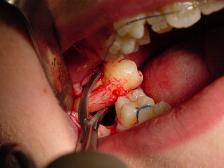
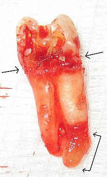
A dental extraction is the removal of teeth from the dental alveolus (socket) in the alveolar bone. Extractions are performed for a wide variety of reasons, but most commonly to remove teeth which have become unrestorable through tooth decay, periodontal disease, or dental trauma, especially when they are associated with toothache. Sometimes impacted wisdom teeth cause recurrent infections of the gum (pericoronitis), and may be removed when other conservative treatments have failed. In orthodontics, if the teeth are crowded, healthy teeth may be extracted to create space so the rest of the teeth can be straightened.

<span class="mw-page-title-main">Pericoronitis</span> Inflammation of the soft tissues surrounding the crown of a partially erupted tooth

Pericoronitis is inflammation of the soft tissues surrounding the crown of a partially erupted tooth, including the gingiva (gums) and the dental follicle. The soft tissue covering a partially erupted tooth is known as an operculum, an area which can be difficult to access with normal oral hygiene methods. The hyponym operculitis technically refers to inflammation of the operculum alone.

A dental emergency is an issue involving the teeth and supporting tissues that are of high importance to be treated by the relevant professional. Dental emergencies do not always involve pain, although this is a common signal that something needs to be looked at. Pain can originate from the tooth, surrounding tissues or can have the sensation of originating in the teeth but be caused by an independent source. Depending on the type of pain experienced an experienced clinician can determine the likely cause and can treat the issue as each tissue type gives different messages in a dental emergency.

<span class="mw-page-title-main">Periapical cyst</span> Medical condition

Commonly known as a dental cyst, the periapical cyst is the most common odontogenic cyst. It may develop rapidly from a periapical granuloma, as a consequence of untreated chronic periapical periodontitis.

<span class="mw-page-title-main">Root canal treatment</span> Dental treatment

Root canal treatment is a treatment sequence for the infected pulp of a tooth which is intended to result in the elimination of infection and the protection of the decontaminated tooth from future microbial invasion. Root canals, and their associated pulp chamber, are the physical hollows within a tooth that are naturally inhabited by nerve tissue, blood vessels and other cellular entities. Together, these items constitute the dental pulp.

Apical periodontitis is typically the body's defense response to the threat of microbial invasion from the root canal. Primary among the members of the host defense mechanism is the polymorphonuclear leukocyte, otherwise known as the neutrophil. The task of the neutrophil is to locate and destroy microbes that intrude into the body – anywhere in the body – and they represent the hallmark of acute inflammation.

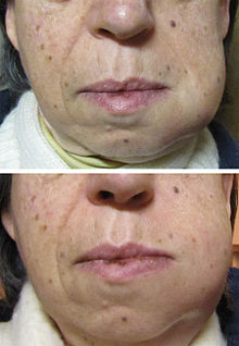
Mouth infections, also known as oral infections, are a group of infections that occur around the oral cavity. They include dental infection, dental abscess, and Ludwig's angina. Mouth infections typically originate from dental caries at the root of molars and premolars that spread to adjacent structures. In otherwise healthy patients, removing the offending tooth to allow drainage will usually resolve the infection. In cases that spread to adjacent structures or in immunocompromised patients, surgical drainage and systemic antibiotics may be required in addition to tooth extraction. Since bacteria that normally reside in the oral cavity cause mouth infections, proper dental hygiene can prevent most cases of infection. As such, mouth infections are more common in populations with poor access to dental care or populations with health-related behaviors that damage one's teeth and oral mucosa. This is a common problem, representing nearly 36% of all encounters within the emergency department related to dental conditions.

<span class="mw-page-title-main">Periodontal abscess</span> Medical condition

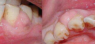
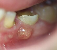
A periodontal abscess, is a localized collection of pus within the tissues of the periodontium. It is a type of dental abscess. A periodontal abscess occurs alongside a tooth, and is different from the more common periapical abscess, which represents the spread of infection from a dead tooth. To reflect this, sometimes the term "lateral (periodontal) abscess" is used. In contrast to a periapical abscess, periodontal abscesses are usually associated with a vital (living) tooth. Abscesses of the periodontium are acute bacterial infections classified primarily by location.

Combined periodontic-endodontic lesions are localized, circumscribed areas of bacterial infection originating from either dental pulp, periodontal tissues surrounding the involved tooth or teeth or both.

A phoenix abscess is an acute exacerbation of a chronic periapical lesion. It is a dental abscess that can occur immediately following root canal treatment. Another cause is due to untreated necrotic pulp. It is also the result of inadequate debridement during the endodontic procedure. Risk of occurrence of a phoenix abscess is minimised by correct identification and instrumentation of the entire root canal, ensuring no missed anatomy.

<span class="mw-page-title-main">Odontogenic infection</span> Infection originating in a tooth

An odontogenic infection is an infection that originates within a tooth or in the closely surrounding tissues. The term is derived from odonto- and -genic. The most common causes for odontogenic infection to be established are dental caries, deep fillings, failed root canal treatments, periodontal disease, and pericoronitis. Odontogenic infection starts as localised infection and may remain localised to the region where it started, or spread into adjacent or distant areas.

Osteomyelitis of the jaws is osteomyelitis which occurs in the bones of the jaws. Historically, osteomyelitis of the jaws was a common complication of odontogenic infection. Before the antibiotic era, it was frequently a fatal condition.

<span class="mw-page-title-main">Periapical periodontitis</span> Medical condition

Periapical periodontitis or apical periodontitis (AP) is an acute or chronic inflammatory lesion around the apex of a tooth root, most commonly caused by bacterial invasion of the pulp of the tooth. It is a likely outcome of untreated dental caries, and in such cases it can be considered a sequela in the natural history of tooth decay, irreversible pulpitis and pulpal necrosis. Other causes can include occlusal trauma due to 'high spots' after restoration work, extrusion from the tooth of root filling material, or bacterial invasion and infection from the gums. Periapical periodontitis may develop into a periapical abscess, where a collection of pus forms at the end of the root, the consequence of spread of infection from the tooth pulp, or into a periapical cyst, where an epithelial lined, fluid-filled structure forms.

There are many circumstances during dental treatment where antibiotics are prescribed by dentists to prevent further infection. The most common antibiotic prescribed by dental practitioners is penicillin in the form of amoxicillin, however many patients are hypersensitive to this particular antibiotic. Therefore, in the cases of allergies, erythromycin is used instead.

In the dental specialty of endodontics, periradicular surgery is surgery to the external root surface. Examples of periradicular surgery include apicoectomy, root resection, repair of root perforation or resorption defects, removal of broken fragments of the tooth or a filling material, and exploratory surgery to look for root fractures.

Periapical granuloma, also sometimes referred to as a radicular granuloma or apical granuloma, is an inflammation at the tip of a dead (nonvital) tooth. It is a lesion or mass that typically starts out as an epithelial lined cyst, and undergoes an inward curvature that results in inflammation of granulation tissue at the root tips of a dead tooth. This is usually due to dental caries or a bacterial infection of the dental pulp. Periapical granuloma is an infrequent disorder that has an occurrence rate between 9.3 to 87.1 percent. Periapical granuloma is not a true granuloma due to the fact that it does not contain granulomatous inflammation; however, periapical granuloma is a common term used.

References

  1. Rapini RP, Bolognia JL, Jorizzo JL (2007). Dermatology: 2-Volume Set. St. Louis: Mosby. ISBN   978-1-4160-2999-1.
  2. "Tooth abscess: MedlinePlus Medical Encyclopedia". medlineplus.gov. Retrieved 18 November 2022.
  3. "Fistulas". medlineplus.gov. Retrieved 18 November 2022.
  4. 1 2 3 4 Fan KF, Jones J (2009). OSCEs for dentistry (2nd, new and updated ed.). 2nd: PasTest Ltd. p. 68. ISBN   978-1-905635-50-4.{{cite book}}: CS1 maint: location (link)
  5. "Dental abscess - Management". Clinical Knowledge Summaries. Archived from the original on 27 December 2011. Retrieved 1 March 2012.
  6. Dar-Odeh NS, Abu-Hammad OA, Al-Omiri MK, Khraisat AS, Shehabi AA (July 2010). "Antibiotic prescribing practices by dentists: a review". Therapeutics and Clinical Risk Management. 6: 301–6. doi: 10.2147/tcrm.s9736 . PMC   2909496 . PMID   20668712.
  7. Oberoi SS, Dhingra C, Sharma G, Sardana D (February 2015). "Antibiotics in dental practice: how justified are we". International Dental Journal. 65 (1): 4–10. doi: 10.1111/idj.12146 . PMC   9376535 . PMID   25510967.
  8. Cope, Anwen L; Francis, Nick; Wood, Fiona; Chestnutt, Ivor G (27 September 2018). Cochrane Oral Health Group (ed.). "Systemic antibiotics for symptomatic apical periodontitis and acute apical abscess in adults". Cochrane Database of Systematic Reviews. 2018 (9): CD010136. doi:10.1002/14651858.CD010136.pub3. PMC   6513530 . PMID   30259968.
  9. Neville BW (1995). Oral and Maxillofacial Pathology (1st ed.). Saunders. pp. 104–5. ISBN   978-1-4160-3435-3.