Pitted keratolysis

Last updated
Pitted keratolysis
Other namesKeratolysis plantare sulcatum, Keratoma plantare sulcatum, Ringed keratolysis, [1] and sweaty sock syndrome [2]
Pitted keratolysis 3.jpg
Right foot affected with pitted keratolysis
Specialty Dermatology   OOjs UI icon edit-ltr-progressive.svg
Symptoms Large, crater-like holes on the foot
Causes Corynebacterium species and other bacteria
Differential diagnosis Athlete's foot (Tinea pedis), erythrasma, hyperhidrosis
PreventionKeeping the feet dry, antiperspirants
TreatmentAntibiotics

Pitted keratolysis (also known as keratolysis plantare sulcatum, [1] keratoma plantare sulcatum, [1] and ringed keratolysis [1] ) is a bacterial skin infection of the foot. [2] The infection is characterized by craterlike pits on the sole of the feet and toes, particularly weight bearing areas.

Contents

The infection is caused by Kytococcus sedentarius . [2] [3] Excessive sweating of the feet and use of occlusive footwear provide an environment in which these bacteria thrive and therefore increase the risk of developing pitted keratolysis. [2] [4]

The condition is fairly common, especially in the military where wet shoes/boots are worn for extended periods of time without removing/cleaning. Skin biopsy specimens are not usually utilized, as the diagnosis of pitted keratolysis is often made by visual examination and recognition of the characteristic odor. Wood's lamp examination results are inconsistent. Treatment of pitted keratolysis requires the application of antibiotics to the skin such as benzoyl peroxide, clindamycin, erythromycin, fusidic acid, or mupirocin. [2] [5] Prevention efforts aim to keep the feet dry by using moisture-wicking shoes and socks as well as antiperspirants. [2]

Signs and symptoms

Pitted keratolysis typically presents with white discoloration of the skin and numerous discrete, "punched-out" pitted lesions or erosions, usually located on the soles of the feet. [2] The pits are typically 1–7 millimeters in diameter. [2] [5] These circular and shallow pits are characteristic of pitted keratolysis, and often overlap to produce larger areas of erosion. The appearance of this condition’s characteristic lesions becomes more pronounced when the affected area is wet or submerged in water. Occasionally these lesions present with a green or brown hue around and within the pits.[ citation needed ]

These superficial erosions are found under the toes and on the soles of the feet, and especially at the pressure bearing points such as the heel. [2] Typically, both feet are equally affected. Rarely, the condition affects the palms.[ citation needed ]

Cause

The most common cause of pitted keratolysis is Kytococcus sedentarius . However, several other bacteria may also cause the condition, particularly Actinomyces keratolytica, Dermatophilus congolensis , and Streptomyces . [2] [5] Less frequently, it is due to Acinetobacter , Clostridium , Klebsiella , and Pseudomonas species.[ citation needed ]

Pathogenesis

Pitted keratolysis is associated with excessive sweating of the palms or soles (palmoplantar hyperhidrosis.) [2] The pits seen in pitted keratolysis are caused by bacteria secreting proteinase enzymes which cause the breakdown of the keratin proteins in the stratum corneum layer of the affected skin. [5] This results in the formation of sulfur compounds which leads to a very strong and foul foot odor. [2] The bacteria that cause pitted keratolysis thrive in warm and humid environments. [5] Irritation is generally minimal, though occasionally burning, itching, and soreness are experienced with pitted keratolysis.[ citation needed ]

Diagnosis

The diagnosis of pitted keratolysis is based primarily on the physical examination, with recognition of the classic pitted lesions and pungent odor. Dermoscopic examination can facilitate visualization of pits and pit walls. A Wood's lamp may show coral red fluorescence, as seen in erythrasma. However, this finding is not uniformly present, as the condition may be caused by bacteria that do not produce fluorescent pigments. Further laboratory testing is not typically required for the diagnosis. However, a potassium hydroxide preparation can help rule out the presence of a fungal infection. Imaging and biopsy are not necessary.[ citation needed ]

Differential diagnosis

Treatment

As of 2014, there is little high-quality evidence to provide guidance and support one treatment method over another. Therefore, the optimal treatment approach for pitted keratolysis remains unclear as the condition has not been well-studied. [5] One review suggested a treatment approach requiring modification of risk factors (e.g., keeping feet clean and dry) and treating the underlying bacterial infection. [5] Effective antibiotic options include clindamycin, erythromycin, mupirocin, and fusidic acid. Topical clindamycin is generally preferred as the first-line choice due to lower cost and better availability. [5] Fusidic acid is preferred over mupirocin in most cases due to less resistance to fusidic acid amongst Methicillin-sensitive staphylococcus aureus and Methicillin-resistant staphylococcus aureus . [5] Benzoyl peroxide is an effective alternative over-the-counter option and is thought to be as effective as topical clindamycin. [5] Clinical cure was typically seen after 2.5–3 weeks. [5] Oral antibiotics are not typically recommended. [5]

Foot hygiene is important. The feet may be washed at least daily with soap and water, and dried thoroughly afterwards. Moisture-wicking socks and shoes may be worn and antiperspirants, such as aluminum chlorohydrate, may help to keep the feet dry. Injections of botulinum toxin have successfully induced cessation of sweating (anhidrosis) of the soles of the feet and led to resolution of pitted keratolysis. [5] These injections are typically reserved for refractory cases of pitted keratolysis that have failed to respond to environmental modifications and antibiotics due to the high cost and pain associated with botulinum toxin injections. [5]

Pitted keratolysis can be reduced and eventually stopped by regularly applying a liberal amount of antiperspirant body powder to the inside of the shoes and socks of the affected person. Regular powder application will greatly reduce foot perspiration and keep the plantar surface of the foot dry therefore creating an environment hostile to the Corynebacterium.[ citation needed ]

Epidemiology

Pitted keratolysis occurs worldwide and in various climates. The infection is more common in people who live in tropical climates and walk barefoot, and those who spend a lot of time wearing occlusive footwear (e.g., tight shoes, rubber boots), [5] such as in the military where wet shoes are worn for a prolonged period of time without removing and proper hygiene. [6]

History

Pitted keratolysis was first named keratoma plantare sulcatum. [7]

See also

Related Research Articles

<span class="mw-page-title-main">Perspiration</span> Fluid secreted from sudoriferous glands

Perspiration, also known as sweat, is the fluid secreted by sweat glands in the skin of mammals.

<span class="mw-page-title-main">Impetigo</span> Human disease (bacterial infection)

Impetigo is a bacterial infection that involves the superficial skin. The most common presentation is yellowish crusts on the face, arms, or legs. Less commonly there may be large blisters which affect the groin or armpits. The lesions may be painful or itchy. Fever is uncommon.

<span class="mw-page-title-main">Plantar wart</span> Medical condition

A plantar wart is a wart occurring on the bottom of the foot or toes. Its color is typically similar to that of the skin. Small black dots often occur on the surface. One or more may occur in an area. They may result in pain with pressure such that walking is difficult.

<span class="mw-page-title-main">Tinea versicolor</span> Skin disease

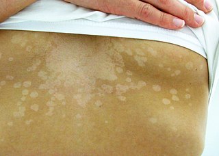
Tinea versicolor is a condition characterized by a skin eruption on the trunk and proximal extremities. The majority of tinea versicolor is caused by the fungus Malassezia globosa, although Malassezia furfur is responsible for a small number of cases. These yeasts are normally found on the human skin and become troublesome only under certain conditions, such as a warm and humid environment, although the exact conditions that cause initiation of the disease process are poorly understood.

<span class="mw-page-title-main">Folliculitis</span> Human and animal disease of follicles

Folliculitis is the infection and inflammation of one or more hair follicles. The condition may occur anywhere on hair-covered skin. The rash may appear as pimples that come to white tips on the face, chest, back, arms, legs, buttocks, or head.

<span class="mw-page-title-main">Hyperhidrosis</span> Excessive sweating

Hyperhidrosis is a condition characterized by abnormally increased sweating, in excess of that required for regulation of body temperature. Although primarily a benign physical burden, hyperhidrosis can deteriorate quality of life from a psychological, emotional, and social perspective. In fact, hyperhidrosis almost always leads to psychological as well as physical and social consequences. People suffering from it present difficulties in the professional field, more than 80% experiencing a moderate to severe emotional impact from the disease and half are subject to depression.

<span class="mw-page-title-main">Athlete's foot</span> Skin infection caused by fungus

Athlete's foot, known medically as tinea pedis, is a common skin infection of the feet caused by a fungus. Signs and symptoms often include itching, scaling, cracking and redness. In rare cases the skin may blister. Athlete's foot fungus may infect any part of the foot, but most often grows between the toes. The next most common area is the bottom of the foot. The same fungus may also affect the nails or the hands. It is a member of the group of diseases known as tinea.

<span class="mw-page-title-main">Fusidic acid</span> Antibiotic

Fusidic acid, sold under the brand name Fucidin among others, is a steroid antibiotic that is often used topically in creams or ointments and eyedrops but may also be given systemically as tablets or injections. As of October 2008, the global problem of advancing antimicrobial resistance has led to a renewed interest in its use.

<span class="mw-page-title-main">Pseudofolliculitis barbae</span> Medical condition

Pseudofolliculitis barbae (PFB) is a type of irritant folliculitis that commonly affects people who have curly or coarse facial hair. It occurs when hair curls back into the skin after shaving, causing inflammation, redness, and bumps. This can lead to ingrown hairs, scarring, and skin discoloration. PFB can be treated with various methods, including changing shaving habits, using topical creams or ointments, and undergoing laser hair removal. Prevention measures include proper shaving techniques, using sharp razors, and avoiding too close a shave.

<span class="mw-page-title-main">Paronychia</span> Medical condition

Paronychia is an inflammation of the skin around the nail, which can occur suddenly, when it is usually due to the bacterium Staphylococcus aureus, or gradually when it is commonly caused by the fungus Candida albicans. The term is from Greek: παρωνυχία from para 'around', onyx 'nail', and the abstract noun suffix -ia.

<i>Staphylococcus epidermidis</i> Species of bacterium

Staphylococcus epidermidis is a Gram-positive bacterium, and one of over 40 species belonging to the genus Staphylococcus. It is part of the normal human microbiota, typically the skin microbiota, and less commonly the mucosal microbiota and also found in marine sponges. It is a facultative anaerobic bacteria. Although S. epidermidis is not usually pathogenic, patients with compromised immune systems are at risk of developing infection. These infections are generally hospital-acquired. S. epidermidis is a particular concern for people with catheters or other surgical implants because it is known to form biofilms that grow on these devices. Being part of the normal skin microbiota, S. epidermidis is a frequent contaminant of specimens sent to the diagnostic laboratory.

<span class="mw-page-title-main">Erythrasma</span> Medical condition

Erythrasma is a superficial skin infection that causes brown, scaly skin patches. It is caused by Corynebacterium minutissimum bacteria, a normal part of skin flora.

<span class="mw-page-title-main">Angular cheilitis</span> Cheilitis characterized by inflammation of one or both of the corners of the mouth

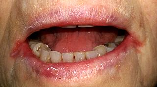
Angular cheilitis (AC) is inflammation of one or both corners of the mouth. Often the corners are red with skin breakdown and crusting. It can also be itchy or painful. The condition can last for days to years. Angular cheilitis is a type of cheilitis.

Keratolysis exfoliativa is a sometimes harmless, sometimes painful skin condition that can affect the focal surface of the fingers and/or the palm or soles of the feet. It is often misdiagnosed as chronic contact dermatitis or psoriasis. It is characterized by dry skin and superficial, air-filled blisters. These blisters can be peeled off very easily and will leave reddish, tender areas. The loss of this corneal layer of the skin, which protects the underlying layers, leaves the skin more vulnerable to dryness and cracking.

<span class="mw-page-title-main">Pimple</span> Type of comedo

A pimple or zit is a kind of comedo that results from excess sebum and dead skin cells getting trapped in the pores of the skin. In its aggravated state, it may evolve into a pustule or papule. Pimples can be treated by acne medications, antibiotics, and anti-inflammatories prescribed by a physician, or various over the counter remedies purchased at a pharmacy.

<span class="mw-page-title-main">Bullous impetigo</span> Medical condition

Bullous impetigo is a bacterial skin infection caused by Staphylococcus aureus that results in the formation of large blisters called bullae, usually in areas with skin folds like the armpit, groin, between the fingers or toes, beneath the breast, and between the buttocks. It accounts for 30% of cases of impetigo, the other 70% being non-bullous impetigo.

<span class="mw-page-title-main">Streptococcal intertrigo</span> Medical condition

Streptococcal intertrigo is a skin condition that is secondary to a streptococcal bacterial infection. It is often seen in infants and young children and can be characterized by a fiery-red color of the skin, foul odor with an absence of satellite lesions, and skin softening in the neck, armpits or folds of the groin. Newborn children and infants commonly develop intertrigo because of physical features such as deep skin folds, short neck, and flexed posture. Prompt diagnosis by a medical professional and treatment with topical and/or oral antibiotics can effectively relieve symptoms.

<i>Kytococcus sedentarius</i> Species of bacterium

Kytococcus sedentarius is a marine dwelling Gram positive bacterium in the genus Kytococcus. It is known for the production of polyketide antibiotics as well as for its role as an opportunistic pathogen. It is strictly aerobic and can only grow when amino acids are provided.

Foot odor or bromodosis is a type of body odor that affects the feet of humans.

References

  1. 1 2 3 4 Rapini, Ronald P.; Bolognia, Jean L.; Jorizzo, Joseph L. (2007). Dermatology: 2-Volume Set. St. Louis: Mosby. ISBN   978-1-4160-2999-1.[ page needed ]
  2. 1 2 3 4 5 6 7 8 9 10 11 12 Hsu, AR; Hsu, JW (July 2012). "Topical review: skin infections in the foot and ankle patient". Foot & Ankle International (Review). 33 (7): 612–619. doi:10.3113/FAI.2012.0612. PMID   22835400.
  3. Makhecha, Meena; Dass, Shreya; Singh, Tishya; Gandhi, Riddhi; Yadav, Tulika; Rathod, Dipali (November 2017). "Pitted keratolysis – a study of various clinical manifestations". International Journal of Dermatology. 56 (11): 1154–1160. doi:10.1111/ijd.13744. ISSN   1365-4632. PMID   28924971.
  4. "Without Proper Treatment, Skin Infections Can Sideline Your Season". American Academy of Dermatology. March 3, 2006.
  5. 1 2 3 4 5 6 7 8 9 10 11 12 13 14 15 Bristow, IR; Lee, YL (March 2014). "Pitted keratolysis: a clinical review". Journal of the American Podiatric Medical Association (Review). 104 (2): 177–182. doi:10.7547/0003-0538-104.2.177. PMID   24725039.
  6. van der Snoek, E.M.; Ekkelenkamp, M.B.; Suykerbuyk, J.C.C.W. (September 2013). "Pitted keratolysis; physicians' treatment and their perceptions in Dutch army personnel: Pitted keratolysis in Dutch army personnel". Journal of the European Academy of Dermatology and Venereology. 27 (9): 1120–1126. doi:10.1111/j.1468-3083.2012.04674.x. PMID   22882561.
  7. James, William D.; Berger, Timothy G.; et al. (2006). Andrews' Diseases of the Skin: clinical Dermatology. Fairly common, especially common in military. Saunders Elsevier. ISBN   0-7216-2921-0.[ page needed ]