
The ileum is the final section of the small intestine in most higher vertebrates, including mammals, reptiles, and birds. In fish, the divisions of the small intestine are not as clear and the terms posterior intestine or distal intestine may be used instead of ileum. Its main function is to absorb vitamin B12, bile salts, and whatever products of digestion that were not absorbed by the jejunum.

In vertebrates, the gallbladder, also known as the cholecyst, is a small hollow organ where bile is stored and concentrated before it is released into the small intestine. In humans, the pear-shaped gallbladder lies beneath the liver, although the structure and position of the gallbladder can vary significantly among animal species. It receives bile, produced by the liver, via the common hepatic duct, and stores it. The bile is then released via the common bile duct into the duodenum, where the bile helps in the digestion of fats.

A bile duct is any of a number of long tube-like structures that carry bile, and is present in most vertebrates. The bile duct is separated into three main parts: the fundus (superior), the body (middle), and the neck (inferior).
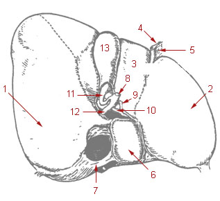
The cystic duct is the duct that (typically) joins the gallbladder and the common hepatic duct; the union of the cystic duct and common hepatic duct forms the bile duct. Its length varies. It is often tortuous.

The yolk sac is a membranous sac attached to an embryo, formed by cells of the hypoblast layer of the bilaminar embryonic disc. This is alternatively called the umbilical vesicle by the Terminologia Embryologica (TE), though yolk sac is far more widely used. In humans, the yolk sac is important in early embryonic blood supply, and much of it is incorporated into the primordial gut during the fourth week of embryonic development.

In medicine or biology, a diverticulum is an outpouching of a hollow structure in the body. Depending upon which layers of the structure are involved, diverticula are described as being either true or false.
The common hepatic duct is the first part of the biliary tract. It joins the cystic duct coming from the gallbladder to form the common bile duct.
The Kocher manoeuvre is a surgical procedure to expose structures in the retroperitoneum behind the duodenum and pancreas. In vascular surgery, it is described as a method to expose the abdominal aorta. It usually has been in contrast to midline laparotomy and right retroperitoneal space dissection. These two procedures have been used for diverse cases, but have approximately equivalent outcomes.
Cholestasis is a condition where the flow of bile from the liver to the duodenum is impaired. The two basic distinctions are:

The foregut in humans is the anterior part of the alimentary canal, from the distal esophagus to the first half of the duodenum, at the entrance of the bile duct. Beyond the stomach, the foregut is attached to the abdominal walls by mesentery. The foregut arises from the endoderm, developing from the folding primitive gut, and is developmentally distinct from the midgut and hindgut. Although the term “foregut” is typically used in reference to the anterior section of the primitive gut, components of the adult gut can also be described with this designation. Pain in the epigastric region, just below the intersection of the ribs, typically refers to structures in the adult foregut.

The cystohepatic triangle is an anatomic space bordered by the cystic duct laterally, the common hepatic duct medially, and the inferior surface of the liver superiorly.

The hepatic plexus is a sympathetic and parasympathetic nerve plexus that provides innervation to the parenchyma of the liver as well as contributing innervation to some other abdominal structures.

The bilaminar embryonic disc, bilaminar blastoderm or embryonic disc is the distinct two-layered structure of cells formed in an embryo. In the development of the human embryo this takes place by day eight. It is formed when the inner cell mass, also known as the embryoblast, forms a bilaminar disc of two layers, an upper layer called the epiblast and a lower layer called the hypoblast, which will eventually form into fetus. These two layers of cells are stretched between two fluid-filled cavities at either end: the primitive yolk sac and the amniotic sac.

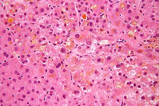
In histology, the lobules of liver, or hepatic lobules, are small divisions of the liver defined at the microscopic scale. The hepatic lobule is a building block of the liver tissue, consisting of a portal triad, hepatocytes arranged in linear cords between a capillary network, and a central vein.

The biliary tract refers to the liver, gallbladder and bile ducts, and how they work together to make, store and secrete bile. Bile consists of water, electrolytes, bile acids, cholesterol, phospholipids and conjugated bilirubin. Some components are synthesized by hepatocytes ; the rest are extracted from the blood by the liver.

Congenital hepatic fibrosis is an inherited fibrocystic liver disease associated with proliferation of interlobular bile ducts within the portal areas and fibrosis that do not alter hepatic lobular architecture. The fibrosis would affect resistance in portal veins leading to portal hypertension.

The liver is a major metabolic organ only found in vertebrate animals, which performs many essential biological functions such as detoxification of the organism, and the synthesis of proteins and biochemicals necessary for digestion and growth. In humans, it is located in the right upper quadrant of the abdomen, below the diaphragm and mostly shielded by the lower right rib cage. Its other metabolic roles include carbohydrate metabolism, the production of hormones, conversion and storage of nutrients such as glucose and glycogen, and the decomposition of red blood cells.
The connecting stalk, or body stalk, is an embryonic structure that is formed by the third week of development and connects the embryo to its shell of trophoblasts. The connecting stalk is derived from the extraembryonic mesoderm. Initially it lies caudally to the trilaminar germ disc, but, with subsequent embryonic folding, the body stalk assume a more ventral position. Progressive expansion of the amnion from the umbilical ring creates a tube with a covering of amniotic membrane with allantois and umbilical vessels as its content and mesoderm of the connecting stalk as the ground substance. This extraembryonic mesodermal ground substance forms the future Wharton's jelly. The amniotic membrane and its contents form the umbilical cord that connects the embryo and the placenta.
The development of the digestive system in the human embryo concerns the epithelium of the digestive system and the parenchyma of its derivatives, which originate from the endoderm. Connective tissue, muscular components, and peritoneal components originate in the mesoderm. Different regions of the gut tube such as the esophagus, stomach, duodenum, etc. are specified by a retinoic acid gradient that causes transcription factors unique to each region to be expressed. Differentiation of the gut and its derivatives depends upon reciprocal interactions between the gut endoderm and its surrounding mesoderm. Hox genes in the mesoderm are induced by a Hedgehog signaling pathway secreted by gut endoderm and regulate the craniocaudal organization of the gut and its derivatives. The gut system extends from the oropharyngeal membrane to the cloacal membrane and is divided into the foregut, midgut, and hindgut.

The human digestive system consists of the gastrointestinal tract plus the accessory organs of digestion. Digestion involves the breakdown of food into smaller and smaller components, until they can be absorbed and assimilated into the body. The process of digestion has three stages: the cephalic phase, the gastric phase, and the intestinal phase.
