Effectiveness
A 2014 Cochrane review found that hip protectors decrease the number of hip fractures among the elderly. [3]
A number of reviews have found that hip protectors are cost-effective, particularly among residents of care homes and for long-stay patients in hospital [4]
A previous review found that the effect for preventing hip fracture among nursing home residents was small and not effective among community dwelling elderly individuals. [5] A 2007 review found a decreased risk of hip fractures in elderly nursing home residents. [6]
However, acceptance and long-term compliance towards them has historically been quite low, [5] mainly because of discomfort, dislike of their appearance by the person wearing it, and disagreement about fracture risk. [7] More modern hip protectors do not suffer from these disadvantages because they are slimmer with a low profile, so less noticeable, have ventilation holes and ducting to keep the skin cool under the pad and are soft and pliable conforming to the contours of the hip. Better independent testing procedures developed by Professor Julian Minns have established a reliable baseline for impact absorption performance. [8]
Research which has found hip protectors to be beneficial found that hard, energy-shunting hip protectors to be superior to soft, energy-absorbing ones. [9] However this research predates the introduction of hip protector pads in 2011/2012 using modern non-Newtonian materials, such as D3o that absorb around 75% of the impact, typically twice that of previous devices that used soft materials such as textiles or foam pads in an airtight bag, but comparable to the best of the energy-shunting devices, which have now largely disappeared from the market because of a slight tendency to cause pelvic fractures when the energy is transferred [10] Another study showed that hip protectors' design and mechanical properties vary drastically among commercially available hip protectors. [11]

Osteoporosis is a systemic skeletal disorder characterized by low bone mass, micro-architectural deterioration of bone tissue leading to more porous bone, and consequent increase in fracture risk. It is the most common reason for a broken bone among the elderly. Bones that commonly break include the vertebrae in the spine, the bones of the forearm, the wrist, and the hip. Until a broken bone occurs there are typically no symptoms. Bones may weaken to such a degree that a break may occur with minor stress or spontaneously. After the broken bone heals, the person may have chronic pain and a decreased ability to carry out normal activities.

Dual-energy X-ray absorptiometry is a means of measuring bone mineral density (BMD) using spectral imaging. Two X-ray beams, with different energy levels, are aimed at the patient's bones. When soft tissue absorption is subtracted out, the bone mineral density (BMD) can be determined from the absorption of each beam by bone. Dual-energy X-ray absorptiometry is the most widely used and most thoroughly studied bone density measurement technology.

Bisphosphonates are a class of drugs that prevent the loss of bone density, used to treat osteoporosis and similar diseases. They are the most commonly prescribed drugs used to treat osteoporosis. They are called bisphosphonates because they have two phosphonate groups. They are thus also called diphosphonates.

Paget's disease of bone is a condition involving cellular remodeling and deformity of one or more bones. The affected bones show signs of dysregulated bone remodeling at the microscopic level, specifically excessive bone breakdown and subsequent disorganized new bone formation. These structural changes cause the bone to weaken, which may result in deformity, pain, fracture or arthritis of associated joints.

A bone fracture is a medical condition in which there is a partial or complete break in the continuity of any bone in the body. In more severe cases, the bone may be broken into several fragments, known as a comminuted fracture. A bone fracture may be the result of high force impact or stress, or a minimal trauma injury as a result of certain medical conditions that weaken the bones, such as osteoporosis, osteopenia, bone cancer, or osteogenesis imperfecta, where the fracture is then properly termed a pathologic fracture.
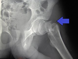
A hip fracture is a break that occurs in the upper part of the femur, at the femoral neck or (rarely) the femoral head. Symptoms may include pain around the hip, particularly with movement, and shortening of the leg. Usually the person cannot walk.

Osteopenia, known as "low bone mass" or "low bone density", is a condition in which bone mineral density is low. Because their bones are weaker, people with osteopenia may have a higher risk of fractures, and some people may go on to develop osteoporosis. In 2010, 43 million older adults in the US had osteopenia. Unlike osteoporosis, osteopenia does not usually cause symptoms, and losing bone density in itself does not cause pain.
Fall prevention includes any action taken to help reduce the number of accidental falls suffered by susceptible individuals, such as the elderly (idiopathic) and people with neurological or orthopedic indications.

Bone density, or bone mineral density, is the amount of bone mineral in bone tissue. The concept is of mass of mineral per volume of bone, although clinically it is measured by proxy according to optical density per square centimetre of bone surface upon imaging. Bone density measurement is used in clinical medicine as an indirect indicator of osteoporosis and fracture risk. It is measured by a procedure called densitometry, often performed in the radiology or nuclear medicine departments of hospitals or clinics. The measurement is painless and non-invasive and involves low radiation exposure. Measurements are most commonly made over the lumbar spine and over the upper part of the hip. The forearm may be scanned if the hip and lumbar spine are not accessible.

Strontium ranelate, a strontium(II) salt of ranelic acid, is a medication for osteoporosis marketed as Protelos or Protos by Servier. Studies indicate it can also slow the course of osteoarthritis of the knee. The drug is unusual in that it both increases deposition of new bone by osteoblasts and reduces the resorption of bone by osteoclasts. It is therefore promoted as a "dual action bone agent" (DABA).
Senile osteoporosis has been recently recognized as a geriatric syndrome with a particular pathophysiology. There are different classification of osteoporosis: primary, in which bone loss is a result of aging and secondary, in which bone loss occurs from various clinical and lifestyle factors. Primary, or involuntary osteoporosis, can further be classified into Type I or Type II. Type I refers to postmenopausal osteoporosis and is caused by the deficiency of estrogen. While senile osteoporosis is categorized as an involuntary, Type II, and primary osteoporosis, which affects both men and women over the age of 70 years. It is accompanied by vitamin D deficiency, body's failure to absorb calcium, and increased parathyroid hormone.

Frailty is a common geriatric syndrome that embodies an elevated risk of catastrophic declines in health and function among older adults. Frailty is a condition associated with ageing, and it has been recognized for centuries. It is a marker of a more widespread syndrome of frailty, with associated weakness, slowing, decreased energy, lower activity, and, when severe, unintended weight loss. As a frequent clinical syndrome in the elderly, various health risks are linked to health deterioration and frailty in older age, such as falls, disability, hospitalization, and mortality. Generally, frailty refers to older adults who lose independence. It also links to the experiences of losing dignity due to social and emotional isolation risk. Frailty has been identified as a risk factor for the development of dementia.
Steroid-induced osteoporosis is osteoporosis arising from the use of glucocorticoids analogous to Cushing's syndrome but involving mainly the axial skeleton. The synthetic glucocorticoid prescription drug prednisone is a main candidate after prolonged intake. Bisphosphonates are beneficial in reducing the risk of vertebral fractures. Some professional guidelines recommend prophylactic calcium and vitamin D supplementation in patients who take the equivalent of more than 30 mg hydrocortisone, especially when this is in excess of three months. The use of thiazide diuretics, and gonadal hormone replacement has also been recommended, with the use of calcitonin, bisphosphonates, sodium fluoride or anabolic steroids also suggested in refractory cases. Alternate day use may not prevent this complication.

Falls in older adults are a significant cause of morbidity and mortality and are a major class of preventable injuries. Falling is one of the most common accidents that cause a loss in the quality of life for older adults, and is usually precipitated by a loss of balance and weakness in the legs. The cause of falling in old age is often multifactorial and may require a multidisciplinary approach both to treat any injuries sustained and to prevent future falls. Falls include dropping from a standing position or from exposed positions such as those on ladders or stepladders. The severity of injury is generally related to the height of the fall. The state of the ground surface onto which the victim falls is also important, harder surfaces causing more severe injury. Falls can be prevented by ensuring that carpets are tacked down, that objects like electric cords are not in one's path, that hearing and vision are optimized, dizziness is minimized, alcohol intake is moderated and that shoes have low heels or rubber soles.

A femoral fracture is a bone fracture that involves the femur. They are typically sustained in high-impact trauma, such as car crashes, due to the large amount of force needed to break the bone. Fractures of the diaphysis, or middle of the femur, are managed differently from those at the head, neck, and trochanter; those are conventionally called hip fractures. Thus, mentions of femoral fracture in medicine usually refer implicitly to femoral fractures at the shaft or distally.
FRAX is a diagnostic tool used to evaluate the 10-year probability of bone fracture risk. It was developed by the University of Sheffield. FRAX integrates clinical risk factors and bone mineral density at the femoral neck to calculate the 10-year probability of hip fracture and the 10-year probability of a major osteoporotic fracture. The models used to develop the FRAX diagnostic tool were derived from studying patient populations in North America, Europe, Latin America, Asia and Australia.

Eldecalcitol is an analog of calcitriol, the active form of vitamin D.
The human skeletal system is a complex organ in constant equilibrium with the rest of the body. In addition to supporting and giving structure to the body, a bone is the major reservoir for many minerals and compounds essential for maintaining a healthy pH balance. The deterioration of the body with age renders the elderly particularly susceptible to and affected by poor bone health. Illnesses like osteoporosis, characterized by weakening of the bone's structural matrix, increases the risk of hip-fractures and other life-changing secondary symptoms. In 2010, over 258,000 people aged 65 and older were admitted to the hospital for hip fractures. Incidence of hip fractures is expected to rise by 12% in America, with a projected 289,000 admissions in the year 2030. Other sources estimate up to 1.5 million Americans will have an osteoporotic-related fracture each year. The cost of treating these people is also enormous, in 1991 Medicare spent an estimated $2.9 billion for treatment and out-patient care of hip fractures, this number can only be expected to rise.
Dual X-ray absorptiometry and laser technique (DXL) in the area of bone density studies for osteoporosis assessment is an improvement to the DXA Technique, adding an exact laser measurement of the thickness of the region scanned. The addition of object thickness adds a third input to the two x-ray energies used by DXA, better solving the equation for bone and excluding more efficiently these soft tissues components.
Locomotive syndrome is a medical condition of decreased mobility due to disorders of the locomotor system. The locomotor system comprises bones, joints, muscles and nerves. It is a concept put forward by three professional medical societies in Japan: the Japanese Society for Musculoskeletal Medicine, the Japanese Orthopaedic Association, and the Japanese Clinical Orthopaedic Association. Locomotive syndrome is generally found in the ageing population as locomotor functions deteriorate with age. Symptoms of locomotive syndrome include limitations in joint mobility, pain, balance disorder, malalignment and gait abnormality. Locomotive syndrome is commonly caused by chronic locomotive organ diseases. Diagnosis and assessment of locomotive syndrome is done using several tests such as the stand-up and two-step tests. The risk of having locomotive syndrome can be decreased via adequate nutrition, attainment of an exercise habit and being active.
