
The lymphatic system, or lymphoid system, is an organ system in vertebrates that is part of the immune system, and complementary to the circulatory system. It consists of a large network of lymphatic vessels, lymph nodes, lymphoid organs, lymphoid tissues and lymph. Lymph is a clear fluid carried by the lymphatic vessels back to the heart for re-circulation. The Latin word for lymph, lympha, refers to the deity of fresh water, "Lympha".

An eye is a sensory organ that allows an organism to perceive visual information. It detects light and converts it into electro-chemical impulses in neurons (neurones). It is part of an organism's visual system.

Floaters or eye floaters are sometimes visible deposits within the eye's vitreous humour, which is normally transparent, or between the vitreous and retina. They can become particularly noticeable when looking at a blank surface or an open monochromatic space, such as blue sky. Each floater can be measured by its size, shape, consistency, refractive index, and motility. They are also called muscae volitantes, or mouches volantes. The vitreous usually starts out transparent, but imperfections may gradually develop as one ages. The common type of floater, present in most people's eyes, is due to these degenerative changes of the vitreous. The perception of floaters, which may be annoying or problematic to some people, is known as myodesopsia, or, less commonly, as myodaeopsia, myiodeopsia, or myiodesopsia. It is not often treated, except in severe cases, where vitrectomy (surgery), laser vitreolysis, and medication may be effective.

The lens, or crystalline lens, is a transparent biconvex structure in most land vertebrate eyes. Along with the cornea, aqueous and vitreous humours it refracts light, focusing it onto the retina. In many land animals the shape of the lens can be altered, effectively changing the focal length of the eye, enabling them to focus on objects at various distances. This adjustment of the lens is known as accommodation. In many fully aquatic vertebrates such as fish other methods of accommodation are used such as changing the lens's position relative to the retina rather than changing lens shape. Accommodation is analogous to the focusing of a photographic camera via changing its lenses. In land vertebrates the lens is flatter on its anterior side than on its posterior side, while in fish the lens is often close to spherical.

The vitreous body is the clear gel that fills the space between the lens and the retina of the eyeball in humans and other vertebrates. It is often referred to as the vitreous humor or simply "the vitreous". Vitreous fluid or "liquid vitreous" is the liquid component of the vitreous gel, found after a vitreous detachment. It is not to be confused with the aqueous humor, the other fluid in the eye that is found between the cornea and lens.

Eye surgery, also known as ophthalmic surgery or ocular surgery, is surgery performed on the eye or its adnexa. Eye surgery is part of ophthalmology and is performed by an ophthalmologist or eye surgeon. The eye is a fragile organ, and requires due care before, during, and after a surgical procedure to minimize or prevent further damage. An eye surgeon is responsible for selecting the appropriate surgical procedure for the patient, and for taking the necessary safety precautions. Mentions of eye surgery can be found in several ancient texts dating back as early as 1800 BC, with cataract treatment starting in the fifth century BC. It continues to be a widely practiced class of surgery, with various techniques having been developed for treating eye problems.
The aqueous humour is a transparent water-like fluid similar to blood plasma, but containing low protein concentrations. It is secreted from the ciliary body, a structure supporting the lens of the eyeball. It fills both the anterior and the posterior chambers of the eye, and is not to be confused with the vitreous humour, which is located in the space between the lens and the retina, also known as the posterior cavity or vitreous chamber. Blood cannot normally enter the eyeball.

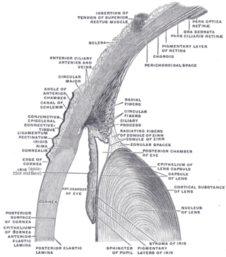
The ciliary body is a part of the eye that includes the ciliary muscle, which controls the shape of the lens, and the ciliary epithelium, which produces the aqueous humor. The aqueous humor is produced in the non-pigmented portion of the ciliary body. The ciliary body is part of the uvea, the layer of tissue that delivers oxygen and nutrients to the eye tissues. The ciliary body joins the ora serrata of the choroid to the root of the iris.

The human eye is an organ of the sensory nervous system that reacts to visible light and allows the use of visual information for various purposes including seeing things, keeping balance, and maintaining circadian rhythm.

Cataract surgery, also called lens replacement surgery, is the removal of the natural lens of the eye that has developed a cataract, an opaque or cloudy area. The eye's natural lens is usually replaced with an artificial intraocular lens (IOL) implant.

Accommodation is the process by which the vertebrate eye changes optical power to maintain a clear image or focus on an object as its distance varies. In this, distances vary for individuals from the far point—the maximum distance from the eye for which a clear image of an object can be seen, to the near point—the minimum distance for a clear image. Accommodation usually acts like a reflex, including part of the accommodation-convergence reflex, but it can also be consciously controlled.

The ciliary muscle is an intrinsic muscle of the eye formed as a ring of smooth muscle in the eye's middle layer, uvea. It controls accommodation for viewing objects at varying distances and regulates the flow of aqueous humor into Schlemm's canal. It also changes the shape of the lens within the eye but not the size of the pupil which is carried out by the sphincter pupillae muscle and dilator pupillae.
The lens capsule is a component of the globe of the eye. It is a clear elastic basement membrane composed of collagen IV laminin etc. a quality that keeps it under constant tension. As a result, the lens naturally tends towards a rounder or more globular configuration, a shape it must assume for the eye to focus at a near distance. The lens capsule is the thickest basement membrane in the body.

A simple eye refers to a form of eye or an optical arrangement composed of a single lens and without an elaborate retina such as occurs in most vertebrates. In this sense "simple eye" is distinct from a multi-lensed "compound eye", and is not necessarily at all simple in the usual sense of the word.

The central retinal artery branches off the ophthalmic artery, running inferior to the optic nerve within its dural sheath to the eyeball.
The hyaloid artery is a branch of the ophthalmic artery, which is itself a branch of the internal carotid artery. It is contained within the optic stalk of the eye and extends from the optic disc through the vitreous humor to the lens. Usually fully regressed before birth, its purpose is to supply nutrients to the developing lens in the growing fetus.
The vitreous membrane is a layer of collagen separating the vitreous humour from the rest of the eye. At least two parts have been identified anatomically. The posterior hyaloid membrane separates the rear of the vitreous from the retina. It is a false anatomical membrane. The anterior hyaloid membrane separates the front of the vitreous from the lens. Bernal et al. describe it "as a delicate structure in the form of a thin layer that runs from the pars plana to the posterior lens, where it shares its attachment with the posterior zonule via Weigert's ligament, also known as Egger's line".

Sodium hyaluronate is the sodium salt of hyaluronic acid, a glycosaminoglycan found in various connective tissue of humans.

Persistent fetal vasculature(PFV), also known as persistent fetal vasculature syndrome (PFVS), and until 1997 known primarily as persistent hyperplastic primary vitreous (PHPV), is a rare congenital anomaly which occurs when blood vessels within the developing eye, known as the embryonic hyaloid vasculature network, fail to regress as they normally would in-utero after the eye is fully developed. Defects which arise from this lack of vascular regression are diverse; as a result, the presentation, symptoms, and prognosis of affected patients vary widely, ranging from clinical insignificance to irreversible blindness. The underlying structural causes of PFV are considered to be relatively common, and the vast majority of cases do not warrant additional intervention. When symptoms do manifest, however, they are often significant, causing detrimental and irreversible visual impairment. Persistent fetal vasculature heightens the lifelong risk of glaucoma, cataracts, intraocular hemorrhages, and Retinal detachments, accounting for the visual loss of nearly 5% of the blind community in the developed world. In diagnosed cases of PFV, approximately 90% of patients with a unilateral disease have associated poor vision in the affected eye.

This glossary of medical terms is a list of definitions about medicine, its sub-disciplines, and related fields.

