Immunoelectrophoresis

Last updated
Crossed immunoelectrophoresis of 2 microlitres of normal human serum. The electrophoresis was performed in thin layers of agarose; the pictured gel is about 7x7 cm. The lower part is the first dimension gel without antibodies, where the serum was applied into the slot at the lower left. The upper part is the second dimension gel with Dako antibodies against human serum proteins. More than 50 major serum proteins can be named. CrossedimmunoelectrophoresisTCBH.jpg
Crossed immunoelectrophoresis of 2 microlitres of normal human serum. The electrophoresis was performed in thin layers of agarose; the pictured gel is about 7x7 cm. The lower part is the first dimension gel without antibodies, where the serum was applied into the slot at the lower left. The upper part is the second dimension gel with Dako antibodies against human serum proteins. More than 50 major serum proteins can be named.
Fused rocket immunoelectrophoresis of an affinity chromatographic separation of human serum proteins on con A. A 15 microlitre sample from each fraction was applied (starting from left) and allowed to diffuse about one hour, then electrophoresis was performed overnight. Peak "a" was not retained, while peak "b" was retained and eluted with methyl-mannose. The arrow indicates some slightly retained proteins. FusedrocketimmunoelectrophoresisHuSeonConA 01.jpg
Fused rocket immunoelectrophoresis of an affinity chromatographic separation of human serum proteins on con A. A 15 microlitre sample from each fraction was applied (starting from left) and allowed to diffuse about one hour, then electrophoresis was performed overnight. Peak "a" was not retained, while peak "b" was retained and eluted with methyl-mannose. The arrow indicates some slightly retained proteins.

Immunoelectrophoresis is a general name for a number of biochemical methods for separation and characterization of proteins based on electrophoresis and reaction with antibodies. All variants of immunoelectrophoresis require immunoglobulins, also known as antibodies, reacting with the proteins to be separated or characterized. The methods were developed and used extensively during the second half of the 20th century. In somewhat chronological order: Immunoelectrophoretic analysis (one-dimensional immunoelectrophoresis ad modum Grabar), crossed immunoelectrophoresis (two-dimensional quantitative immunoelectrophoresis ad modum Clarke and Freeman or ad modum Laurell), rocket-immunoelectrophoresis (one-dimensional quantitative immunoelectrophoresis ad modum Laurell), fused rocket immunoelectrophoresis ad modum Svendsen and Harboe, affinity immunoelectrophoresis ad modum Bøg-Hansen.

Contents

Methods

Immunoelectrophoresis is a general term describing many combinations of the principles of electrophoresis and reaction of antibodies, also known as immunodiffusion. [1]

Agarose as 1% gel slabs of about 1 mm thickness buffered at high pH (around 8.6) is traditionally preferred for electrophoresis and the reaction with antibodies. The agarose was chosen as the gel matrix because it has large pores allowing free passage and separation of proteins but provides an anchor for the immunoprecipitates of protein and specific antibodies. The high pH was chosen because antibodies are practically immobile at high pH. Electrophoresis equipment with a horizontal cooling plate was normally recommended for the electrophoresis.

Immunoprecipitates are visible in the wet agarose gel, but are stained with protein stains like Coomassie brilliant blue in the dried gel. In contrast to SDS-gel electrophoresis, the electrophoresis in agarose allows native conditions, preserving the native structure and activities of the proteins under investigation, therefore immunoelectrophoresis allows characterization of enzyme activities and ligand binding etc. in addition to electrophoretic separation.

Plasmodium Glutamate dehydrogenase (pGluDH) separated by Counterimmunoelectrophoresis Plasmodium glutamate dehydrogenase precipitation.jpg
Plasmodium Glutamate dehydrogenase (pGluDH) separated by Counterimmunoelectrophoresis

Counterimmunoelectrophoresis is the combination of immunodiffusion with electrophoresis. In essence electrophoresis speeds up the process of moving the reactants together.

The immunoelectrophoretic analysis ad modum Grabar is the classical method of immunoelectrophoresis. Proteins are separated by electrophoresis, then antibodies are applied in a trough next to the separated proteins and immunoprecipitates are formed after a period of diffusion of the separated proteins and antibodies against each other. The introduction of the immunoelectrophoretic analysis gave a great boost to protein chemistry, some of the first results were the resolution of proteins in biological fluids and biological extracts. Among the important observations made were the great number of different proteins in serum, the existence of several immunoglobulin classes and their electrophoretic heterogeneity.

Crossed immunoelectrophoresis is also called two-dimensional quantitative immunoelectrophoresis ad modum Clarke and Freeman or ad modum Laurell. In this method the proteins are first separated during the first dimension electrophoresis, then instead of the diffusion towards the antibodies, the proteins are electrophoresed into an antibody-containing gel in the second dimension. Immunoprecipitation will take place during the second dimension electrophorsis and the immunoprecipitates have a characteristic bell-shape, each precipitate representing one antigen, the position of the precipitate being dependent on the amount of protein as well as the amount of specific antibody in the gel, so relative quantification can be performed. The sensitivity and resolving power of crossed immunoelectrophoresis is than that of the classical immunoelectrophoretic analysis and there are multiple variations of the technique useful for various purposes. Crossed immunoelectrophoresis has been used for studies of proteins in biological fluids, particularly human serum, and biological extracts.

Rocket immunoelectrophoresis is one-dimensional quantitative immunoelectrophoresis. The method has been used for quantitation of human serum proteins before automated methods became available.

Fused rocket immunoelectrophoresis is a modification of one-dimensional quantitative immunoelectrophorsis used for detailed measurement of proteins in fractions from protein separation experiments.

Affinity immunoelectrophoresis is based on changes in the electrophoretic pattern of proteins through specific interaction or complex formation with other macromolecules or ligands. Affinity immunoelectrophoresis has been used for estimation of binding constants, as for instance with lectins or for characterization of proteins with specific features like glycan content or ligand binding. Some variants of affinity immunoelectrophoresis are similar to affinity chromatography by use of immobilized ligands.

Binding of ligands. The open structure of the immunoprecipitate in the agarose gel will allow additional binding of radioactively labeled antibodies and other ligands to reveal specific proteins. Application of this possibility has been used for instance for identification of allergens through reaction with immunoglobulin E (IgE) and for identification of glycoproteins with lectins.

General comments. Two factors determine that immunoelectrophoretic methods are not widely used. First they are rather work intensive and require some manual expertise. Second they require rather large amounts of polyclonal antibodies. Today gel electrophoresis followed by electroblotting is the preferred method for protein characterization because its ease of operation, its high sensitivity, and its low requirement for specific antibodies. In addition proteins are separated by gel electrophoresis on the basis of their apparent molecular weight, which is not accomplished by immunoelectrophoresis, but nevertheless immunoelectrophoretic methods are still useful when non-reducing conditions are needed.

Applications

Counter-immunoelectrophoresis and its modification

In comparison to other conventional methods of diagnosis e.g. for viral infection testing, counter-immunoelectrophoresis is a highly specific, simple, and speedy method that does not require sophisticated, expensive tools, input materials, or long-term capacity building. Considering the high informativeness of counter-immunoelectrophoresis, the results in practice can be dubious at times. As a result, by using a manufactured amphiphilic fluorescein-containing copolymer to increase the antigen and antibody interaction, counter-immunoelectrophoresis procedures can be improved. The use of the fluorescein copolymer-antigen mixture improved the association with plasma levels antibodies of animals immunized against hemorrhage illness and enhanced protein concentration in the precipitated zone, according to the findings. The capability of the amphiphilic fluorescein copolymer to boost antigen-antibody association and see the fluorescent accumulation domain may improve the efficiency of counter-immunoelectrophoresis for infectious disease rapid diagnosis. [3]

Immunomethods

The terminologies, immune-methods and immune-chemical techniques refer to a variety of immunoelectrophoresis processes whose results are identified using antibodies and immunological methodologies. [4] As a result, immunomethods' great sensitivity is a beneficial compared to the great expense of utilizing antibodies. Many different types of agarose electrophoresis are used to see how proteins travel under diverse circumstances. Proteins are recognized after the timer has expired by incubating gels with certain antibodies, which are then stained with Comassie blue. [5]

Radial immunodiffusion

The radial immunodiffusion is an immunoassay technique for determining the concentration of a particular protein in a mixture including other modules. It is made up of an agarose gel, just like the others. Furthermore, in this procedure, the materials are placed into round wells in the gel's core part and disperse through it, generating a deposition ring with a diameter relation to the number of unbound protein that has diffused. [4]

Identification of nanomaterial interaction with C3 protein complement and 2D immunoelectrophoresis

2D immunoelectrophoresis is a potential method that can be used for a range of functions involving protein flow of migrants, such as the deep examination of protein opsonization, in succession of first dimension as an activity of protein molar mass and the second dimension as a role of the isoelectric point. Despite the fact that it contains a large number of proteins, each spot on the 2D gel will symbolize a particular protein with a specific molecular mass and feature. [5]

2D immunoelectrophoresis is also provided as a valuable implement for examining the stimulation of the signal transduction pathway, which is an essential factor in researching nanoparticles before in vivo delivery, because it will impact nanoparticle longevity, destination, and bio-distribution. This method employs two-dimensional horizontally agarose protein electrophoresis to specifically identify the association of nanoparticles with the C3 protein. Proteins can be separated in the first dimension according to their molecular mass (the shorter the protein, the far it drifts), and in the second dimension according to their abundance [6]

Some limitations of immunoelectrophoresis

Though immunoelectrophoresis has a number of benefits, it also has certain drawbacks, such as when compared to other methods of electrophoresis, such as immunofixation, this method is sluggish and less precise. It can be difficult to interpret the results. Several tiny monoclonal proteins may be harder to identify. The accessibility of particular antibodies limits its utility in analytical techniques. Traditional (classical or conventional) immunoelectrophoresis has a number of drawbacks, including the fact that it is time consuming and the protocol might take up to 3 days to finish, has limited specificity and sensitivity, and the results can be difficult to read. As a result, newer immunoelectrophoresis techniques have largely supplanted the conventional immunoelectrophoresis.

Related Research Articles

<span class="mw-page-title-main">Agarose gel electrophoresis</span> Method for separation and analysis of biomolecules using agarose gel

Agarose gel electrophoresis is a method of gel electrophoresis used in biochemistry, molecular biology, genetics, and clinical chemistry to separate a mixed population of macromolecules such as DNA or proteins in a matrix of agarose, one of the two main components of agar. The proteins may be separated by charge and/or size, and the DNA and RNA fragments by length. Biomolecules are separated by applying an electric field to move the charged molecules through an agarose matrix, and the biomolecules are separated by size in the agarose gel matrix.

<span class="mw-page-title-main">Agarose</span> Heteropolysaccharide found in red algae

Agarose is a heteropolysaccharide, generally extracted from certain red algae. It is a linear polymer made up of the repeating unit of agarobiose, which is a disaccharide made up of D-galactose and 3,6-anhydro-L-galactopyranose. Agarose is one of the two principal components of agar, and is purified from agar by removing agar's other component, agaropectin.

<span class="mw-page-title-main">Gel electrophoresis</span> Method for separation and analysis of biomolecules

Gel electrophoresis is a method for separation and analysis of biomacromolecules and their fragments, based on their size and charge. It is used in clinical chemistry to separate proteins by charge or size and in biochemistry and molecular biology to separate a mixed population of DNA and RNA fragments by length, to estimate the size of DNA and RNA fragments or to separate proteins by charge.

<span class="mw-page-title-main">Western blot</span> Analytical technique used in molecular biology

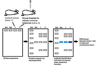
The western blot, or western blotting, is a widely used analytical technique in molecular biology and immunogenetics to detect specific proteins in a sample of tissue homogenate or extract. Besides detecting the proteins, this technique is also utilized to visualize, distinguish, and quantify the different proteins in a complicated protein combination.

<span class="mw-page-title-main">Electrophoresis</span> Motion of charged particles in electric field

In chemistry, electrophoresis is the motion of charged dispersed particles or dissolved charged molecules relative to a fluid under the influence of a spatially uniform electric field. As a rule, these are zwitterions. Electrophoresis of positively charged particles or molecules (cations) is sometimes called cataphoresis, while electrophoresis of negatively charged particles or molecules (anions) is sometimes called anaphoresis..

<span class="mw-page-title-main">Immunostaining</span> Biochemical technique

In biochemistry, immunostaining is any use of an antibody-based method to detect a specific protein in a sample. The term "immunostaining" was originally used to refer to the immunohistochemical staining of tissue sections, as first described by Albert Coons in 1941. However, immunostaining now encompasses a broad range of techniques used in histology, cell biology, and molecular biology that use antibody-based staining methods.

Protein purification is a series of processes intended to isolate one or a few proteins from a complex mixture, usually cells, tissues or whole organisms. Protein purification is vital for the specification of the function, structure and interactions of the protein of interest. The purification process may separate the protein and non-protein parts of the mixture, and finally separate the desired protein from all other proteins. Ideally, to study a protein of interest, it must be separated from other components of the cell so that contaminants will not interfere in the examination of the protein of interest's structure and function. Separation of one protein from all others is typically the most laborious aspect of protein purification. Separation steps usually exploit differences in protein size, physico-chemical properties, binding affinity and biological activity. The pure result may be termed protein isolate.

<span class="mw-page-title-main">Gel electrophoresis of proteins</span> Technique for separating proteins

Protein electrophoresis is a method for analysing the proteins in a fluid or an extract. The electrophoresis may be performed with a small volume of sample in a number of alternative ways with or without a supporting medium, namely agarose or polyacrylamide. Variants of gel electrophoresis include SDS-PAGE, free-flow electrophoresis, electrofocusing, isotachophoresis, affinity electrophoresis, immunoelectrophoresis, counterelectrophoresis, and capillary electrophoresis. Each variant has many subtypes with individual advantages and limitations. Gel electrophoresis is often performed in combination with electroblotting or immunoblotting to give additional information about a specific protein.

Affinity chromatography is a method of separating a biomolecule from a mixture, based on a highly specific macromolecular binding interaction between the biomolecule and another substance. The specific type of binding interaction depends on the biomolecule of interest; antigen and antibody, enzyme and substrate, receptor and ligand, or protein and nucleic acid binding interactions are frequently exploited for isolation of various biomolecules. Affinity chromatography is useful for its high selectivity and resolution of separation, compared to other chromatographic methods.

Immunoprecipitation (IP) is the technique of precipitating a protein antigen out of solution using an antibody that specifically binds to that particular protein. This process can be used to isolate and concentrate a particular protein from a sample containing many thousands of different proteins. Immunoprecipitation requires that the antibody be coupled to a solid substrate at some point in the procedure.

Protein methods are the techniques used to study proteins. There are experimental methods for studying proteins. Computational methods typically use computer programs to analyze proteins. However, many experimental methods require computational analysis of the raw data.

Electrophoresis is the motion of charged dispersed particles or dissolved charged molecules relative to a fluid under the influence of a spatially uniform electric field.

<span class="mw-page-title-main">Electrophoretic mobility shift assay</span>

An electrophoretic mobility shift assay (EMSA) or mobility shift electrophoresis, also referred as a gel shift assay, gel mobility shift assay, band shift assay, or gel retardation assay, is a common affinity electrophoresis technique used to study protein–DNA or protein–RNA interactions. This procedure can determine if a protein or mixture of proteins is capable of binding to a given DNA or RNA sequence, and can sometimes indicate if more than one protein molecule is involved in the binding complex. Gel shift assays are often performed in vitro concurrently with DNase footprinting, primer extension, and promoter-probe experiments when studying transcription initiation, DNA gang replication, DNA repair or RNA processing and maturation, as well as pre-mRNA splicing. Although precursors can be found in earlier literature, most current assays are based on methods described by Garner and Revzin and Fried and Crothers.

<span class="mw-page-title-main">Ouchterlony double immunodiffusion</span>

Ouchterlony double immunodiffusion is an immunological technique used in the detection, identification and quantification of antibodies and antigens, such as immunoglobulins and extractable nuclear antigens. The technique is named after Örjan Ouchterlony, the Swedish physician who developed the test in 1948 to evaluate the production of diphtheria toxins from isolated bacteria.

DNA footprinting is a method of investigating the sequence specificity of DNA-binding proteins in vitro. This technique can be used to study protein-DNA interactions both outside and within cells.

<span class="mw-page-title-main">Immunoproteomics</span> Study of large set of protein

Immunoproteomics is the study of large sets of proteins (proteomics) involved in the immune response.

<span class="mw-page-title-main">Immunofixation</span>

Immunofixation permits the detection and typing of monoclonal antibodies or immunoglobulins in serum or urine. It is of great importance for the diagnosis and monitoring of certain blood related diseases such as myeloma.

<span class="mw-page-title-main">Electroblotting</span>

Electroblotting is a method in molecular biology/biochemistry/immunogenetics to transfer proteins or nucleic acids onto a membrane by using PVDF or nitrocellulose, after gel electrophoresis. The protein or nucleic acid can then be further analyzed using probes such as specific antibodies, ligands like lectins, or stains. This method can be used with all polyacrylamide and agarose gels. An alternative technique for transferring proteins from a gel is capillary blotting.

<span class="mw-page-title-main">Affinity electrophoresis</span>

Affinity electrophoresis is a general name for many analytical methods used in biochemistry and biotechnology. Both qualitative and quantitative information may be obtained through affinity electrophoresis. Cross electrophoresis, the first affinity electrophoresis method, was created by Nakamura et al. Enzyme-substrate complexes have been detected using cross electrophoresis. The methods include the so-called electrophoretic mobility shift assay, charge shift electrophoresis and affinity capillary electrophoresis. The methods are based on changes in the electrophoretic pattern of molecules through biospecific interaction or complex formation. The interaction or binding of a molecule, charged or uncharged, will normally change the electrophoretic properties of a molecule. Membrane proteins may be identified by a shift in mobility induced by a charged detergent. Nucleic acids or nucleic acid fragments may be characterized by their affinity to other molecules. The methods have been used for estimation of binding constants, as for instance in lectin affinity electrophoresis or characterization of molecules with specific features like glycan content or ligand binding. For enzymes and other ligand-binding proteins, one-dimensional electrophoresis similar to counter electrophoresis or to "rocket immunoelectrophoresis", affinity electrophoresis may be used as an alternative quantification of the protein. Some of the methods are similar to affinity chromatography by use of immobilized ligands.

Pierre Grabar was a French biochemist and immunologist, born in Russia. He was the founding president of the Société Française d'Immunologie. He studied antigen-antibody reactions and developed a "carrier" theory of antibody function. His award-winning development of Immunoelectrophoresis made it possible to identify specific bodily proteins, opening new avenues in medical research.

References

  1. Homburger, Henry A.; Singh, Ravinder Jit (2008). "96 - Assessment of proteins of the immune system". In Rich, Robert R.; Fleisher, Thomas A.; Shearer, William T.; Schroeder Jr., Harry W.; Frew, Anthony J.; Weyand, Cornelia M. (eds.). Clinical Immunology: Principles and Practice (3rd ed.). Elsevier. pp. 1419–1434. doi:10.1016/b978-0-323-04404-2.10096-x. ISBN   9780323044042.
  2. Ling IT.; Cooksley S.; Bates PA.; Hempelmann E.; Wilson RJM. (1986). "Antibodies to the glutamate dehydrogenase of Plasmodium falciparum" (PDF). Parasitology. 92 (2): 313–324. doi:10.1017/s0031182000064088. PMID   3086819. S2CID   16953840 . Retrieved 10 November 2022.
  3. Ostapiv, D.D.; Kuz’mina, N.V.; Kozak, М.R.; Hu, Shan; Vlizlo, V.V.; Kotsiumbas, I.Ya.; Varvarenko, S.M.; Samaryk, V.Ya.; Nosova, N.G.; Yakoviv, M.V. (2021). "Application of fluorescein copolymer to improve the efficiency of counter-immunoelectrophoresis for diagnostics of animal infectious diseases". The Ukrainian Biochemical Journal. 93 (1): 104–112. doi: 10.15407/ubj93.01.104 . S2CID   233915465.
  4. 1 2 Vauthier, Christine; Bouchemal, Kawthar (2008-12-24). "Methods for the Preparation and Manufacture of Polymeric Nanoparticles". Pharmaceutical Research. 26 (5): 1025–1058. doi:10.1007/s11095-008-9800-3. ISSN   0724-8741. PMID   19107579. S2CID   22199493.
  5. 1 2 Fornaguera, Cristina; Solans, Conxita (2017-01-27). "Methods for the In Vitro Characterization of Nanomedicines—Biological Component Interaction". Journal of Personalized Medicine. 7 (1): 2. doi: 10.3390/jpm7010002 . ISSN   2075-4426. PMC   5374392 . PMID   28134833.
  6. Bertholon, Isabelle; Vauthier, Christine; Labarre, Denis (2006-05-25). "Complement Activation by Core–Shell Poly(isobutylcyanoacrylate)–Polysaccharide Nanoparticles: Influences of Surface Morphology, Length, and Type of Polysaccharide". Pharmaceutical Research. 23 (6): 1313–1323. doi:10.1007/s11095-006-0069-0. ISSN   0724-8741. PMID   16715369. S2CID   30404102.