Standard anatomical terms of location are used to unambiguously describe the anatomy of animals, including humans. The terms, typically derived from Latin or Greek roots, describe something in its standard anatomical position. This position provides a definition of what is at the front ("anterior"), behind ("posterior") and so on. As part of defining and describing terms, the body is described through the use of anatomical planes and anatomical axes.

The brachialis, also known as the Teichmann muscle, is a muscle in the upper arm that flexes the elbow. It lies beneath the biceps brachii, and makes up part of the floor of the region known as the cubital fossa. It originates from the anterior aspect of the distal humerus; it inserts onto the tuberosity of the ulna. It is innervated by the musculocutaneous nerve, and commonly also receives additional innervation from the radial nerve. The brachialis is the prime mover of elbow flexion generating about 50% more power than the biceps.

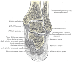
In human anatomy, the fibularis longus is a superficial muscle in the lateral compartment of the leg. It acts to tilt the sole of the foot away from the midline of the body (eversion) and to extend the foot downward away from the body at the ankle.

The tibia, also known as the shinbone or shankbone, is the larger, stronger, and anterior (frontal) of the two bones in the leg below the knee in vertebrates ; it connects the knee with the ankle. The tibia is found on the medial side of the leg next to the fibula and closer to the median plane. The tibia is connected to the fibula by the interosseous membrane of leg, forming a type of fibrous joint called a syndesmosis with very little movement. The tibia is named for the flute tibia. It is the second largest bone in the human body, after the femur. The leg bones are the strongest long bones as they support the rest of the body.

The fibula or calf bone is a leg bone on the lateral side of the tibia, to which it is connected above and below. It is the smaller of the two bones and, in proportion to its length, the most slender of all the long bones. Its upper extremity is small, placed toward the back of the head of the tibia, below the knee joint and excluded from the formation of this joint. Its lower extremity inclines a little forward, so as to be on a plane anterior to that of the upper end; it projects below the tibia and forms the lateral part of the ankle joint.

In human anatomy, the metacarpal bones or metacarpus form the intermediate part of the skeletal hand located between the phalanges of the fingers and the carpal bones of the wrist, which forms the connection to the forearm. The metacarpal bones are analogous to the metatarsal bones in the foot.
The ankle, or the talocrural region, or the jumping bone (informal) is the area where the foot and the leg meet. The ankle includes three joints: the ankle joint proper or talocrural joint, the subtalar joint, and the inferior tibiofibular joint. The movements produced at this joint are dorsiflexion and plantarflexion of the foot. In common usage, the term ankle refers exclusively to the ankle region. In medical terminology, "ankle" can refer broadly to the region or specifically to the talocrural joint.
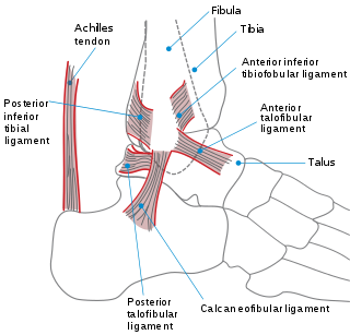
Pott's fracture, also known as Pott's syndrome I and Dupuytren fracture, is an archaic term loosely applied to a variety of bimalleolar ankle fractures. The injury is caused by a combined abduction external rotation from an eversion force. This action strains the sturdy medial (deltoid) ligament of the ankle, often tearing off the medial malleolus due to its strong attachment. The talus then moves laterally, shearing off the lateral malleolus or, more commonly, breaking the fibula superior to the tibiofibular syndesmosis. If the tibia is carried anteriorly, the posterior margin of the distal end of the tibia is also sheared off by the talus. A fractured fibula in addition to detaching the medial malleolus will tear the tibiofibular syndesmosis. The combined fracture of the medial malleolus, lateral malleolus, and the posterior margin of the distal end of the tibia is known as a "trimalleolar fracture".

The Maisonneuve fracture is a spiral fracture of the proximal third of the fibula associated with a tear of the distal tibiofibular syndesmosis and the interosseous membrane. There is an associated fracture of the medial malleolus or rupture of the deep deltoid ligament of the ankle. This type of injury can be difficult to detect.

The talus, talus bone, astragalus, or ankle bone is one of the group of foot bones known as the tarsus. The tarsus forms the lower part of the ankle joint. It transmits the entire weight of the body from the lower legs to the foot.

In human anatomy, the fibularis brevis is a muscle that lies underneath the fibularis longus within the lateral compartment of the leg. It acts to tilt the sole of the foot away from the midline of the body (eversion) and to extend the foot downward away from the body at the ankle.

The annular ligament is a strong band of fibers that encircles the head of the radius, and retains it in contact with the radial notch of the ulna.
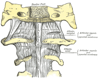
The atlanto-axial joint is a joint in the upper part of the neck between the atlas bone and the axis bone, which are the first and second cervical vertebrae. It is a pivot joint.

The anterior ligament of the lateral malleolus is a flat, trapezoidal band of fibers, broader below than above, which extends obliquely downward and lateralward between the adjacent margins of the tibia and fibula, on the front aspect of the syndesmosis.
The inferior transverse ligament of the tibiofibular syndesmosis is a connective tissue structure in the lower leg that lies in front of the posterior ligament. It is a strong, thick band, of yellowish fibers which passes transversely across the back of the ankle joint, from the lateral malleolus to the posterior border of the articular surface of the tibia, almost as far as its malleolar process.
The posterior ligament of the lateral malleolus is smaller than the anterior ligament of the lateral malleolus and is disposed in a similar manner on the posterior surface of the syndesmosis. It connects the tibia and fibular on the inferior part of both bones.

A malleolus is the bony prominence on each side of the human ankle.

The cruciate ligament of the atlas is a ligament in the neck. It forms part of the atlanto-axial joint. The ligament is named after its cross shape. It consists of transverse and longitudinal components. The posterior longitudinal band may be absent in some people. The cruciate ligament of the atlas prevents abnormal movement of the atlanto-axial joint. It may be torn, such as by fractures of the atlas bone.

The Danis–Weber classification is a method of describing ankle fractures. It has three categories:
Anatomical terminology is a form of scientific terminology used by anatomists, zoologists, and health professionals such as doctors.