Influenza D virus

Last updated
Influenza D virus
3D Influenza virus.png
Virus classification OOjs UI icon edit-ltr.svg
(unranked): Virus
Realm: Riboviria
Kingdom: Orthornavirae
Phylum: Negarnaviricota
Class: Insthoviricetes
Order: Articulavirales
Family: Orthomyxoviridae
Genus: Deltainfluenzavirus
Species:
Influenza D virus

Influenza D virus is a species in the virus genus Deltainfluenzavirus, in the family Orthomyxoviridae , that causes influenza.

Contents

Influenza D viruses are known to infect pigs and cattle; no human infections from this virus have been observed. [1] First isolated from pigs in 2011, the virus was categorized as a new genus of Orthomyxoviridae in 2016, distinct from the previously-known Influenzavirus C genus; [1] [2] before then, Influenza D virus was thought to be a subtype of Influenzavirus C. [1]

Cases of infections from the Type D virus are rare compared to Types A, B, and C. Similar to Type C, Type D has 7 RNA segments and encodes 9 proteins, while Types A and B have 8 RNA segments and encode at least 10 proteins. [3]

Influenza D virus

The influenza viruses are members of the family Orthomyxoviridae . [1] Influenza viruses A, B, C, and D represent the four antigenic types of influenza viruses. [4] Of the four antigenic types, influenza A virus is the most severe, influenza B virus is less severe but can still cause outbreaks, and influenza C virus is usually only associated with minor symptoms. [5] Influenzavirus D is less common than the other antigenic types, and it is not known to cause any human infections. No samples of influenza D virus were detected in serum samples from humans; however, hemagglutination-inhibiting antibodies against influenza D virus have been detected in humans, with an estimated occurrence of 1.3% in the general population, suggesting that this virus may infect humans as well. However, those antibodies may have been produced after an infection by influenza C virus, the antibodies for which cross-react with the Type D virus. More studies are needed to conclude whether or not the Type D virus can infect humans. [1]

Influenza D virus is 50% similar in amino acid composition to influenza C virus, similar to the level of divergence between types A and B, while types C and D have a much greater level of divergence from types A and B. [1] [6] Influenzaviruses C and D were estimated to have diverged from a single ancestor over 1,500 years ago, around 482 AD. Influenzavirus D itself currently has two lineages, which were estimated to have emerged over 45 years ago, around 1972 AD. [1] Influenza viruses A and B are estimated to have diverged from a single ancestor around 4,000 years ago, while the ancestor of influenza viruses A and B and the ancestor of influenza viruses C and D are estimated to have diverged from a common ancestor around 8,000 years ago. [7] Metatranscriptomics studies have also identified closely related "Influenza C and D-like" viruses in a number of amphibian species. [8]

Influenza A virus can infect a variety of animals as well as humans, and its natural host or reservoir is birds, whereas influenza viruses B, C, and D do not have animal reservoirs. [5] [9] [1] Influenza viruses C and D are not as easily isolated so less information is known about these types, but studies show that they occur worldwide. [1] [6]

This virus may be spread through respiratory droplets or by fomites (non-living material) due to its ability to survive on surfaces for short durations. [5] Influenza viruses have a relatively short incubation period (lapse of time from exposure to pathogen to the appearance of symptoms) of 18–72 hours and infect the epithelial cells of the respiratory tract. [5]

In cell culture, influenza D virus has demonstrated an ability to replicate well at 37°C, the normal lung temperature, and can also replicate better and in more types of cells than the Type C virus. This study suggests that influenza D virus may be only a few genetic changes away from being able to invade the lower lung, even though the virus does not actively spread among humans and has a much slower mutation rate than the other influenza viruses. [1]

Structure and Variation

Influenza viruses, like all viruses in the family Orthomyxoviridae, are enveloped RNA viruses with single stranded genomes. [1] [10] The antigens, matrix protein (M1) and nucleoprotein (NP), are used to determine if an influenza virus is type A, B, C, or D. [5] The M1 protein is required for virus assembly and NP functions in transcription and replication. [11] [12] These viruses also contain proteins on the surface of the cell membrane called glycoproteins. Type A and B have two glycoproteins: hemagglutinin (HA) and neuraminidase (NA). Types C and D have only one glycoprotein: hemagglutinin-esterase fusion (HEF). [5] [13] [1] These glycoproteins allow for attachment and fusion of viral and cellular membranes. Fusion of these membranes allows the viral proteins and genome to be released into the host cell, which then causes the infection. [14] Types C and D are the only influenza viruses to express the enzyme esterase. This enzyme is similar to the enzyme neuraminidase produced by Types A and B in that they both function in destroying the host cell receptors. [15] [1] Glycoproteins may undergo mutations (antigenic drift) or reassortment in which a new HA or NA is produced (antigenic shift). Influenza viruses C and D are only capable of antigenic drift whereas Type A undergoes antigenic shift, as well. When either of these processes occur, the antibodies formed by the immune system no longer protect against these altered glycoproteins. Because of this, viruses continually cause infections. [5]

Identification

Influenza viruses C and D are different from Types A and B in their growth requirements. Because of this, Influenzavirus D is not isolated and identified as frequently. Diagnosis is by virus isolation, serology, and other tests. [16] Hemagglutination inhibition (HI) is one method of serology that detects antibodies for diagnostic purposes. [17] Western blot (immunoblot assay) and enzyme-linked immunosorbent assay (ELISA) are two other methods used to detect proteins (or antigens) in serum. In each of these techniques, the antibodies for the protein of interest are added and the presence of the specific protein is indicated by a color change. [18] ELISA was shown to have higher sensitivity to the HEF than the HI test. [9] Because only Influenza viruses C and D produce esterase, In Situ Esterase Assays provide a quick and inexpensive method of detecting just Types C and D. [15]

Vaccination

Because influenza virus A has an animal reservoir that contains all the known subtypes and can undergo antigenic shift, this type of influenza virus is capable of producing pandemics. [9] Influenza viruses A and B also cause seasonal epidemics every year due to their ability to antigenic shift. [4] Influenza viruses C and D do not have this capability, and they have not been implicated in any pandemics; thus, there are currently no human vaccines available for Influenza viruses C or D. [6] An inactivated Influenzavirus D vaccine was developed for cattle; however, the vaccine only provided partial protection in challenge experiments. [1]

Related Research Articles

<i>Influenza A virus</i> Species of virus

Influenza A virus (IAV) is a pathogen that causes the flu in birds and some mammals, including humans. It is an RNA virus whose subtypes have been isolated from wild birds. Occasionally, it is transmitted from wild to domestic birds, and this may cause severe disease, outbreaks, or human influenza pandemics.

<span class="mw-page-title-main">Hemagglutinin (influenza)</span> Hemagglutinin of influenza virus

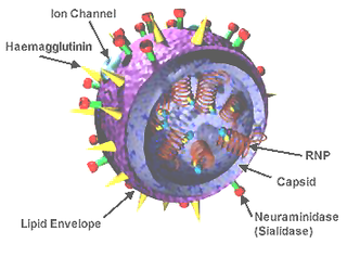
Influenza hemagglutinin (HA) or haemagglutinin[p] is a homotrimeric glycoprotein found on the surface of influenza viruses and is integral to its infectivity.

<i>Orthomyxoviridae</i> Family of RNA viruses including the influenza viruses

Orthomyxoviridae is a family of negative-sense RNA viruses. It includes seven genera: Alphainfluenzavirus, Betainfluenzavirus, Gammainfluenzavirus, Deltainfluenzavirus, Isavirus, Thogotovirus, and Quaranjavirus. The first four genera contain viruses that cause influenza in birds and mammals, including humans. Isaviruses infect salmon; the thogotoviruses are arboviruses, infecting vertebrates and invertebrates. The Quaranjaviruses are also arboviruses, infecting vertebrates (birds) and invertebrates (arthropods).

Antigenic drift is a kind of genetic variation in viruses, arising from the accumulation of mutations in the virus genes that code for virus-surface proteins that host antibodies recognize. This results in a new strain of virus particles that is not effectively inhibited by the antibodies that prevented infection by previous strains. This makes it easier for the changed virus to spread throughout a partially immune population. Antigenic drift occurs in both influenza A and influenza B viruses.

<span class="mw-page-title-main">Hemagglutinin esterase</span> Glycoprotein present in some enveloped viruses

Hemagglutinin esterase (HEs) is a glycoprotein that certain enveloped viruses possess and use as an invading mechanism. HEs helps in the attachment and destruction of certain sialic acid receptors that are found on the host cell surface. Viruses that possess HEs include influenza C virus, toroviruses, and coronaviruses of the subgenus Embecovirus. HEs is a dimer transmembrane protein consisting of two monomers, each monomer is made of three domains. The three domains are: membrane fusion, esterase, and receptor binding domains.

<span class="mw-page-title-main">Envelope glycoprotein GP120</span> Glycoprotein exposed on the surface of the HIV virus

Envelope glycoprotein GP120 is a glycoprotein exposed on the surface of the HIV envelope. It was discovered by Professors Tun-Hou Lee and Myron "Max" Essex of the Harvard School of Public Health in 1984. The 120 in its name comes from its molecular weight of 120 kDa. Gp120 is essential for virus entry into cells as it plays a vital role in attachment to specific cell surface receptors. These receptors are DC-SIGN, Heparan Sulfate Proteoglycan and a specific interaction with the CD4 receptor, particularly on helper T-cells. Binding to CD4 induces the start of a cascade of conformational changes in gp120 and gp41 that lead to the fusion of the viral membrane with the host cell membrane. Binding to CD4 is mainly electrostatic although there are van der Waals interactions and hydrogen bonds.

<i>Thogotovirus</i> Genus of viruses

Thogotovirus is a genus of enveloped RNA viruses, one of seven genera in the virus family Orthomyxoviridae. Their single-stranded, negative-sense RNA genome has six or seven segments. Thogotoviruses are distinguished from most other orthomyxoviruses by being arboviruses – viruses that are transmitted by arthropods, in this case usually ticks. Thogotoviruses can replicate in both tick cells and vertebrate cells; one subtype has also been isolated from mosquitoes. A consequence of being transmitted by blood-sucking vectors is that the virus must spread systemically in the vertebrate host – unlike influenza viruses, which are transmitted by respiratory droplets and are usually confined to the respiratory system.

<i>Influenza B virus</i> Species of virus

Influenza B virus is the only species in the genus Betainfluenzavirus in the virus family Orthomyxoviridae.

<i>Influenza C virus</i> Genus of viruses in the family Orthomyxoviridae

Influenza C virus is the only species in the genus Gammainfluenzavirus, in the virus family Orthomyxoviridae, which like other influenza viruses, causes influenza.

<span class="mw-page-title-main">H5N1 genetic structure</span>

H5N1 genetic structure is the molecular structure of the H5N1 virus's RNA.

<i>Murine coronavirus</i> Species of virus

Murine coronavirus (M-CoV) is a virus in the genus Betacoronavirus that infects mice. Belonging to the subgenus Embecovirus, murine coronavirus strains are enterotropic or polytropic. Enterotropic strains include mouse hepatitis virus (MHV) strains D, Y, RI, and DVIM, whereas polytropic strains, such as JHM and A59, primarily cause hepatitis, enteritis, and encephalitis. Murine coronavirus is an important pathogen in the laboratory mouse and the laboratory rat. It is the most studied coronavirus in animals other than humans, and has been used as an animal disease model for many virological and clinical studies.

Antigenic variation or antigenic alteration refers to the mechanism by which an infectious agent such as a protozoan, bacterium or virus alters the proteins or carbohydrates on its surface and thus avoids a host immune response, making it one of the mechanisms of antigenic escape. It is related to phase variation. Antigenic variation not only enables the pathogen to avoid the immune response in its current host, but also allows re-infection of previously infected hosts. Immunity to re-infection is based on recognition of the antigens carried by the pathogen, which are "remembered" by the acquired immune response. If the pathogen's dominant antigen can be altered, the pathogen can then evade the host's acquired immune system. Antigenic variation can occur by altering a variety of surface molecules including proteins and carbohydrates. Antigenic variation can result from gene conversion, site-specific DNA inversions, hypermutation, or recombination of sequence cassettes. The result is that even a clonal population of pathogens expresses a heterogeneous phenotype. Many of the proteins known to show antigenic or phase variation are related to virulence.

<i>Murine respirovirus</i> Sendai virus, virus of rodents

Murine respirovirus, formerly Sendai virus (SeV) and previously also known as murine parainfluenza virus type 1 or hemagglutinating virus of Japan (HVJ), is an enveloped, 150-200 nm–diameter, negative sense, single-stranded RNA virus of the family Paramyxoviridae. It typically infects rodents and it is not pathogenic for humans or domestic animals

<span class="mw-page-title-main">Spike protein</span> Glycoprotein spike on a viral capsid or viral envelope

In virology, a spike protein or peplomer protein is a protein that forms a large structure known as a spike or peplomer projecting from the surface of an enveloped virus. The proteins are usually glycoproteins that form dimers or trimers.

<span class="mw-page-title-main">Influenza</span> Infectious disease, often just "the flu"

Influenza, commonly known as "the flu", is an infectious disease caused by influenza viruses. Symptoms range from mild to severe and often include fever, runny nose, sore throat, muscle pain, headache, coughing, and fatigue. These symptoms begin from one to four days after exposure to the virus and last for about 2–8 days. Diarrhea and vomiting can occur, particularly in children. Influenza may progress to pneumonia, which can be caused by the virus or by a subsequent bacterial infection. Other complications of infection include acute respiratory distress syndrome, meningitis, encephalitis, and worsening of pre-existing health problems such as asthma and cardiovascular disease.

<span class="mw-page-title-main">Viral neuraminidase</span>

Viral neuraminidase is a type of neuraminidase found on the surface of influenza viruses that enables the virus to be released from the host cell. Neuraminidases are enzymes that cleave sialic acid groups from glycoproteins. Viral neuraminidase was discovered by Alfred Gottschalk at the Walter and Eliza Hall Institute in 1957. Neuraminidase inhibitors are antiviral agents that inhibit influenza viral neuraminidase activity and are of major importance in the control of influenza.

<span class="mw-page-title-main">Hemagglutinin</span>

In molecular biology, hemagglutinins are receptor-binding membrane fusion glycoproteins produced by viruses in the Paramyxoviridae and Orthomyxoviridae families. Hemagglutinins are responsible for binding to receptors on red blood cells to initiate viral attachment and infection. The agglutination of red cells occurs when antibodies on one cell bind to those on others, causing amorphous aggregates of clumped cells.

A neutralizing antibody (NAb) is an antibody that defends a cell from a pathogen or infectious particle by neutralizing any effect it has biologically. Neutralization renders the particle no longer infectious or pathogenic. Neutralizing antibodies are part of the humoral response of the adaptive immune system against viruses, intracellular bacteria and microbial toxin. By binding specifically to surface structures (antigen) on an infectious particle, neutralizing antibodies prevent the particle from interacting with its host cells it might infect and destroy.

Virus quantification is counting or calculating the number of virus particles (virions) in a sample to determine the virus concentration. It is used in both research and development (R&D) in academic and commercial laboratories as well as in production situations where the quantity of virus at various steps is an important variable that must be monitored. For example, the production of virus-based vaccines, recombinant proteins using viral vectors, and viral antigens all require virus quantification to continually monitor and/or modify the process in order to optimize product quality and production yields and to respond to ever changing demands and applications. Other examples of specific instances where viruses need to be quantified include clone screening, multiplicity of infection (MOI) optimization, and adaptation of methods to cell culture.

<span class="mw-page-title-main">Universal flu vaccine</span> Vaccine that prevents infection from all strains of the flu

A universal flu vaccine is a flu vaccine that is effective against all influenza strains regardless of the virus sub type, antigenic drift or antigenic shift. Hence it should not require modification from year to year. As of 2021 no universal flu vaccine had been approved for general use, several were in development, and one was in clinical trial.

References

  1. 1 2 3 4 5 6 7 8 9 10 11 12 13 14 Shuo Su; Xinliang Fu; Gairu Li; Fiona Kerlin; Michael Veit (25 August 2017). "Novel Influenza D virus: Epidemiology, pathology, evolution and biological characteristics". Virulence. 8 (8): 1580–1591. doi:10.1080/21505594.2017.1365216. PMC   5810478 . PMID   28812422.
  2. "New virus gets official name, influenza D". ScienceDaily. 1 September 2016. Retrieved 28 September 2018.
  3. Skelton, Raegan M.; Huber, Victor C. (May 2022). "Comparing Influenza Virus Biology for Understanding Influenza D Virus". Viruses. 14 (5): 1036. doi: 10.3390/v14051036 . ISSN   1999-4915. PMC   9147167 . PMID   35632777.
  4. 1 2 "Types of Influenza Viruses". Influenza (Flu). Centers for Disease Control and Prevention. November 2, 2021. Archived from the original on 2021-11-03. Retrieved 2022-02-22.
  5. 1 2 3 4 5 6 7 Margaret Hunt (2009). "Microbiology and Immunology On-line". University of South Carolina School of Medicine.
  6. 1 2 3 "Influenza C and Influenza D Viruses" (PDF). 2016. Retrieved 28 September 2018.
  7. Yoshiyuki Suzuki; Masatoshi Nei (April 2001). "Origin and Evolution of Influenza Virus Hemagglutinin Genes". Molecular Biology and Evolution . Ocford Academic. 19 (4): 501–509. doi: 10.1093/oxfordjournals.molbev.a004105 . PMID   11919291.
  8. Parry R, Wille M, Turnbull OM, Geoghegan JL, Holmes EC (2020). "Divergent Influenza-Like Viruses of Amphibians and Fish Support an Ancient Evolutionary Association". Viruses. 12 (9): 1042. doi: 10.3390/v12091042 . PMC   7551885 . PMID   32962015.
  9. 1 2 3 World Health Organization (2006). "Review of latest available evidence on potential transmission of avian influenza (H5H1) through water and sewage and ways to reduce the risks to human health" (PDF).
  10. Pattison; McMullin; Bradbury; Alexander (2008). Poultry Diseases (6th ed.). Elsevier. pp.  317. ISBN   978-0-7020-28625.
  11. Ali A, Avalos RT, Ponimaskin E, Nayak DP (2000). "Influenza virus assembly: effect of influenza virus glycoproteins on the membrane association of M1 protein". J. Virol. 74 (18): 8709–19. doi:10.1128/jvi.74.18.8709-8719.2000. PMC   116382 . PMID   10954572.
  12. Portela A, Digard P (2002). "The influenza virus nucleoprotein: a multifunctional RNA-binding protein pivotal to virus replication". J. Gen. Virol. 83 (Pt 4): 723–34. doi: 10.1099/0022-1317-83-4-723 . PMID   11907320.
  13. Gao Q, Brydon EW, Palese P (2008). "A seven-segmented influenza A virus expressing the influenza C virus glycoprotein HEF". J. Virol. 82 (13): 6419–26. doi:10.1128/JVI.00514-08. PMC   2447078 . PMID   18448539.
  14. Weissenhorn W, Dessen A, Calder LJ, Harrison SC, Skehel JJ, Wiley DC (1999). "Structural basis for membrane fusion by enveloped viruses". Mol. Membr. Biol. 16 (1): 3–9. doi: 10.1080/096876899294706 . PMID   10332732.
  15. 1 2 Wagaman PC, Spence HA, O'Callaghan RJ (May 1989). "Detection of influenza C virus by using an in situ esterase assay". J. Clin. Microbiol. 27 (5): 832–36. doi:10.1128/JCM.27.5.832-836.1989. PMC   267439 . PMID   2745694.
  16. Matsuzaki Y, Katsushima N, Nagai Y, Shoji M, Itagaki T, Sakamoto M, Kitaoka S, Mizuta K, Nishimura H (2006). "Clinical features of influenza C virus infection in children". J. Infect. Dis. 193 (9): 1229–35. doi: 10.1086/502973 . PMID   16586359.
  17. Manuguerra JC, Hannoun C, Sáenz Mdel C, Villar E, Cabezas JA (1994). "Sero-epidemiological survey of influenza C virus infection in Spain". Eur. J. Epidemiol. 10 (1): 91–94. doi:10.1007/bf01717459. PMID   7957798. S2CID   13204506.
  18. Nelson, DL; Cox, MM (2013). Principles of Biochemistry (6th ed.). p. 179. ISBN   978-1-4292-3414-6.

Further reading