
The chorion is the outermost fetal membrane around the embryo in mammals, birds and reptiles (amniotes). It develops from an outer fold on the surface of the yolk sac, which lies outside the zona pellucida, known as the vitelline membrane in other animals. In insects it is developed by the follicle cells while the egg is in the ovary.

The blastocyst is a structure formed in the early development of mammals. It possesses an inner cell mass (ICM) which subsequently forms the embryo. The outer layer of the blastocyst consists of cells collectively called the trophoblast. This layer surrounds the inner cell mass and a fluid-filled cavity known as the blastocoel. The trophoblast gives rise to the placenta. The name "blastocyst" arises from the Greek βλαστός blastos and κύστις kystis. In other animals this is called a blastula.

Leydig cells, also known as interstitial cells of Leydig, are found adjacent to the seminiferous tubules in the testicle. They produce testosterone in the presence of luteinizing hormone (LH). Leydig cells are polyhedral in shape, and have a large prominent nucleus, an eosinophilic cytoplasm and numerous lipid-filled vesicles.

An adenoma is a benign tumor of epithelial tissue with glandular origin, glandular characteristics, or both. Adenomas can grow from many glandular organs, including the adrenal glands, pituitary gland, thyroid, prostate, and others. Some adenomas grow from epithelial tissue in nonglandular areas but express glandular tissue structure. Although adenomas are benign, they should be treated as pre-cancerous. Over time adenomas may transform to become malignant, at which point they are called adenocarcinomas. Most adenomas do not transform. However, even though benign, they have the potential to cause serious health complications by compressing other structures and by producing large amounts of hormones in an unregulated, non-feedback-dependent manner. Some adenomas are too small to be seen macroscopically but can still cause clinical symptoms.
Trophoblasts are cells that form the outer layer of a blastocyst, and are present four days post fertilization in humans. They provide nutrients to the embryo and develop into a large part of the placenta. They form during the first stage of pregnancy and are the first cells to differentiate from the fertilized egg to become extraembryonic structures and do not directly contribute to the embryo. After gastrulation, the trophoblast is contiguous with the ectoderm of the embryo, and is referred to as the trophectoderm. After the first differentiation, the cells in the human embryo lose their totipotency and are no longer totipotent stem cells because they cannot form a trophoblast. They are now pluripotent stem cells.

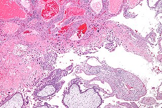
Choriocarcinoma is a malignant, trophoblastic cancer, usually of the placenta. It is characterized by early hematogenous spread to the lungs. It belongs to the malignant end of the spectrum in gestational trophoblastic disease (GTD). It is also classified as a germ cell tumor and may arise in the testis or ovary.

Anaplastic large-cell lymphoma (ALCL) is a form of cancer. It is a type of non-Hodgkin lymphoma involving aberrant T cells or null lymphocytes. The term anaplastic large-cell lymphoma (ALCL) encompasses at least four different clinical entities with the same name, which on histological examination share the presence of large pleomorphic cells that express CD30 and T-cell markers. Two types of ALCL present as systemic disease and are considered as aggressive lymphomas, while two types present as localized disease and may progress locally. Anaplastic large cell lymphoma is associated with various types of medical implants.
Gestational trophoblastic disease (GTD) is a term used for a group of pregnancy-related tumours. These tumours are rare, and they appear when cells in the womb start to proliferate uncontrollably. The cells that form gestational trophoblastic tumours are called trophoblasts and come from tissue that grows to form the placenta during pregnancy.

A seminoma is a germ cell tumor of the testicle or, more rarely, the mediastinum or other extra-gonadal locations. It is a malignant neoplasm and is one of the most treatable and curable cancers, with a survival rate above 95% if discovered in early stages.

An oncocytoma is a tumor made up of oncocytes, epithelial cells characterized by an excessive amount of mitochondria, resulting in an abundant acidophilic, granular cytoplasm. The cells and the tumor that they compose are often benign but sometimes may be premalignant or malignant.
Jaagsiekte sheep retrovirus (JSRV) is a betaretrovirus which is the causative agent of a contagious lung cancer in sheep, called ovine pulmonary adenocarcinoma.

The cytotrophoblast is the inner layer of the trophoblast. It is interior to the syncytiotrophoblast and external to the wall of the blastocyst in a developing embryo.

In humans, implantation is the stage of pregnancy at which the embryo adheres to the wall of the uterus. At this stage of prenatal development, the conceptus is called a blastocyst. It is by this adhesion that the embryo receives oxygen and nutrients from the mother to be able to grow.

Spermatocytic tumor, previously called spermatocytic seminoma, is a neoplasm of the testis, and classified as a germ cell tumour.

Placental site trophoblastic tumor is a form of gestational trophoblastic disease, which is thought to arise from intermediate trophoblast.
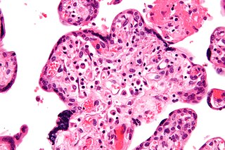
Villitis of unknown etiology (VUE), also known as chronic villitis, is a placental injury. VUE is an inflammatory condition involving the chorionic villi. VUE is a recurrent condition and can be associated with intrauterine growth restriction (IUGR). IUGR involves the poor growth of the foetus, stillbirth, miscarriage, and premature delivery. VUE recurs in about 1/3 of subsequent pregnancies.
Amitosis, also called 'karyostenosis' or direct cell division or binary fission. It is cell proliferation that does not occur by mitosis, the mechanism usually identified as essential for cell division in eukaryotes. The polyploid macronucleus found in ciliates divides amitotically. While normal mitosis results in a precise division of parental alleles, amitosis results in a random distribution of parental alleles. Ploidy levels of >1000 in some species means both parental alleles can be maintained over many generations, while species with fewer numbers of each chromosome will tend to become homozygous for one or the other parental allele through a process known as phenotypic or allelic assortment.
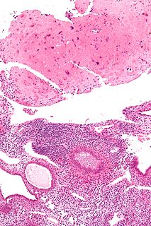
A placental site nodule (PSN) is benign remnant from a previous pregnancy.
Epithelioid trophoblastic tumor (ETT) is a gestational trophoblastic disease with about 110 case reports in the literature. It is a trophoblastic tumor of neoplastic chorionic type associated with the intermediate trophoblast.

The histopathology of colorectal cancer of the adenocarcinoma type involves analysis of tissue taken from a biopsy or surgery. A pathology report contains a description of the microscopical characteristics of the tumor tissue, including both tumor cells and how the tumor invades into healthy tissues and finally if the tumor appears to be completely removed. The most common form of colon cancer is adenocarcinoma, constituting between 95% to 98% of all cases of colorectal cancer. Other, rarer types include lymphoma, adenosquamous and squamous cell carcinoma. Some subtypes have been found to be more aggressive.

