Kuru (disease)

Last updated
Kuru
PMC4235695 pathogens-02-00472-g009.png
A Fore child with advanced kuru. He is unable to walk or sit upright without assistance and is severely malnourished.
Pronunciation
  • IPA: kuru
Specialty Neuropathology, infectious disease
Symptoms Body tremors, random outbursts of laughter, gradual loss of coordination
Complications Infection and pneumonia during the terminal stage.
Usual onsetOften takes years or even decades for symptoms to appear after exposure
Duration11–14 month life expectancy after onset of symptoms [1]
CausesTransmission of infected prion proteins
Risk factors Cannibalism
Diagnostic method Autopsy
Differential diagnosis Creutzfeldt–Jakob disease
PreventionAvoiding practices of cannibalism
TreatmentSupportive care
Prognosis Fatal
FrequencyRare
DeathsApproximately 2,700 as of 2005

Kuru is a rare, incurable, and fatal neurodegenerative disorder that was formerly common among the Fore people of Papua New Guinea. Kuru is a form of transmissible spongiform encephalopathy (TSE) caused by the transmission of abnormally folded proteins (prions), which leads to symptoms such as tremors and loss of coordination from neurodegeneration.

Contents

The term kuru derives from the Fore word kuria or guria ("to shake"), [2] due to the body tremors that are a classic symptom of the disease. Kúru itself means "trembling". [3] It is also known as the "laughing sickness" due to the pathologic bursts of laughter which are a symptom of the disease. It is now widely accepted that kuru was transmitted among members of the Fore tribe of Papua New Guinea via funerary cannibalism. Deceased family members were traditionally cooked and eaten, which was thought to help free the spirit of the dead. [4] Women and children usually consumed the brain, the organ in which infectious prions were most concentrated, thus allowing for transmission of kuru. The disease was therefore more prevalent among women and children.

The epidemic likely started when a villager developed sporadic Creutzfeldt–Jakob disease and died. When villagers ate the brain, they contracted the disease and then spread it to other villagers who ate their infected brains. [5]

While the Fore people stopped consuming human meat in the early 1960s, when it was first speculated to be transmitted via endocannibalism, the disease lingered due to kuru's long incubation period of anywhere from 10 to over 50 years. [6] The epidemic finally declined sharply after half a century, from 200 deaths per year in 1957 to no deaths from at least 2010 onwards, with sources disagreeing on whether the last known kuru victim died in 2005 or 2009. [7] [8] [9] [10]

Signs and symptoms

Kuru, a transmissible spongiform encephalopathy, is a disease of the nervous system that causes physiological and neurological effects which ultimately lead to death. It is characterized by progressive cerebellar ataxia, or loss of coordination and control over muscle movements. [11] [12]

The preclinical or asymptomatic phase, also called the incubation period, averages 10–13 years, but can be as short as five and has been estimated to last as long as 50 years or more after initial exposure. [13]

The clinical stage, which begins at the first onset of symptoms, lasts an average of 12 months. The clinical progression of kuru is divided into three specific stages: the ambulant, sedentary and terminal stages. While there is some variation in these stages from individual to individual, they are highly conserved among the affected population. [11] Before the onset of clinical symptoms, an individual can also present with prodromal symptoms including headache and joint pain in the legs. [14]

Ambulant stage

In the ambulant stage, the infected individual may exhibit unsteady stance and gait, decreased muscle control, difficulty pronouncing words (dysarthria), and tremors (titubation). This stage is named the ambulant because the individual is still able to walk around despite symptoms. [14]

Sedentary stage

In the sedentary stage, the infected individual is incapable of walking without support and experiences ataxia and severe tremors. Furthermore, the individual shows signs of emotional instability and depression, yet exhibits uncontrolled and sporadic laughter. Despite the other neurological symptoms, tendon reflexes are still intact at this stage of the disease. [14]

Terminal stage

In the terminal stage, the infected individual's existing symptoms, like ataxia, progress to the point where it is no longer possible to sit up without support. New symptoms also emerge: the individual develops dysphagia, which can lead to severe malnutrition, and may also become incontinent, lose the ability or will to speak, and become unresponsive to their surroundings despite maintaining consciousness. [14] Towards the end of the terminal stage, patients often develop chronic decubitus ulcerated wounds that can be easily infected. An infected person usually dies within three months to two years after the first terminal stage symptoms, often because of aspiration pneumonia [15] or other secondary infections. [16]

Causes

Kuru is largely localized to the Fore people and people with whom they intermarried. [17] The Fore people ritualistically cooked and consumed body parts of their family members following their death to incorporate "the body of the dead person into the bodies of living relatives, thus helping to free the spirit of the dead". [18] Because the brain is the organ enriched in the infectious prion, women and children, who consumed brain, had a much higher likelihood of being infected than men, who preferentially consumed muscles. [19]

Normally folded prion protein PrP subdomain-Residues 125-228. Note the presence of alpha helices (blue). Prion subdomain-colored sec structure.png
Normally folded prion protein PrP subdomain-Residues 125–228. Note the presence of alpha helices (blue).

Prion

Cryoelectron microscopy model of the misfolded PrP protein, enriched in beta sheets (center) Model of a three dimensional structure for PrPSc.jpg
Cryoelectron microscopy model of the misfolded PrP protein, enriched in beta sheets (center)

The infectious agent is a misfolded form of a host-encoded protein called prion (PrP). Prion proteins are encoded by the Prion Protein Gene (PRNP). [21] The two forms of prion are designated as PrPc, which is a normally folded protein, and PrPsc, a misfolded form which gives rise to the disease. The two forms do not differ in their amino acid sequence; however, the pathogenic PrPsc isoform differs from the normal PrPc form in its secondary and tertiary structure. The PrPsc isoform is more enriched in beta sheets, while the normal PrPc form is enriched in alpha helices. [19] The differences in conformation allow PrPsc to aggregate and be extremely resistant to protein degradation by enzymes or by other chemical and physical means. The normal form, on the other hand, is susceptible to complete proteolysis and soluble in non-denaturing detergents. [14]

It has been suggested that pre-existing or acquired PrPsc can promote the conversion of PrPc into PrPsc, which goes on to convert other PrPc. This initiates a chain reaction that allows for its rapid propagation, resulting in the pathogenesis of prion diseases. [14]

Transmission

In 1961, Australian medical researcher Michael Alpers conducted extensive field studies among the Fore accompanied by anthropologist Shirley Lindenbaum. [9] Their historical research suggested the epidemic may have originated around 1900 from a single individual who lived on the edge of Fore territory and who is thought to have spontaneously developed some form of Creutzfeldt–Jakob disease. [22] Alpers and Lindenbaum's research conclusively demonstrated that kuru spread easily and rapidly in the Fore people due to their endocannibalistic funeral practices, in which relatives consumed the bodies of the dead to return the person's "life force" to the hamlet, a Fore social subunit. [23] Corpses of family members were often buried for days, then exhumed once the corpses were colonized by insect larvae, at which point the corpse would be dismembered and served with the larvae as a side dish. [24]

The demographic distribution evident in the infection rates – kuru was eight to nine times more prevalent in women and children than in men at its peak – is because Fore men considered consuming human flesh to weaken them in times of conflict or battle, while the women and children were more likely to eat the bodies of the deceased, including the brain, where the prion particles were particularly concentrated. Also, the strong possibility exists that it was passed on to women and children more easily because they took on the task of cleaning relatives after death and might have had open sores and cuts on their hands. [19]

Although ingestion of the prion particles can lead to the disease, [25] a high degree of transmission occurred if the prion particles could reach the subcutaneous tissue. With elimination of cannibalism because of Australian colonial law enforcement and the local Christian missionaries' efforts, Alpers' research showed that kuru was already declining among the Fore by the mid‑1960s. However, the mean incubation period of the disease is 14 years, and seven cases were reported with latencies of 40 years or more for those who were most genetically resilient, continuing to appear for several more decades. Sources disagree on whether the last person with kuru died in 2005 or 2009. [9] [10] [7] [8]

Diagnosis

Kuru is diagnosed by reviewing the patient's history of cerebellar signs and symptoms, performing neurological exams, and excluding other neurological diseases during exams. [15] The symptoms evaluated are typically coordination issues and involuntary muscle movements, but these markers can be confused with other diseases that affect the nervous and muscle system; physical scans are often required to differentiate Kuru from other disorders. [26] [27] There is no laboratory test to determine the presence of Kuru, except for postmortem evaluation of central nervous system (CNS) tissues, so diagnoses are achieved by eliminating other possible disorders.

Electroencephalogram (EEG) is used to distinguish kuru from Creutzfeldt–Jakob disease, a similar encephalopathy (any disease that affects the structure of the brain). [27] EEGs search for electrical activity in the patient's brain and measure the frequency of each wave to determine if there is an issue with the brain's activity. [28] Periodic complexes (PC), reoccurring patterns with spike wave-complexes occurring at intervals, are recorded frequently in some diseases but are not presented in the kuru readings. [29] Exams and testing, like EEG, MRIs, blood test, and scans, can be used to determine if the infected person is dealing with Kuru disease or another encephalopathy. However, testing over periods of time can be difficult.

Immunity

Cerebellum of a kuru patient Cerebellum of kuru victim.png
Cerebellum of a kuru patient

In 2009, researchers at the Medical Research Council discovered a naturally occurring variant of a prion protein in a population from Papua New Guinea that confers strong resistance to kuru. In the study, which began in 1996, [30] researchers assessed over 3,000 people from the affected and surrounding Eastern Highland populations, and identified a variation in the prion protein G127. [31] G127 polymorphism is the result of a missense mutation, and is highly geographically restricted to regions where the kuru epidemic was the most widespread. Researchers believe that the PrnP variant occurred very recently, estimating that the most recent common ancestor lived 10 generations ago. [31]

The findings of the study could help researchers better understand and develop treatments for other related prion diseases, such as Creutzfeldt–Jakob disease [30] and other neurodegenerate diseases like Alzheimer's disease. [32]

History

Kuru was first described in official reports by Australian officers patrolling the Eastern Highlands of Papua New Guinea in the early 1950s. [33] Some unofficial accounts place kuru in the region as early as 1910. [7] In 1951, Arthur Carey was the first to use the term kuru in a report to describe a new disease afflicting the Fore tribes of Papua New Guinea (PNG). In his report, Carey noted that kuru mostly affected Fore women, eventually killing them. Kuru was noted in the Fore, Yate and Usurufa people in 1952–1953 by anthropologists Ronald Berndt and Catherine Berndt. [7] In 1953, kuru was observed by patrol officer John McArthur, who provided a description of the disease in his report. McArthur believed that kuru was merely a psychosomatic episode resulting from the sorcery practices of the tribal people in the region. [33] After the disease had progressed into a larger epidemic, the tribal people asked Charles Pfarr, a Lutheran medical officer, to come to the area to report the disease to Australian authorities. [7]

Initially, the Fore people believed the causes of kuru to be sorcery or witchcraft. [34] They also thought that the magic causing kuru was contagious. It was also called negi-nagi, which meant foolish person as the victims laughed at spontaneous intervals. [35] This disease, the Fore people believed, was caused by ghosts, because of the shaking and strange behaviour that comes with kuru. Attempting to cure this, they would feed victims casuarina bark. [36]

When kuru disease had become an epidemic, Daniel Carleton Gajdusek, M.D., a virologist, and Vincent Zigas, another medical doctor, started research on the disease. In 1957, Zigas and Gajdusek published a report in the Medical Journal of Australia that suggested that kuru had a genetic origin, and that "any ethnic-environmental variables that are operating in kuru pathogenesis have not yet been determined." [37]

Cannibalism was suspected as a possible cause from the very beginning but was not formally put forth as a hypothesis until 1967 by Glasse and more formally in 1968 by Mathews, Glasse, and Lindenbaum. [35]

Even before anthropophagy had been linked to kuru, cannibalism was banned by the Australian administration of Papua New Guinea, and the practice was nearly eliminated by 1960. While the number of cases of kuru was decreasing, medical researchers were finally able to properly investigate kuru, which eventually led to the modern understanding of prions as its cause. [38]

In an effort to understand the pathology of kuru disease, Gajdusek established the first experimental tests on chimpanzees for kuru at the National Institutes of Health (NIH). [7] Michael Alpers, an Australian doctor, collaborated with Gajdusek by providing samples of brain tissues he had taken from an 11-year-old Fore girl who had died of kuru. [39] In his work, Gajdusek was also the first to compile a bibliography of kuru disease. [40] Joe Gibbs joined Gajdusek to monitor and record the behavior of the apes at the NIH and conduct their autopsies. Within two years, one of the chimps, Daisy, had developed kuru, demonstrating that an unknown disease factor was transmitted through infected biomaterial and that it was capable of crossing the species barrier to other primates. After Elisabeth Beck confirmed that this experiment had brought about the first experimental transmission of kuru, the finding was deemed a very important advance in human medicine, leading to the award of the Nobel Prize in Physiology or Medicine to Gajdusek in 1976. [7]

Subsequently, E. J. Field spent large parts of the late 1960s and early 1970s in New Guinea investigating the disease, [41] connecting it to scrapie and multiple sclerosis. [42] He noted the disease's interactions with glial cells, including the critical observation that the infectious process may depend on the structural rearrangement of the host's molecules. [43] This was an early observation of what was to later become the prion hypothesis. [44]

Related Research Articles

<span class="mw-page-title-main">Creutzfeldt–Jakob disease</span> Degenerative neurological disorder

Creutzfeldt–Jakob disease (CJD), also known as subacute spongiform encephalopathy or neurocognitive disorder due to prion disease, is a fatal degenerative brain disorder. Early symptoms include memory problems, behavioral changes, poor coordination, and visual disturbances. Later symptoms include dementia, involuntary movements, blindness, weakness, and coma. About 70% of people die within a year of diagnosis. The name Creutzfeldt–Jakob disease was introduced by Walther Spielmeyer in 1922, after the German neurologists Hans Gerhard Creutzfeldt and Alfons Maria Jakob.

<span class="mw-page-title-main">Prion</span> Pathogenic type of misfolded protein

A prion is a misfolded protein that can induce misfolding of normal variants of the same protein and trigger cellular death. Prions cause prion diseases known as transmissible spongiform encephalopathies (TSEs) that are transmissible, fatal neurodegenerative diseases in humans and animals. The proteins may misfold sporadically, due to genetic mutations, or by exposure to an already misfolded protein. The consequent abnormal three-dimensional structure confers on them the ability to cause misfolding of other proteins.

<span class="mw-page-title-main">Stanley B. Prusiner</span> American neurologist and biochemist

Stanley Ben Prusiner is an American neurologist and biochemist. He is the director of the Institute for Neurodegenerative Diseases at University of California, San Francisco (UCSF). Prusiner discovered prions, a class of infectious self-reproducing pathogens primarily or solely composed of protein, a scientific theory considered by many as a heretical idea when first proposed. He received the Albert Lasker Award for Basic Medical Research in 1994 and the Nobel Prize in Physiology or Medicine in 1997 for research on prion diseases developed by him and his team of experts beginning in the early 1970s.

<span class="mw-page-title-main">Scrapie</span> Degenerative disease that affects sheep and goats

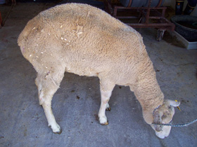
Scrapie is a fatal, degenerative disease affecting the nervous systems of sheep and goats. It is one of several transmissible spongiform encephalopathies (TSEs), and as such it is thought to be caused by a prion. Scrapie has been known since at least 1732 and does not appear to be transmissible to humans. However, it has been found to be experimentally transmissible to humanised transgenic mice and non-human primates.

<span class="mw-page-title-main">Transmissible spongiform encephalopathy</span> Group of brain diseases induced by prions

Transmissible spongiform encephalopathies (TSEs) also known as prion diseases, are a group of progressive, incurable, and fatal conditions that are associated with prions and affect the brain and nervous system of many animals, including humans, cattle, and sheep. According to the most widespread hypothesis, they are transmitted by prions, though some other data suggest an involvement of a Spiroplasma infection. Mental and physical abilities deteriorate and many tiny holes appear in the cortex causing it to appear like a sponge when brain tissue obtained at autopsy is examined under a microscope. The disorders cause impairment of brain function, including memory changes, personality changes and problems with movement that worsen chronically.

<span class="mw-page-title-main">Chronic wasting disease</span> Prion disease affecting the deer family

Chronic wasting disease (CWD), sometimes called zombie deer disease, is a transmissible spongiform encephalopathy (TSE) affecting deer. TSEs are a family of diseases thought to be caused by misfolded proteins called prions and include similar diseases such as BSE in cattle, Creutzfeldt–Jakob disease (CJD) in humans and scrapie in sheep. Natural infection causing CWD affects members of the deer family. In the United States, CWD affects mule deer, white-tailed deer, red deer, sika deer, elk, caribou, and moose. The transmission of CWD to other species such as squirrel monkeys and humanized mice has been observed in experimental settings.

<span class="mw-page-title-main">Daniel Carleton Gajdusek</span> American medical researcher and Nobel Prize laureate

Daniel Carleton Gajdusek was an American physician and medical researcher who was the co-recipient of the Nobel Prize in Physiology or Medicine in 1976 for work on the transmissibility of kuru, implying the existence of an infectious agent, which he named an 'unconventional virus'.

The Fore people live in the Okapa District of the Eastern Highlands Province, Papua New Guinea. There are approximately 20,000 Fore who are separated by the Wanevinti Mountains into the North Fore and South Fore regions. Their main form of subsistence is slash-and-burn farming. The Fore language has three distinct dialects and is the southernmost member of the East Central Family, East New Guinea Highlands Stock, Trans–New Guinea phylum of Papuan languages.

<span class="mw-page-title-main">Gerstmann–Sträussler–Scheinker syndrome</span> Human neurodegenerative disease

Gerstmann–Sträussler–Scheinker syndrome (GSS) is an extremely rare, always fatal neurodegenerative disease that affects patients from 20 to 60 years in age. It is exclusively heritable, and is found in only a few families all over the world. It is, however, classified with the transmissible spongiform encephalopathies (TSE) due to the causative role played by PRNP, the human prion protein. GSS was first reported by the Austrian physicians Josef Gerstmann, Ernst Sträussler and Ilya Scheinker in 1936.

Transmissible mink encephalopathy (TME) is a rare sporadic disease that affects the central nervous system of ranch-raised adult mink. It is a transmissible spongiform encephalopathy, caused by proteins called prions.

<span class="mw-page-title-main">Major prion protein</span> Protein involved in multiple prion diseases

Major prion protein (PrP) is encoded in the human body by the PRNP gene also known as CD230. Expression of the protein is most predominant in the nervous system but occurs in many other tissues throughout the body.

Laura Manuelidis is a physician and neuropathologist at Yale University.

<span class="mw-page-title-main">Variant Creutzfeldt–Jakob disease</span> Degenerative brain disease caused by prions

Variant Creutzfeldt–Jakob disease (vCJD), commonly referred to as "mad cow disease" or "human mad cow disease" to distinguish it from its BSE counterpart, is a fatal type of brain disease within the transmissible spongiform encephalopathy family. Initial symptoms include psychiatric problems, behavioral changes, and painful sensations. In the later stages of the illness, patients may exhibit poor coordination, dementia and involuntary movements. The length of time between exposure and the development of symptoms is unclear, but is believed to be years to decades. Average life expectancy following the onset of symptoms is 13 months.

Endocannibalism is a practice of cannibalism in one's own locality or community. In most cases this refers to the consumption of the remains of the deceased in a mortuary context.

<span class="mw-page-title-main">Bovine spongiform encephalopathy</span> Fatal neurodegenerative disease of cattle

Bovine spongiform encephalopathy (BSE), commonly known as mad cow disease, is an incurable and invariably fatal neurodegenerative disease of cattle. Symptoms include abnormal behavior, trouble walking, and weight loss. Later in the course of the disease, the cow becomes unable to function normally. There is conflicting information about the time between infection and onset of symptoms. In 2002, the World Health Organization (WHO) suggested it to be approximately four to five years. Time from onset of symptoms to death is generally weeks to months. Spread to humans is believed to result in variant Creutzfeldt–Jakob disease (vCJD). As of 2018, a total of 231 cases of vCJD had been reported globally.

<span class="mw-page-title-main">Variably protease-sensitive prionopathy</span> Medical condition

Variably protease-sensitive prionopathy (VPSPr) is a sporadic prion protein disease first described in an abstract for a conference on prions in 2006, and this study was published in a 2008 report on 11 cases. The study was conducted by Gambetti P., Zou W.Q., and coworkers from the United States National Prion Disease Pathology Surveillance Center. It was first identified as a distinct disease in 2010 by Zou W.Q. and coworkers from the United States National Prion Disease Pathology Surveillance Center.

Frank O. Bastian is an American physician and neuropathologist, who previously worked at Louisiana State University, moved to a university in New Orleans in 2019. He specializes in the transmissible spongiform encephalopathies (TSEs), which include, but are not limited to, Bovine spongiform encephalopathy (BSE) "Mad cow disease" in cattle, scrapie in sheep and goats, and Creutzfeldt–Jakob disease (CJD) in humans.

Shirley Inglis Lindenbaum is an Australian anthropologist notable for her medical anthropology work on kuru in Papua New Guinea, HIV/AIDS in the United States of America, and cholera in Bangladesh.

<span class="mw-page-title-main">United Kingdom BSE outbreak</span> Mad cow disease outbreak in the 1980s and 90s

The United Kingdom was afflicted with an outbreak of Bovine spongiform encephalopathy, and its human equivalent variant Creutzfeldt–Jakob disease (vCJD), in the 1980s and 1990s. Over four million head of cattle were slaughtered in an effort to contain the outbreak, and 178 people died after contracting vCJD through eating infected beef. A political and public health crisis resulted, and British beef was banned from export to numerous countries around the world, with some bans remaining in place until as late as 2019.

Michael Coulthart is a Canadian microbiologist who is employed as the head of the Canadian Creutzfeldt–Jakob Disease Surveillance System (CJDSS) within the Public Health Agency of Canada (PHAC), which terms CJD a zoonotic and infectious disease. In 2006, a working group named "classic CJD" as well as Variant Creutzfeldt–Jakob disease as two notifiable diseases. It is unknown whether PHAC tracks in an official capacity other transmissible spongiform encephalopathies (TSE), but Coulthart is on the Advisory Committee of the Center for Infectious Disease Research and Policy for Chronic Wasting Disease of cervidae.

References

  1. "The epidemiology of kuru in the period 1987 to 1995", Department of Health (Australia) , retrieved February 5, 2019
  2. Hoskin, J.O.; Kiloh, L.G.; Cawte, J.E. (April 1969). "Epilepsy and guria: The shaking syndromes of New Guinea". Social Science & Medicine. 3 (1): 39–48. doi:10.1016/0037-7856(69)90037-7. PMID   5809623.
  3. Scott, Graham (1978). The Fore Language of Papua New Guinea. Pacific Linguistics. pp. 2, 6. doi:10.15144/PL-B47. hdl:1885/146489. ISBN   978-0-85883-173-5.
  4. Whitfield, Jerome T; Pako, Wandagi H; Collinge, John; Alpers, Michael P (27 November 2008). "Mortuary rites of the South Fore and kuru". Philosophical Transactions of the Royal Society B: Biological Sciences. 363 (1510): 3721–3724. doi:10.1098/rstb.2008.0074. PMC   2581657 . PMID   18849288.
  5. Bichell, Rae Ellen (September 6, 2016). "When People Ate People, A Strange Disease Emerged". NPR.org . Retrieved 2018-04-08.
  6. "Kuru". MedlinePlus Medical Encyclopedia. Retrieved 2016-11-14.
  7. 1 2 3 4 5 6 7 Alpers, MP (2007). "A history of kuru". Papua and New Guinea Medical Journal. 50 (1–2): 10–9. PMID   19354007.
  8. 1 2 Rense, Sarah (September 7, 2016). "Here's What Happens to Your Body When You Eat Human Meat". Esquire .
  9. 1 2 3 "A life of determination". Monash University — Faculty of Medicine, Nursing and Health Sciences. 2009-02-23. Archived from the original on 2015-12-10. Retrieved 20 January 2016.
  10. 1 2 Collinge, John; Whitfield, Jerome; McKintosh, Edward; Beck, John; Mead, Simon; Thomas, Dafydd J; Alpers, Michael P (June 2006). "Kuru in the 21st century—an acquired human prion disease with very long incubation periods". The Lancet. 367 (9528): 2068–2074. doi:10.1016/S0140-6736(06)68930-7. PMID   16798390. S2CID   11506094.
  11. 1 2 Alpers, Michael P (December 2005). "The epidemiology of kuru in the period 1987 to 1995". Communicable Diseases Intelligence. 29 (4): 391–399. PMID   16465931 . Retrieved 2016-11-10.
  12. Liberski, Pawel P.; Sikorska, Beata; Lindenbaum, Shirley; Goldfarb, Lev G.; McLean, Catriona; Hainfellner, Johannes A.; Brown, Paul (February 2012). "Kuru". Journal of Neuropathology & Experimental Neurology. 71 (2): 92–103. doi:10.1097/NEN.0b013e3182444efd. PMC   5120877 . PMID   22249461.
  13. Collinge, John; Whitfield, Jerome; McKintosh, Edward; Frosh, Adam; Mead, Simon; Hill, Andrew F; Brandner, Sebastian; Thomas, Dafydd; Alpers, Michael P (27 November 2008). "A clinical study of kuru patients with long incubation periods at the end of the epidemic in Papua New Guinea". Philosophical Transactions of the Royal Society B: Biological Sciences. 363 (1510): 3725–3739. doi:10.1098/rstb.2008.0068. PMC   2581654 . PMID   18849289.
  14. 1 2 3 4 5 6 Imran, Muhammad; Mahmood, Saqib (24 December 2011). "An overview of human prion diseases". Virology Journal. 8 (1): 559. doi: 10.1186/1743-422X-8-559 . PMC   3296552 . PMID   22196171.
  15. 1 2 Mahat, Sanjay; Asuncion, Ria Monica D. (2023), "Kuru", StatPearls, Treasure Island (FL): StatPearls Publishing, PMID   32644529 , retrieved 2023-10-19
  16. Wadsworth, Jonathan D. F.; Joiner, Susan; Linehan, Jacqueline M.; Desbruslais, Melanie; Fox, Katie; Cooper, Sharon; Cronier, Sabrina; Asante, Emmanuel A.; Mead, Simon; Brandner, Sebastian; Hill, Andrew F.; Collinge, John (11 March 2008). "Kuru prions and sporadic Creutzfeldt–Jakob disease prions have equivalent transmission properties in transgenic and wild-type mice". Proceedings of the National Academy of Sciences of the United States of America. 105 (10): 3885–3890. Bibcode:2008PNAS..105.3885W. doi: 10.1073/pnas.0800190105 . PMC   2268835 . PMID   18316717.
  17. Lindenbaum, Shirley (2001-01-01). "Kuru, Prions, and Human Affairs: Thinking About Epidemics". Annual Review of Anthropology . 30 (1): 363–385. doi:10.1146/annurev.anthro.30.1.363. S2CID   162196301.
  18. Whitfield, Jerome T.; Pako, Wandagi H.; Collinge, John; Alpers, Michael P. (2008-11-27). "Mortuary rites of the South Fore and kuru". Philosophical Transactions of the Royal Society B: Biological Sciences. 363 (1510): 3721–3724. doi:10.1098/rstb.2008.0074. ISSN   0962-8436. PMC   2581657 . PMID   18849288.
  19. 1 2 3 Kuru at eMedicine
  20. Kupfer, L.; Hinrichs, W.; Groschup, M. (1 September 2009). "Prion Protein Misfolding". Current Molecular Medicine. 9 (7): 826–835. doi:10.2174/156652409789105543. PMC   3330701 . PMID   19860662.
  21. Linden, Rafael; Martins, Vilma R.; Prado, Marco A. M.; Cammarota, Martín; Izquierdo, Iván; Brentani, Ricardo R. (April 2008). "Physiology of the Prion Protein". Physiological Reviews. 88 (2): 673–728. doi:10.1152/physrev.00007.2007. PMID   18391177.
  22. Kuru: The Science and the Sorcery (Siamese Films, 2010)
  23. Diamond J.M. (1997). Guns, germs, and steel: the fates of human societies. New York: W.W. Norton. p. 208. ISBN   978-0-393-03891-0.[ page needed ]
  24. Liberski, P.P.; Brown, P. (January 2009). "Kuru: Its ramifications after fifty years" (PDF). Experimental Gerontology. 44 (1–2): 63–69. doi:10.1016/j.exger.2008.05.010. PMID   18606515. S2CID   28215397.
  25. Gibbs, Clarence J.; Amyx, Herbert L.; Bacote, Alfred; Masters, Colin L.; Gajdnsek, D. Carleton (August 1980). "Oral Transmission of Kuru, Creutzfeldt-Jakob Disease, and Scrapie to Nonhuman Primates". The Journal of Infectious Diseases. 142 (2): 205–208. doi:10.1093/infdis/142.2.205. PMID   6997404.
  26. "Kuru: MedlinePlus Medical Encyclopedia". medlineplus.gov. Retrieved 2023-10-19.
  27. 1 2 "Kuru: What Is It, Causes, Signs and Symptoms, and More | Osmosis". www.osmosis.org. Retrieved 2023-10-19.
  28. Services, Department of Health & Human. "EEG test". www.betterhealth.vic.gov.au. Retrieved 2023-10-19.
  29. Smith, S. J. M. (2005-06-01). "EEG in neurological conditions other than epilepsy: when does it help, what does it add?". Journal of Neurology, Neurosurgery & Psychiatry. 76 (suppl 2): ii8–ii12. doi: 10.1136/jnnp.2005.068486 . ISSN   0022-3050. PMC   1765689 . PMID   15961870.
  30. 1 2 "Brain disease 'resistance gene' evolves in Papua New Guinea community; could offer insights into CJD". ScienceDaily. 2009-11-21. Retrieved 2016-11-12.
  31. 1 2 Mead, Simon; Whitfield, Jerome; Poulter, Mark; Shah, Paresh; Uphill, James; Campbell, Tracy; Al-Dujaily, Huda; Hummerich, Holger; Beck, Jon; Mein, Charles A.; Verzilli, Claudio; Whittaker, John; Alpers, Michael P.; Collinge, John (19 November 2009). "A Novel Protective Prion Protein Variant that Colocalizes with Kuru Exposure" (PDF). New England Journal of Medicine. 361 (21): 2056–2065. doi:10.1056/NEJMoa0809716. PMID   19923577.
  32. "Natural genetic variation gives complete resistance in prion diseases". Ucl.ac.uk. 2015-06-11. Retrieved 2016-11-12.
  33. 1 2 Shirley Lindenbaum (14 Apr 2015). "An annotated history of kuru". Medicine Anthropology Theory. Archived from the original on 15 September 2018. Retrieved 12 November 2016.
  34. "Kuru". Transmissible Spongiform Encephalopathies. Archived from the original on 2016-11-21. Retrieved 2016-11-21.
  35. 1 2 Liberski, Paweł P.; Gajos, Agata; Sikorska, Beata; Lindenbaum, Shirley (7 March 2019). "Kuru, the First Human Prion Disease". Viruses. 11 (3): 232. doi: 10.3390/v11030232 . PMC   6466359 . PMID   30866511.
  36. Liberski, Paweł; Gajos, Agata; Sikorska, Beata; Lindenbaum, Shirley (2019-03-07). "Kuru, the First Human Prion Disease". Viruses. 11 (3): 232. doi: 10.3390/v11030232 . ISSN   1999-4915. PMC   6466359 . PMID   30866511.
  37. Zigas, Vincent; Gajdusek, Daniel (23 November 1957). "Kuru: Clinical study of a new syndrome resembling paralysis agitans of the eastern highlands of Australian New Guinea". Medical Journal of Australia. 2 (21): 745–754. doi:10.5694/j.1326-5377.1957.tb60287.x. PMID   13492772. S2CID   38647011.
  38. Kennedy, John (15 May 2012). "Kuru Among the Foré — The Role of Medical Anthropology in Explaining Aetiology and Epidermiology". ArcJohn.wordpress.com. Retrieved 2016-11-21.
  39. "Revisiting the history of kuru - Hektoen International". hekint.org. 30 May 2019. Retrieved 2023-09-14.
  40. Liberski, P. P.; Brown, P. (2004). "Kuru: A half-opened window onto the landscape of neurodegenerative diseases". Folia Neuropathologica. 42 Suppl A: 3–14. PMID   15449456.
  41. "Kuru - To Tremble with Fear". Horizon. Season 8. Episode 6. 22 February 1971. BBC2.
  42. Field, EJ (7 Dec 1967). "The significance of astroglial hypertrophy in Scrapie, Kuru, Multiple Sclerosis and old age together with a note on the possible nature of the scrapie agent". Journal of Neurology. 192 (3): 265–274. doi:10.1007/bf00244170. S2CID   8855940.
  43. Field, EJ (Feb 1978). "Immunological assessment of ageing: emergence of scrapie-like antigens". Age Ageing. 7 (1): 28–39. doi:10.1093/ageing/7.1.28. PMID   416662.
  44. Peat, Ray. "BSE - mad cow - scrapie, etc.: Stimulated amyloid degeneration and the toxic fats". RayPeat.com.
  45. Holub, Miroslav (1990). Vanishing Lung Syndrome. Translated by David Young and Dana Habova. Oberlin College Press. ISBN   0-932440-52-5.; (Faber and Faber, 1990). ISBN   0-571-14339-3 [ page needed ]
  46. "The X-Files: "First Person Shooter" / "Theef"". The A.V. Club. 2013-01-19. Retrieved 2024-01-30.
  47. Estrada, Aura (2020-06-01). "Dead Island: Riptide - Tips & Tricks For New Players". ScreenRant. Retrieved 2024-01-30.
  48. Lerman, Elizabeth (2020-07-25). "We Are What We Are: Ending & Cannibal Family Traditions Explained". ScreenRant. Retrieved 2024-01-30.
  49. Vasundhra (2023-06-18). "What is Cannibalism in Anthropology? | Anthroholic" . Retrieved 2024-01-30.

Further reading