| Laminar organization | |
|---|---|
 | |
| Anatomical terminology |
A laminar organization describes the way certain tissues, such as bone membrane, skin, or brain tissues, are arranged in layers.
| Laminar organization | |
|---|---|
| | |
| Anatomical terminology |
A laminar organization describes the way certain tissues, such as bone membrane, skin, or brain tissues, are arranged in layers.
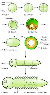
The earliest forms of laminar organization are shown in the diploblastic and triploblastic formation of the germ layers in the embryo. In the first week of human embryogenesis two layers of cells have formed, an external epiblast layer (the primitive ectoderm), and an internal hypoblast layer (primitive endoderm). This gives the early bilaminar disc. [1] In the third week in the stage of gastrulation epiblast cells invaginate to form endoderm, and a third layer of cells known as mesoderm. Cells that remain in the epiblast become ectoderm. This is the trilaminar disc and the epiblast cells have given rise to the three germ layers. [2]
In the brain a laminar organization is evident in the arrangement of the three meninges, the membranes that cover the brain and spinal cord. These membranes are the dura mater, arachnoid mater, and pia mater. The dura mater has two layers a periosteal layer near to the bone of the skull, and a meningeal layer next to the other meninges. [3]
The cerebral cortex, the outer neural sheet covering the cerebral hemispheres can be described by its laminar organization, due to the arrangement of cortical neurons into six distinct layers.
The eye in mammals has an extensive laminar organization. There are three main layers – the outer fibrous tunic, the middle uvea, and the inner retina. [4] These layers have sublayers with the retina having ten ranging from the outer choroid to the inner vitreous humor and including the retinal nerve fiber layer.

The human skin has a dense laminar organization. The outer epidermis has four or five layers.

In all bilaterian animals, the mesoderm is one of the three primary germ layers in the very early embryo. The other two layers are the ectoderm and endoderm, with the mesoderm as the middle layer between them.

The amniotic sac, commonly called the bag of waters, sometimes the membranes, is the sac in which the fetus develops in amniotes. It is a thin but tough transparent pair of membranes that hold a developing embryo until shortly before birth. The inner of these fetal membranes, the amnion, encloses the amniotic cavity, containing the amniotic fluid and the fetus. The outer membrane, the chorion, contains the amnion and is part of the placenta. On the outer side, the amniotic sac is connected to the yolk sac, the allantois, and via the umbilical cord, the placenta.

Gastrulation is a phase early in the embryonic development of most animals, during which the single-layered blastula is reorganized into a multilayered structure known as the gastrula. Before gastrulation, the embryo is a continuous epithelial sheet of cells; by the end of gastrulation, the embryo has begun differentiation to establish distinct cell lineages, set up the basic axes of the body, and internalized one or more cell types including the prospective gut.

Ectoderm is one of the three primary germ layers in the very early embryo. The other two layers are the mesoderm and endoderm, with the ectoderm as the most exterior layer. It emerges and originates from the outer layer of germ cells. The word ectoderm comes from the Greek ektos meaning "outside", and derma, meaning "skin."
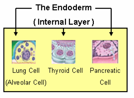
Endoderm is one of the three primary germ layers in the very early embryo. The other two layers are the ectoderm and mesoderm, with the endoderm being the innermost layer. Cells migrating inward along the archenteron form the inner layer of the gastrula, which develops into the endoderm.

The meninges are the three membranes that envelop the brain and spinal cord. In mammals, the meninges are the dura mater, the arachnoid mater, and the pia mater. Cerebrospinal fluid is located in the subarachnoid space between the arachnoid mater and the pia mater. The primary function of the meninges is to protect the central nervous system.

The blastocyst is a structure formed in the early development of mammals. It possesses an inner cell mass (ICM) which subsequently forms the embryo. The outer layer of the blastocyst consists of cells collectively called the trophoblast. This layer surrounds the inner cell mass and a fluid-filled cavity known as the blastocoel. The trophoblast gives rise to the placenta. The name "blastocyst" arises from the Greek βλαστός blastos and κύστις kystis. In other animals this is called a blastula.

Pia mater, often referred to as simply the pia, is the delicate innermost layer of the meninges, the membranes surrounding the brain and spinal cord. Pia mater is medieval Latin meaning "tender mother". The other two meningeal membranes are the dura mater and the arachnoid mater. Both the pia and arachnoid mater are derivatives of the neural crest while the dura is derived from embryonic mesoderm. The pia mater is a thin fibrous tissue that is permeable to water and small solutes. The pia mater allows blood vessels to pass through and nourish the brain. The perivascular space between blood vessels and pia mater is proposed to be part of a pseudolymphatic system for the brain. When the pia mater becomes irritated and inflamed the result is meningitis.

Dura mater is a thick membrane made of dense irregular connective tissue that surrounds the brain and spinal cord. It is the outermost of the three layers of membrane called the meninges that protect the central nervous system. The other two meningeal layers are the arachnoid mater and the pia mater. The dura surrounds the brain and the spinal cord. It envelops the arachnoid mater, which is responsible for keeping in the cerebrospinal fluid. It is derived primarily from the neural crest cell population, with postnatal contributions of the paraxial mesoderm.
A germ layer is a primary layer of cells that forms during embryonic development. The three germ layers in vertebrates are particularly pronounced; however, all eumetazoans produce two or three primary germ layers. Some animals, like cnidarians, produce two germ layers making them diploblastic. Other animals such as chordates produce a third layer between these two layers, making them triploblastic. Germ layers eventually give rise to all of an animal’s tissues and organs through the process of organogenesis.
Embryonic development also embryogenesis is the process by which the embryo forms and develops. In mammals, the term refers chiefly to early stages of prenatal development, whereas the terms fetus and fetal development describe later stages.
The arachnoid mater is one of the three meninges, the protective membranes that cover the brain and spinal cord. The arachnoid mater is a derivative of the neural crest mesectoderm in the embryo.

The cranial cavity, also known as intracranial space, is the space within the skull. The space inside the skull is formed by eight cranial bones known as the neurocranium. The neurocranium is the upper back part that forms the protective case around the brain. The skull cap of the neurocranium covers the cranial cavity and the remainder of the skull is called the facial skeleton. The skull is also known as the cranium, and contains the brain. Meninges are protective membranes that surround the brain to minimize damage of the brain when there is head trauma. Meningitis is the inflammation of meninges caused by bacterial or viral infections.

Lateral plate mesoderm is a type of mesoderm that is found at the periphery of the embryo.
A trilaminar embryo is an early stage in the development of triploblastic organisms, which include humans and many other animals.

In amniote animal embryology, the epiblast is one of two distinct layers arising from the inner cell mass in the mammalian blastocyst or from the blastodisc in reptiles and birds. It derives the embryo proper through its differentiation into the three primary germ layers, ectoderm, mesoderm and endoderm, during gastrulation. The amnionic ectoderm and extraembryonic mesoderm also originate from the epiblast.
Bilaminar blastocyst or bilaminar disc refers to the epiblast and the hypoblast, evolved from the embryoblast. These two layers are sandwiched between two balloons: the primitive yolk sac and the amniotic cavity.

Human embryonic development, or human embryogenesis, refers to the development and formation of the human embryo. It is characterised by the processes of cell division and cellular differentiation of the embryo that occurs during the early stages of development. In biological terms, the development of the human body entails growth from a one-celled zygote to an adult human being. Fertilisation occurs when the sperm cell successfully enters and fuses with an egg cell (ovum). The genetic material of the sperm and egg then combine to form a single cell called a zygote and the germinal stage of development commences. Embryonic development in the human, covers the first eight weeks of development; at the beginning of the ninth week the embryo is termed a fetus. Human embryology is the study of this development during the first eight weeks after fertilisation. The normal period of gestation (pregnancy) is about nine months or 40 weeks.
The hypoblast is a tissue type that forms from the inner cell mass during early embryonic development. It lies beneath the epiblast and consists of small cuboidal cells. The hypoblast gives rise to the yolk sac, which in turn gives rise to the chorion. The epiblast on the other hand gives rise to the embryo itself, through the three germ layers, the endoderm, mesoderm and ectoderm.
The development of the digestive system concerns the epithelium of the digestive system and the parenchyma of its derivatives, which originate from the endoderm. Connective tissue, muscular components, and peritoneal components originate in the mesoderm. Different regions of the gut tube such as the esophagus, stomach, duodenum, etc. are specified by a retinoic acid gradient that causes transcription factors unique to each region to be expressed. Differentiation of the gut and its derivatives depends upon reciprocal interactions between the gut endoderm and its surrounding mesoderm. Hox genes in the mesoderm are induced by a Hedgehog signaling pathway secreted by gut endoderm and regulate the craniocaudal organization of the gut and its derivatives. The gut system extends from the oropharyngeal membrane to the cloacal membrane and is divided into the foregut, midgut, and hindgut.