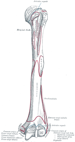
The humerus is a long bone in the arm that runs from the shoulder to the elbow. It connects the scapula and the two bones of the lower arm, the radius and ulna, and consists of three sections. The humeral upper extremity consists of a rounded head, a narrow neck, and two short processes. The body is cylindrical in its upper portion, and more prismatic below. The lower extremity consists of 2 epicondyles, 2 processes, and 3 fossae. As well as its true anatomical neck, the constriction below the greater and lesser tubercles of the humerus is referred to as its surgical neck due to its tendency to fracture, thus often becoming the focus of surgeons.

The brachioradialis is a muscle of the forearm that flexes the forearm at the elbow. It is also capable of both pronation and supination, depending on the position of the forearm. It is attached to the distal styloid process of the radius by way of the brachioradialis tendon, and to the lateral supracondylar ridge of the humerus.

The radial nerve is a nerve in the human body that supplies the posterior portion of the upper limb. It innervates the medial and lateral heads of the triceps brachii muscle of the arm, as well as all 12 muscles in the posterior osteofascial compartment of the forearm and the associated joints and overlying skin.

The extensor carpi radialis longus is one of the five main muscles that control movements at the wrist. This muscle is quite long, starting on the lateral side of the humerus, and attaching to the base of the second metacarpal bone.

The axillary nerve or the circumflex nerve is a nerve of the human body, that originates from the brachial plexus at the level of the axilla (armpit) and carries nerve fibers from C5 and C6. The axillary nerve travels through the quadrangular space with the posterior circumflex humeral artery and vein to innervate the deltoid and teres minor.
In human anatomy, the ulnar nerve is a nerve that runs near the ulna bone. The ulnar collateral ligament of elbow joint is in relation with the ulnar nerve. The nerve is the largest in the human body unprotected by muscle or bone, so injury is common. This nerve is directly connected to the little finger, and the adjacent half of the ring finger, innervating the palmar aspect of these fingers, including both front and back of the tips, perhaps as far back as the fingernail beds.

The radius or radial bone is one of the two large bones of the forearm, the other being the ulna. It extends from the lateral side of the elbow to the thumb side of the wrist and runs parallel to the ulna. The ulna is longer than the radius, but the radius is thicker. The radius is a long bone, prism-shaped and slightly curved longitudinally.

The quadriceps femoris muscle is a large muscle group that includes the four prevailing muscles on the front of the thigh. It is the sole extensor muscle of the knee, forming a large fleshy mass which covers the front and sides of the femur. The name derives from Latin four-headed muscle of the femur.

The coccygeus muscle or ischiococcygeus is a muscle of the pelvic floor located posterior to levator ani and anterior to the sacrospinous ligament.

The olecranon, is a large, thick, curved bony process on the proximal, posterior end of the ulna. It forms the protruding part of the elbow and is opposite to the cubital fossa or elbow pit. The olecranon serves as a lever for the extensor muscles that straighten the elbow joint.

The teres major muscle is a muscle of the upper limb. It attaches to the scapula and the humerus and is one of the seven scapulohumeral muscles. It is a thick but somewhat flattened muscle.

In human anatomy, the fibularis tertius is a muscle in the anterior compartment of the leg. It acts to tilt the sole of the foot away from the midline of the body (eversion) and to pull the foot upward toward the body (dorsiflexion).

The lateral circumflex femoral artery is an artery in the upper thigh. It is usually a branch of the profunda femoris artery, and produces three branches. It is mostly distributed to the muscles of the lateral thigh, supplying arterial blood to muscles of the knee extensor group.

The lower subscapular nerve, also known as the inferior subscapular nerve, is the third branch of the posterior cord of the brachial plexus. It innervates the inferior portion of the subscapularis muscle and the teres major muscle.

The retromandibular vein is a major vein of the face. It is formed within the parotid gland by the confluence of the maxillary vein, and superficial temporal vein. It descends in the gland and splits into two branches upon emerging from the gland. Its anterior branch then joins the (anterior) facial vein forming the common facial vein, while its posterior branch joins the posterior auricular vein forming the external jugular vein.

The bicipital aponeurosis is a broad aponeurosis of the biceps brachii, which is located in the cubital fossa of the elbow. It separates superficial from deep structures in much of the fossa.

The radial styloid process is a projection of bone on the lateral surface of the distal radius bone.
The common extensor tendon is a tendon that attaches to the lateral epicondyle of the humerus.

The elbow is the region between the upper arm and the forearm that surrounds the elbow joint. The elbow includes prominent landmarks such as the olecranon, the cubital fossa, and the lateral and the medial epicondyles of the humerus. The elbow joint is a hinge joint between the arm and the forearm; more specifically between the humerus in the upper arm and the radius and ulna in the forearm which allows the forearm and hand to be moved towards and away from the body. The term elbow is specifically used for humans and other primates, and in other vertebrates it is not used. In those cases, forelimb plus joint is used.

A supracondylar humerus fracture is a fracture of the distal humerus just above the elbow joint. The fracture is usually transverse or oblique and above the medial and lateral condyles and epicondyles. This fracture pattern is relatively rare in adults, but is the most common type of elbow fracture in children. In children, many of these fractures are non-displaced and can be treated with casting. Some are angulated or displaced and are best treated with surgery. In children, most of these fractures can be treated effectively with expectation for full recovery. Some of these injuries can be complicated by poor healing or by associated blood vessel or nerve injuries with serious complications.