Related Research Articles

The cerebral cortex, also known as the cerebral mantle, is the outer layer of neural tissue of the cerebrum of the brain in humans and other mammals. The cerebral cortex mostly consists of the six-layered neocortex, with just 10% consisting of allocortex. It is separated into two cortices, by the longitudinal fissure that divides the cerebrum into the left and right cerebral hemispheres. The two hemispheres are joined beneath the cortex by the corpus callosum. The cerebral cortex is the largest site of neural integration in the central nervous system. It plays a key role in attention, perception, awareness, thought, memory, language, and consciousness. The cerebral cortex is part of the brain responsible for cognition.

The limbic system, also known as the paleomammalian cortex, is a set of brain structures located on both sides of the thalamus, immediately beneath the medial temporal lobe of the cerebrum primarily in the forebrain.

The parietal lobe is one of the four major lobes of the cerebral cortex in the brain of mammals. The parietal lobe is positioned above the temporal lobe and behind the frontal lobe and central sulcus.

The neocortex, also called the neopallium, isocortex, or the six-layered cortex, is a set of layers of the mammalian cerebral cortex involved in higher-order brain functions such as sensory perception, cognition, generation of motor commands, spatial reasoning and language. The neocortex is further subdivided into the true isocortex and the proisocortex.
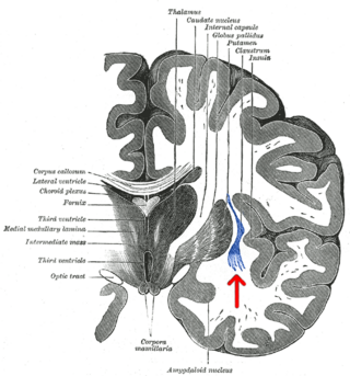
The claustrum is a thin, bilateral collection of neurons and supporting glial cells, that connects to cortical and subcortical regions of the brain. It is located between the insula laterally and the putamen medially, separated by the extreme and external capsules respectively. The blood supply to the claustrum is fulfilled via the middle cerebral artery. It is considered to be the most densely connected structure in the brain, allowing for integration of various cortical inputs into one experience rather than singular events. The claustrum is difficult to study given the limited number of individuals with claustral lesions and the poor resolution of neuroimaging.

The triune brain is a model of the evolution of the vertebrate forebrain and behavior, proposed by the American physician and neuroscientist Paul D. MacLean in the 1960s. The triune brain consists of the reptilian complex, the paleomammalian complex, and the neomammalian complex (neocortex), viewed each as independently conscious, and as structures sequentially added to the forebrain in the course of evolution. According to the model, the basal ganglia are in charge of our primal instincts, the limbic system is in charge of our emotions and the neocortex is responsible for objective or rational thoughts.

A cortical column is a group of neurons forming a cylindrical structure through the cerebral cortex of the brain perpendicular to the cortical surface. The structure was first identified by Mountcastle in 1957. He later identified minicolumns as the basic units of the neocortex which were arranged into columns. Each contains the same types of neurons, connectivity, and firing properties. Columns are also called hypercolumn, macrocolumn, functional column or sometimes cortical module. Neurons within a minicolumn (microcolumn) encode similar features, whereas a hypercolumn "denotes a unit containing a full set of values for any given set of receptive field parameters". A cortical module is defined as either synonymous with a hypercolumn (Mountcastle) or as a tissue block of multiple overlapping hypercolumns.

In psycholinguistics, language processing refers to the way humans use words to communicate ideas and feelings, and how such communications are processed and understood. Language processing is considered to be a uniquely human ability that is not produced with the same grammatical understanding or systematicity in even human's closest primate relatives.

The commissural fibers or transverse fibers are axons that connect the two hemispheres of the brain. In contrast to commissural fibers, association fibers connect regions within the same hemisphere of the brain, and projection fibers connect each region to other parts of the brain or to the spinal cord.

Pasko Rakic is a Yugoslav-born American neuroscientist, who presently works in the Yale School of Medicine Department of Neuroscience in New Haven, Connecticut. His main research interest is in the development and evolution of the human brain. He was the founder and served as Chairman of the Department of Neurobiology at Yale, and was founder and Director of the Kavli Institute for Neuroscience. He is best known for elucidating the mechanisms involved in development and evolution of the cerebral cortex. In 2008, Rakic shared the inaugural Kavli Prize in Neuroscience. He is currently the Dorys McConell Duberg Professor of Neuroscience, leads an active research laboratory, and serves on Advisory Boards and Scientific Councils of a number of Institutions and Research Foundations.
Theresa A. Jones is a researcher and professor at the University of Texas at Austin and the Institute for Neuroscience. Her interests are in neural plasticity across the lifespan, motor skill learning, mechanisms of brain and behavioral adaptation to brain damage, and glial-neuronal interactions. Her research is on the brain changes following stroke, in particular rehabilitation strategies and the brain changes associated with them. She primarily tests rats and uses the Endothelin-1 stroke model. Her most recent work has expanded into the field of microstimulation mapping of the rat cortex.

Animal consciousness, or animal awareness, is the quality or state of self-awareness within a non-human animal, or of being aware of an external object or something within itself. In humans, consciousness has been defined as: sentience, awareness, subjectivity, qualia, the ability to experience or to feel, wakefulness, having a sense of selfhood, and the executive control system of the mind. Despite the difficulty in definition, many philosophers believe there is a broadly shared underlying intuition about what consciousness is.

There is much to be discovered about the evolution of the brain and the principles that govern it. While much has been discovered, not everything currently known is well understood. The evolution of the brain has appeared to exhibit diverging adaptations within taxonomic classes such as Mammalia and more vastly diverse adaptations across other taxonomic classes. Brain to body size scales allometrically. This means as body size changes, so do other physiological, anatomical, and biochemical constructs connecting the brain to the body. Small bodied mammals have relatively large brains compared to their bodies whereas large mammals have a smaller brain to body ratios. If brain weight is plotted against body weight for primates, the regression line of the sample points can indicate the brain power of a primate species. Lemurs for example fall below this line which means that for a primate of equivalent size, we would expect a larger brain size. Humans lie well above the line indicating that humans are more encephalized than lemurs. In fact, humans are more encephalized compared to all other primates. This means that human brains have exhibited a larger evolutionary increase in its complexity relative to its size. Some of these evolutionary changes have been found to be linked to multiple genetic factors such as, proteins and other organelles.

The neural correlates of consciousness (NCC) refer to the relationships between mental states and neural states and constitute the minimal set of neuronal events and mechanisms sufficient for a specific conscious percept. Neuroscientists use empirical approaches to discover neural correlates of subjective phenomena; that is, neural changes which necessarily and regularly correlate with a specific experience. The set should be minimal because, under the materialist assumption that the brain is sufficient to give rise to any given conscious experience, the question is which of its components is necessary to produce it.

In brain anatomy, the lunate sulcus or simian sulcus, also known as the sulcus lunatus, is a fissure in the occipital lobe variably found in humans and more often larger when present in apes and monkeys. The lunate sulcus marks the transition between V1 and V2.

Vivien Alice Casagrande was a professor in the Department of Cell and Developmental Biology at the Vanderbilt University Medical Center.
Cajal–Retzius cells are a heterogeneous population of morphologically and molecularly distinct reelin-producing cell types in the marginal zone/layer I of the developmental cerebral cortex and in the immature hippocampus of different species and at different times during embryogenesis and postnatal life.
Richard Edward Passingham is a British neuroscientist. He is an international authority on the frontal lobe mechanisms for decision making and executive control. He is amongst the most highly cited neuroscientists.
Anna Wang Roe is an American neuroscientist, the director of the Interdisciplinary Institute of Neuroscience and Technology (ZIINT), and full-time professor at the Zhejiang University, Hangzhou, China. She is known for her studies on the functional organization and connectivity of cerebral cortex and for bringing interdisciplinary approaches to address questions in systems neuroscience.
David C. Van Essen is an American neuroscientist specializing in neurobiology and studies the structure, function, development, connectivity and evolution of the cerebral cortex of humans and nonhuman relatives. After over two decades of teaching at the Washington University in St. Louis School of Medicine, he currently serves as an Alumni Endowed Professor of Neuroscience and maintains an active laboratory. Van Essen has held numerous positions, including Editor-in-Chief of the Journal of Neuroscience, Secretary of the Society for Neuroscience, and the President of the Society for Neuroscience from 2006 to 2007. Additionally, Van Essen has received numerous awards for his efforts in education and science, including the Krieg Cortical Discoverer Award from the Cajal Club in 2002, the Peter Raven Lifetime Achievement Award from St. Louis Academy of Science in 2007, and the Second Century Award in 2015 and the Distinguished Educator Award in 2017, both from Washington University School of Medicine.
References
- 1 2 "Psychology. Leah Krubitzer". University of California Davis. Archived from the original on September 25, 2009. Retrieved May 2, 2010.
- ↑ "Krubitzer Lab - Laboratory of Evolutionary Biology". UC Davis. Archived from the original on January 27, 2010. Retrieved May 2, 2010.
- ↑ "NIH Director's Pioneer Award - 2009 Reviewers (Phase One)". Archived from the original on 2010-05-28. Retrieved 2010-05-02.
- ↑ "Leah Krubitzer | CARTA". carta.anthropogeny.org.
- 1 2 3 4 "Research | Evolutionary Neurobiology". krubitzer.faculty.ucdavis.edu.
- ↑ Kaas, Jon H.; Krubitzer, Leah A. (October 1992). "Area 17 lesions deactivate area MT in owl monkeys". Visual Neuroscience. 9 (3–4): 399–407. doi:10.1017/s0952523800010804. PMID 1390397. S2CID 22174525.
- 1 2 3 4 "Leah Krubitzer — People in the Division of Social Sciences at UC Davis". psychology.ucdavis.edu.