The term bioarchaeology has been attributed to British archaeologist Grahame Clark who, in 1972, defined it as the study of animal and human bones from archaeological sites. Redefined in 1977 by Jane Buikstra, bioarchaeology in the United States now refers to the scientific study of human remains from archaeological sites, a discipline known in other countries as osteoarchaeology, osteology or palaeo-osteology. Compared to bioarchaeology, osteoarchaeology is the scientific study that solely focus on the human skeleton. The human skeleton is used to tell us about health, lifestyle, diet, mortality and physique of the past. Furthermore, palaeo-osteology is simple the study of ancient bones.

Tooth development or odontogenesis is the complex process by which teeth form from embryonic cells, grow, and erupt into the mouth. For human teeth to have a healthy oral environment, all parts of the tooth must develop during appropriate stages of fetal development. Primary (baby) teeth start to form between the sixth and eighth week of prenatal development, and permanent teeth begin to form in the twentieth week. If teeth do not start to develop at or near these times, they will not develop at all, resulting in hypodontia or anodontia.

The enthesis is the connective tissue between tendon or ligament and bone.

Tooth development or odontogenesis is the process in which teeth develop and grow into the mouth. Tooth development varies among species.

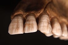
Shovel-shaped incisors are incisors whose lingual surfaces are scooped as a consequence of lingual marginal ridges, crown curvature, or basal tubercles, either alone or in combination.

Enamel hypoplasia is a defect of the teeth in which the enamel is deficient in quantity, caused by defective enamel matrix formation during enamel development, as a result of inherited and acquired systemic condition(s). It can be identified as missing tooth structure and may manifest as pits or grooves in the crown of the affected teeth, and in extreme cases, some portions of the crown of the tooth may have no enamel, exposing the dentin. It may be generalized across the dentition or localized to a few teeth. Defects are categorized by shape or location. Common categories are pit-form, plane-form, linear-form, and localised enamel hypoplasia. Hypoplastic lesions are found in areas of the teeth where the enamel was being actively formed during a systemic or local disturbance. Since the formation of enamel extends over a long period of time, defects may be confined to one well-defined area of the affected teeth. Knowledge of chronological development of deciduous and permanent teeth makes it possible to determine the approximate time at which the developmental disturbance occurred. Enamel hypoplasia varies substantially among populations and can be used to infer health and behavioural impacts from the past. Defects have also been found in a variety of non-human animals.

Odontometrics is the measurement and study of tooth size. It is used in biological anthropology and bioarchaeology to study human phenotypic variation. The rationale for use is similar to that of the study of dentition, the structure and arrangement of teeth. There are a number of features that can be observed in human teeth through the use of odontometrics.

Amelogenesis imperfecta (AI) is a congenital disorder which presents with a rare abnormal formation of the enamel or external layer of the crown of teeth, unrelated to any systemic or generalized conditions. Enamel is composed mostly of mineral, that is formed and regulated by the proteins in it. Amelogenesis imperfecta is due to the malfunction of the proteins in the enamel as a result of abnormal enamel formation via amelogenesis.

Tooth wear refers to loss of tooth substance by means other than dental caries. Tooth wear is a very common condition that occurs in approximately 97% of the population. This is a normal physiological process occurring throughout life; but with increasing lifespan of individuals and increasing retention of teeth for life, the incidence of non-carious tooth surface loss has also shown a rise. Tooth wear varies substantially between people and groups, with extreme attrition and enamel fractures common in archaeological samples, and erosion more common today.

Tooth pathology is any condition of the teeth that can be congenital or acquired. Sometimes a congenital tooth disease is called a tooth abnormality. These are among the most common diseases in humans The prevention, diagnosis, treatment and rehabilitation of these diseases are the base to the dentistry profession, in which are dentists and dental hygienists, and its sub-specialties, such as oral medicine, oral and maxillofacial surgery, and endodontics. Tooth pathology is usually separated from other types of dental issues, including enamel hypoplasia and tooth wear.

Medieval Bioarchaeology is the study of human remains recovered from medieval archaeological sites. Bioarchaeology aims to understand populations through the analysis of human skeletal remains and this application of bioarchaeology specifically aims to understand medieval populations. There is an interest in the Medieval Period when it comes to bioarchaeology, because of how differently people lived back then as opposed to now, in regards to not only their everyday life, but during times of war and famine as well. The biology and behavior of those that lived in the Medieval Period can be analyzed by understanding their health and lifestyle choices.
Cementochronology is a method for assessing age at death and determining season at death. This technique is employed as accurate indicator of age among wildlife biologists on present and archaeological populations but is increasingly used in forensic anthropology and physical anthropology.
An enamel fracture, or chip, is a complete fracture of the tooth enamel without the involvement of the dentine and pulp. A fracture occurs when a tooth contacts a hard object with enough force to break a section of enamel. Chips form with minimal plastic deformation since enamel is strong but brittle. A fracture typically occurs as an irregular break on the occlusal edge of the enamel, and is therefore different to other forms of tooth wear that leave smooth surfaces. Pulp sensibility testing is recommended to confirm pulpal health. Treatment depends on the size of the fractures. If a tooth fragment is still available, it can be bonded to the tooth. For small or minor fractures, it can be smoothed to remove rough margins and edges. For a larger or major fractures, dental composite resin can be used to mask the defective enamel for aesthetic purpose. In archaeological samples enamel fractures can give insight into the diet and behaviour of past populations.

Enamel hypoplasia can take a variety of forms, but all types are associated with a reduction of enamel formation due to disruption in ameloblast production. One of the most common types, Pitting Enamel Hypoplasia (PEH), ranges from small circular pinpricks to larger irregular depressions. Pits also vary in how they occur on a tooth surface, some forming rows and others more randomly scattered. PEH can be associated with other types of hypoplasia, but it is often the only defect observed. Causes of PEH can range from genetic conditions to environmental factors, and the frequency of occurrence varies substantially between populations and species, likely due to environmental, genetic and health differences. The most striking example of this is in Paranthropus robustus, with half of all primary molars, and a quarter of permanent molars, displaying PEH defects, thought to be caused by a specific genetic condition, amelogenesis imperfecta.

Plane-form enamel hypoplasia is often seen as the most severe type of enamel hypoplasia, and results from enamel matrix formation stopping, resulting in areas of crown with little or no dental enamel deposition. A relatively short period of severe stress can potentially lead to a very large defect. Plane-form enamel hypoplasia can be caused by a variety of factors, including severe illness/malnutrition, as well as specific conditions such as amelogenesis imperfecta and congenital syphilis. In severe cases enamel can be completely missing from areas of the crown, exposing the underlying dentine.
Mortuary archaeology is the study of human remains in their archaeological context. This is a known sub-field of bioarchaeology, which is a field that focuses on gathering important information based on the skeleton of an individual. Bioarchaeology stems from the practice of human osteology which is the anatomical study of skeletal remains. Mortuary archaeology, as well as the overarching field it resides in, aims to generate an understanding of disease, migration, health, nutrition, gender, status, and kinship among past populations. Ultimately, these topics help to produce a picture of the daily lives of past individuals. Mortuary archaeologists draw upon the humanities, as well as social and hard sciences to have a full understanding of the individual.
Mary Lewis is Professor of Bioarchaeology at the University of Reading. After completing a PhD in bioarchaeology at the University of Bradford in 1999, Lewis went on to lecture at Bournemouth University (2000–2004) before moving to the University of Reading in 2004. She conducted the first osteological study of a body which has been hanged, drawn, and quartered. Lewis has held editorial roles with the International Journal of Osteoarchaeology, International Journal of Paleopathology, and the American Journal of Biological Anthropology.
Anterior teeth are one of the most scrutinized teeth, the size and shape and color of the anterior upper teeth plays an important role in dental aesthetics and smile aesthetics. A few aesthetic anterior problems could be solved with composite restorations. For example, dental caries, tooth fracture, enamel defects and diastemas. Composite restoration can also improve aesthetic by changing shape, color, length and alignment of teeth.
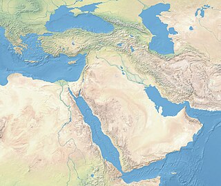
Near Eastern bioarchaeology covers the study of human skeletal remains from archaeological sites in Cyprus, Egypt, Levantine coast, Jordan, Turkey, Iran, Saudi Arabia, Qatar, Kuwait, Bahrain, United Arab Emirates, Oman, and Yemen.
The analysis of dental remains is a valuable tool to archaeologists. Teeth are hard, highly mineralised and chemically stable, so therefore preserve well and are one of the most commonly found animals remains. Analysis of these remains also yields a wealth of information. It can not only be used to determine the sex and age of the individual whose mandibular or dental remains have been found, but can also shed light on their diet, pathology, and even their geographic origins through isotope analysis.