Lipid bilayer fusion

Last updated
Illustration of lipid vesicles fusing showing two possible outcomes: hemifusion and full fusion. In hemifusion only the outer bilayer leaflets mix. In full fusion both leaflets as well as the internal contents mix. Lipid bilayer fusion.svg
Illustration of lipid vesicles fusing showing two possible outcomes: hemifusion and full fusion. In hemifusion only the outer bilayer leaflets mix. In full fusion both leaflets as well as the internal contents mix.

In membrane biology, fusion is the process by which two initially distinct lipid bilayers merge their hydrophobic cores, resulting in one interconnected structure. If this fusion proceeds completely through both leaflets of both bilayers, an aqueous bridge is formed and the internal contents of the two structures can mix. Alternatively, if only one leaflet from each bilayer is involved in the fusion process, the bilayers are said to be hemifused. In hemifusion, the lipid constituents of the outer leaflet of the two bilayers can mix, but the inner leaflets remain distinct. The aqueous contents enclosed by each bilayer also remain separated.

Contents

Fusion is involved in many cellular processes, particularly in eukaryotes since the eukaryotic cell is extensively sub-divided by lipid bilayer membranes. Exocytosis, fertilization of an egg by sperm and transport of waste products to the lysosome are a few of the many eukaryotic processes that rely on some form of fusion. Fusion is also an important mechanism for transport of lipids from their site of synthesis to the membrane where they are needed. Even the entry of pathogens can be governed by fusion, as many bilayer-coated viruses have dedicated fusion proteins to gain entry into the host cell.

Lipid mechanism

There are four fundamental steps in the fusion process, although each of these steps actually represents a complex sequence of events. [1] First, the involved membranes must aggregate, approaching each other to within several nanometers. Second, the two bilayers must come into very close contact (within a few angstroms). To achieve this close contact, the two surfaces must become at least partially dehydrated, as the bound surface water normally present causes bilayers to strongly repel at this distance. Third, a destabilization must develop at one point between the two bilayers, inducing a highly localized rearrangement of the two bilayers. Finally, as this point defect grows, the components of the two bilayers mix and diffuse away from the site of contact. Depending on whether hemifusion or full fusion occurs, the internal contents of the membranes may mix at this point as well.

Schematic illustration of the process of fusion through stalk formation. Membrane fusion via stalk formation.jpg
Schematic illustration of the process of fusion through stalk formation.

The exact mechanisms behind this complex sequence of events are still a matter of debate. To simplify the system and allow more definitive study, many experiments have been performed in vitro with synthetic lipid vesicles. These studies have shown that divalent cations play a critical role in the fusion process by binding to negatively charged lipids such as phosphatidylserine, phosphatidylglycerol and cardiolipin. [2] One role on these ions in the fusion process is to shield the negative charge on the surface of the bilayer, diminishing electrostatic repulsion and allowing the membranes to approach each other. This is clearly not the only role, however, since there is an extensively documented difference in the ability of Mg2+ versus Ca2+ to induce fusion. Although Mg2+ will induce extensive aggregation it will not induce fusion, while Ca2+ induces both. [3] It has been proposed that this discrepancy is due to a difference in extent of dehydration. Under this theory, calcium ions bind more strongly to charged lipids, but less strongly to water. The resulting displacement of calcium for water destabilizes the lipid-water interface and promotes intimate interbilayer contact. [4] A recently proposed alternative hypothesis is that the binding of calcium induces a destabilizing lateral tension. [5] Whatever the mechanism of calcium-induced fusion, the initial interaction is clearly electrostatic, since zwitterionic lipids are not susceptible to this effect. [6] [7]

In the fusion process, the lipid head group is not only involved in charge density, but can affect dehydration and defect nucleation. These effects are independent of the effects of ions. The presence of the uncharged headgroup phosphatidylethanolamine (PE) increases fusion when incorporated into a phosphatidylcholine bilayer. This phenomenon has been explained by some as a dehydration effect similar to the influence of calcium. [8] The PE headgroup binds water less tightly than PC and therefore may allow close apposition more easily. An alternate explanation is that the physical rather than chemical nature of PE may help induce fusion. According to the stalk hypothesis of fusion, a highly curved bridge must form between the two bilayers for fusion to occur. [9] Since PE has a small headgroup and readily forms inverted micelle phases it should, according to the stalk model, promote the formation of these stalks. [10] Further evidence cited in favor of this theory is the fact that certain lipid mixtures have been shown to only support fusion when raised above the transition temperature of these inverted phases. [11] [12] This topic also remains controversial, and even if there is a curved structure present in the fusion process, there is debate in the literature over whether it is a cubic, hexagonal or more exotic extended phase. [13]

Fusion proteins

Diagram of the action of SNARE proteins docking a vesicle for exocytosis. Complementary versions of the protein on the vesicle and the target membrane bind and wrap around each other, drawing the two bilayers close together in the process. Exocytosis-machinery.jpg
Diagram of the action of SNARE proteins docking a vesicle for exocytosis. Complementary versions of the protein on the vesicle and the target membrane bind and wrap around each other, drawing the two bilayers close together in the process.

The situation is further complicated when considering fusion in vivo since biological fusion is almost always regulated by the action of membrane-associated proteins. The first of these proteins to be studied were the viral fusion proteins, which allow an enveloped virus to insert its genetic material into the host cell (enveloped viruses are those surrounded by a lipid bilayer; some others have only a protein coat). Broadly, there are two classes of viral fusion proteins: acidic and pH-independent. [1] pH independent fusion proteins can function under neutral conditions and fuse with the plasma membrane, allowing viral entry into the cell. Viruses utilizing this scheme included HIV, measles and herpes. Acidic fusion proteins such as those found on influenza are only activated when in the low pH of acidic endosomes and must first be endocytosed to gain entry into the cell.

Eukaryotic cells use entirely different classes of fusion proteins, the best studied of which are the SNAREs. SNARE proteins are used to direct all vesicular intracellular trafficking. Despite years of study, much is still unknown about the function of this protein class. In fact, there is still an active debate regarding whether SNAREs are linked to early docking or participate later in the fusion process by facilitating hemifusion. [15] Even once the role of SNAREs or other specific proteins is illuminated, a unified understanding of fusion proteins is unlikely as there is an enormous diversity of structure and function within these classes, and very few themes are conserved. [16]

Fusion in laboratory practice

In studies of molecular and cellular biology it is often desirable to artificially induce fusion. Although this can be accomplished with the addition of calcium as discussed earlier, this procedure is often not feasible because calcium regulates many other biochemical processes and its addition would be a strong confound. Also, as mentioned, calcium induces massive aggregation as well as fusion. The addition of polyethylene glycol (PEG) causes fusion without significant aggregation or biochemical disruption. This procedure is now used extensively, for example by fusing B-cells with myeloma cells. [17] The resulting “hybridoma” from this combination expresses a desired antibody as determined by the B-cell involved, but is immortalized due to the myeloma component. The mechanism of PEG fusion has not been definitively identified, but some researchers believe that the PEG, by binding a large number of water molecules, effectively decreases the chemical activity of the water and thus dehydrates the lipid headgroups. [18] Fusion can also be artificially induced through electroporation in a process known as electrofusion. It is believed that this phenomenon results from the energetically active edges formed during electroporation, which can act as the local defect point to nucleate stalk growth between two bilayers. [19]

Alternatively, SNARE-inspired model systems can be used to induce membrane fusion of lipid vesicles. In those systems membrane anchored complementary DNA, [20] [21] [22] PNA, [23] peptides, [24] or other molecules [25] "zip" together and pull the membranes into proximity. Such systems could have practical applications in the future, for example in drug delivery. [26] The probably best investigated system [27] consists of coiled-coil forming peptides of complementary charge (one is typically carrying an excess of positively charged lysins and is thus termed peptide K, and one negatively charged glutamic acids called peptide E). [28] Interestingly, it was discovered that not only the coiled-coil formation between the two peptides is necessary for membrane fusion to occur, but also that the peptide K interacts with the membrane surface and cause local defects. [29]

Assays to measure membrane fusion

There are two levels of fusion: mixing of membrane lipids and mixing of contents. Assays of membrane fusion report either the mixing of membrane lipids or the mixing of the aqueous contents of the fused entities.

Assays for measuring lipid mixing

Assays evaluating lipid mixing make use of concentration dependent effects such as nonradiative energy transfer, fluorescence quenching and pyrene excimer formation.

(1.) Illustration of lipid mixing assay based on Forster resonance energy transfer. Fret.svg
(1.) Illustration of lipid mixing assay based on Förster resonance energy transfer.
(3.) Illustration of lipid mixing assay based on Fluorescence self-quenching. Self-quenching.jpg
(3.) Illustration of lipid mixing assay based on Fluorescence self-quenching.
  1. NBD-Rhodamine Energy Transfer: [30] In this method, membrane labeled with both NBD (donor) and Rhodamine (acceptor) combine with unlabeled membrane. When NBD and Rhodamine are within a certain distance, the Förster resonance energy transfer (FRET) happens. After fusion, resonance energy transfer (FRET) decreases when the average distance between probes increases, while NBD fluorescence increases.
  2. Pyrene Excimer Formation: Pyrene monomer and excimer emission wavelengths are different. The emission wavelength of monomer is around 400 nm and that of excimer is around 470 nm. In this method, membrane labeled with Pyrene combines with unlabeled membrane. Pyrene self associates in membrane and then excited pyrene excites other pyrene. Before fusion, the majority of the emission is excimer emission. After fusion, the distance between probes increases and the ratio of excimer emission decreases.[ citation needed ]
  3. Octadecyl Rhodamine B Self-Quenching: [31] This assay is based on self-quenching of octadecyl rhodamine B. Octadecyl rhodamine B self-quenching occurs when the probe is incorporated into membrane lipids at concentrations of 1–10 mole percent [32] because Rhodamine dimers quench fluorescence. In this method, membrane labeled Rhodamine combines with unlabeled membrane. Fusion with unlabeled membranes resulting in dilution of the probe, which is accompanied by increasing fluorescence. [33] [34] The major problem of this assay is spontaneous transfer.

Assays for measuring content mixing

Mixing of aqueous contents from vesicles as a result of lysis, fusion or physiological permeability can be detected fluorometrically using low molecular weight soluble tracers.

(1.) Illustration of content mixing assay based on fluorescence quencing pair ANTS/DPX. Quenching.svg
(1.) Illustration of content mixing assay based on fluorescence quencing pair ANTS/DPX.
(2.)Illustration of content mixing assay based on fluorescence enhancement pair Tb /DPA. Content mixing assay based on fluorescence enhancement pair Tb3+-DPA.svg
(2.)Illustration of content mixing assay based on fluorescence enhancement pair Tb /DPA.
  1. Fluorescence quenching assays with ANTS/DPX: [35] [36] ANTS is a polyanionic fluorophore, while DPX is a cationic quencher. The assay is based on the collisional quenching of them. Separate vesicle populations are loaded with ANTS or DPX, respectively. When content mixing happens, ANTS and DPX collide and fluorescence of ANTS monitored at 530 nm, with excitation at 360 nm is quenched. This method is performed at acidic pH and high concentration.
  2. Fluorescence enhancement assays with Tb3+/DPA: [37] [38] This method is based on the fact that chelate of Tb3+/DPA is 10,000 times more fluorescent than Tb3+ alone. In the Tb3+/DPA assay, separate vesicle populations are loaded with TbCl3 or DPA. The formation of Tb3+/DPA chelate can be used to indicate vesicle fusion. This method is good for protein free membranes.[ citation needed ]
  3. Single molecule DNA assay. [39] A DNA hairpin composed of 5 base pair stem and poly-thymidine loop that is labeled with a donor (Cy3) and an acceptor (Cy5) at the ends of the stem was encapsulated in the v-SNARE vesicle. We separately encapsulated multiple unlabeled poly-adenosine DNA strands in the t-SNARE vesicle. If the two vesicles, both ~100 nm in diameter, dock and a large enough fusion pore forms between them, the two DNA molecules should hybridize, opening up the stem region of the hairpin and switching the Förster resonance energy transfer (FRET) efficiency (E) between Cy3 and Cy5 from a high to a low value.

See also

Related Research Articles

<span class="mw-page-title-main">Vesicle (biology and chemistry)</span> Any small, fluid-filled, spherical organelle enclosed by a membrane

In cell biology, a vesicle is a structure within or outside a cell, consisting of liquid or cytoplasm enclosed by a lipid bilayer. Vesicles form naturally during the processes of secretion (exocytosis), uptake (endocytosis), and the transport of materials within the plasma membrane. Alternatively, they may be prepared artificially, in which case they are called liposomes. If there is only one phospholipid bilayer, the vesicles are called unilamellar liposomes; otherwise they are called multilamellar liposomes. The membrane enclosing the vesicle is also a lamellar phase, similar to that of the plasma membrane, and intracellular vesicles can fuse with the plasma membrane to release their contents outside the cell. Vesicles can also fuse with other organelles within the cell. A vesicle released from the cell is known as an extracellular vesicle.

<span class="mw-page-title-main">Exocytosis</span> Active transport and bulk transport in which a cell transports molecules out of the cell

Exocytosis is a form of active transport and bulk transport in which a cell transports molecules out of the cell. As an active transport mechanism, exocytosis requires the use of energy to transport material. Exocytosis and its counterpart, endocytosis, are used by all cells because most chemical substances important to them are large polar molecules that cannot pass through the hydrophobic portion of the cell membrane by passive means. Exocytosis is the process by which a large amount of molecules are released; thus it is a form of bulk transport. Exocytosis occurs via secretory portals at the cell plasma membrane called porosomes. Porosomes are permanent cup-shaped lipoprotein structure at the cell plasma membrane, where secretory vesicles transiently dock and fuse to release intra-vesicular contents from the cell.

<span class="mw-page-title-main">Pardaxin</span> Type of peptide

Pardaxin is a peptide produced by the Red Sea sole and the Pacific Peacock sole that is used as a shark repellent. It causes lysis of mammalian and bacterial cells, similar to melittin.

<span class="mw-page-title-main">Lipid bilayer</span> Membrane of two layers of lipid molecules

The lipid bilayer is a thin polar membrane made of two layers of lipid molecules. These membranes are flat sheets that form a continuous barrier around all cells. The cell membranes of almost all organisms and many viruses are made of a lipid bilayer, as are the nuclear membrane surrounding the cell nucleus, and membranes of the membrane-bound organelles in the cell. The lipid bilayer is the barrier that keeps ions, proteins and other molecules where they are needed and prevents them from diffusing into areas where they should not be. Lipid bilayers are ideally suited to this role, even though they are only a few nanometers in width, because they are impermeable to most water-soluble (hydrophilic) molecules. Bilayers are particularly impermeable to ions, which allows cells to regulate salt concentrations and pH by transporting ions across their membranes using proteins called ion pumps.

<span class="mw-page-title-main">Liposome</span> Composite structures made of phospholipids and may contain small amounts of other molecules

A liposome is a small artificial vesicle, spherical in shape, having at least one lipid bilayer. Due to their hydrophobicity and/or hydrophilicity, biocompatibility, particle size and many other properties, liposomes can be used as drug delivery vehicles for administration of pharmaceutical drugs and nutrients, such as lipid nanoparticles in mRNA vaccines, and DNA vaccines. Liposomes can be prepared by disrupting biological membranes.

<span class="mw-page-title-main">Synaptic vesicle</span> Neurotransmitters that are released at the synapse

In a neuron, synaptic vesicles store various neurotransmitters that are released at the synapse. The release is regulated by a voltage-dependent calcium channel. Vesicles are essential for propagating nerve impulses between neurons and are constantly recreated by the cell. The area in the axon that holds groups of vesicles is an axon terminal or "terminal bouton". Up to 130 vesicles can be released per bouton over a ten-minute period of stimulation at 0.2 Hz. In the visual cortex of the human brain, synaptic vesicles have an average diameter of 39.5 nanometers (nm) with a standard deviation of 5.1 nm.

Phosphatidic acids are anionic phospholipids important to cell signaling and direct activation of lipid-gated ion channels. Hydrolysis of phosphatidic acid gives rise to one molecule each of glycerol and phosphoric acid and two molecules of fatty acids. They constitute about 0.25% of phospholipids in the bilayer.

<span class="mw-page-title-main">Annexin</span> Protein family

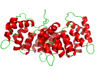
Annexin is a common name for a group of cellular proteins. They are mostly found in eukaryotic organisms.

<span class="mw-page-title-main">SNARE protein</span> Protein family

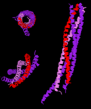
SNARE proteins – "SNAPREceptors" – are a large protein family consisting of at least 24 members in yeasts, more than 60 members in mammalian cells, and some numbers in plants. The primary role of SNARE proteins is to mediate the fusion of vesicles with the target membrane; this notably mediates exocytosis, but can also mediate the fusion of vesicles with membrane-bound compartments. The best studied SNAREs are those that mediate the release of synaptic vesicles containing neurotransmitters in neurons. These neuronal SNAREs are the targets of the neurotoxins responsible for botulism and tetanus produced by certain bacteria.

<span class="mw-page-title-main">Synaptobrevin</span>

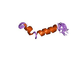
Synaptobrevins are small integral membrane proteins of secretory vesicles with molecular weight of 18 kilodalton (kDa) that are part of the vesicle-associated membrane protein (VAMP) family.

<span class="mw-page-title-main">Cationic liposome</span>

Cationic liposomes are spherical structures that contain positively charged lipids. Cationic liposomes can vary in size between 40 nm and 500 nm, and they can either have one lipid bilayer (monolamellar) or multiple lipid bilayers (multilamellar). The positive charge of the phospholipids allows cationic liposomes to form complexes with negatively charged nucleic acids through ionic interactions. Upon interacting with nucleic acids, cationic liposomes form clusters of aggregated vesicles. These interactions allow cationic liposomes to condense and encapsulate various therapeutic and diagnostic agents in their aqueous compartment or in their lipid bilayer. These cationic liposome-nucleic acid complexes are also referred to as lipoplexes. Due to the overall positive charge of cationic liposomes, they interact with negatively charged cell membranes more readily than classic liposomes. This positive charge can also create some issues in vivo, such as binding to plasma proteins in the bloodstream, which leads to opsonization. These issues can be reduced by optimizing the physical and chemical properties of cationic liposomes through their lipid composition. Cationic liposomes are increasingly being researched for use as delivery vectors in gene therapy due to their capability to efficiently transfect cells. A common application for cationic liposomes is cancer drug delivery.

Calcein, also known as fluorexon, fluorescein complex, is a fluorescent dye with excitation and emission wavelengths of 495 and 515 nm, respectively, and has the appearance of orange crystals. Calcein self-quenches at concentrations above 70 mM and is commonly used as an indicator of lipid vesicle leakage. It has also been traditionally used as a complexometric indicator for titration of calcium ions with EDTA, and for fluorometric determination of calcium.

<span class="mw-page-title-main">STX6</span> Protein-coding gene in the species Homo sapiens

Syntaxin-6 is a protein that in humans is encoded by the STX6 gene.

Protein–lipid interaction is the influence of membrane proteins on the lipid physical state or vice versa.

<span class="mw-page-title-main">Axon terminal</span> Nerve fiber part

Axon terminals are distal terminations of the branches of an axon. An axon, also called a nerve fiber, is a long, slender projection of a nerve cell that conducts electrical impulses called action potentials away from the neuron's cell body in order to transmit those impulses to other neurons, muscle cells or glands. In the central nervous system, most presynaptic terminals are actually formed along the axons, not at their ends.

A model lipid bilayer is any bilayer assembled in vitro, as opposed to the bilayer of natural cell membranes or covering various sub-cellular structures like the nucleus. They are used to study the fundamental properties of biological membranes in a simplified and well-controlled environment, and increasingly in bottom-up synthetic biology for the construction of artificial cells. A model bilayer can be made with either synthetic or natural lipids. The simplest model systems contain only a single pure synthetic lipid. More physiologically relevant model bilayers can be made with mixtures of several synthetic or natural lipids.

Vesicle fusion is the merging of a vesicle with other vesicles or a part of a cell membrane. In the latter case, it is the end stage of secretion from secretory vesicles, where their contents are expelled from the cell through exocytosis. Vesicles can also fuse with other target cell compartments, such as a lysosome. Exocytosis occurs when secretory vesicles transiently dock and fuse at the base of cup-shaped structures at the cell plasma membrane called porosome, the universal secretory machinery in cells. Vesicle fusion may depend on SNARE proteins in the presence of increased intracellular calcium (Ca2+) concentration.

<span class="mw-page-title-main">Cell membrane</span> Biological membrane that separates the interior of a cell from its outside environment

The cell membrane is a biological membrane that separates and protects the interior of a cell from the outside environment. The cell membrane consists of a lipid bilayer, made up of two layers of phospholipids with cholesterols interspersed between them, maintaining appropriate membrane fluidity at various temperatures. The membrane also contains membrane proteins, including integral proteins that span the membrane and serve as membrane transporters, and peripheral proteins that loosely attach to the outer (peripheral) side of the cell membrane, acting as enzymes to facilitate interaction with the cell's environment. Glycolipids embedded in the outer lipid layer serve a similar purpose. The cell membrane controls the movement of substances in and out of a cell, being selectively permeable to ions and organic molecules. In addition, cell membranes are involved in a variety of cellular processes such as cell adhesion, ion conductivity, and cell signalling and serve as the attachment surface for several extracellular structures, including the cell wall and the carbohydrate layer called the glycocalyx, as well as the intracellular network of protein fibers called the cytoskeleton. In the field of synthetic biology, cell membranes can be artificially reassembled.

Membrane vesicle trafficking in eukaryotic animal cells involves movement of biochemical signal molecules from synthesis-and-packaging locations in the Golgi body to specific release locations on the inside of the plasma membrane of the secretory cell. It takes place in the form of Golgi membrane-bound micro-sized vesicles, termed membrane vesicles (MVs).

A unilamellar liposome is a spherical liposome, a vesicle, bounded by a single bilayer of an amphiphilic lipid or a mixture of such lipids, containing aqueous solution inside the chamber. Unilamellar liposomes are used to study biological systems and to mimic cell membranes, and are classified into three groups based on their size: small unilamellar liposomes/vesicles (SUVs) that with a size range of 20–100 nm, large unilamellar liposomes/vesicles (LUVs) with a size range of 100–1000 nm and giant unilamellar liposomes/vesicles (GUVs) with a size range of 1–200 µm. GUVs are mostly used as models for biological membranes in research work. Animal cells are 10–30 µm and plant cells are typically 10–100 µm. Even smaller cell organelles such as mitochondria are typically 1–2 µm. Therefore, a proper model should account for the size of the specimen being studied. In addition, the size of vesicles dictates their membrane curvature which is an important factor in studying fusion proteins. SUVs have a higher membrane curvature and vesicles with high membrane curvature can promote membrane fusion faster than vesicles with lower membrane curvature such as GUVs.

References

  1. 1 2 Yeagle, P. L. (1993). The Membranes of Cells (2nd ed.). San Diego: Academic Press. ISBN   9780127690414.[ page needed ]
  2. Papahadjopoulos, Demetrios; Nir, Shlomo; Düzgünes, Nejat (1990). "Molecular mechanisms of calcium-induced membrane fusion". Journal of Bioenergetics and Biomembranes. 22 (2): 157–79. doi:10.1007/BF00762944. PMID   2139437. S2CID   1465571.
  3. Leventis, Rania; Gagne, Jeannine; Fuller, Nola; Rand, R.; Silvius, J. (1986). "Divalent Cation Induced Fusion and Lipid Lateral Segregation in Phosphatidylcholine-Phosphatidic Acid Vesicles". Biochemistry. 25 (22): 6978–87. doi:10.1021/bi00370a600. PMID   3801406.
  4. Wilschut, Jan; Duezguenes, Nejat; Papahadjopoulos, Demetrios (1981). "Calcium/magnesium specificity in membrane fusion: Kinetics of aggregation and fusion of phosphatidylserine vesicles and the role of bilayer curvature". Biochemistry. 20 (11): 3126–33. doi:10.1021/bi00514a022. PMID   7248275.
  5. Chanturiya, A; Scaria, P; Woodle, MC (2000). "The Role of Membrane Lateral Tension in Calcium-Induced Membrane Fusion". The Journal of Membrane Biology. 176 (1): 67–75. doi:10.1007/s00232001076. PMID   10882429. S2CID   2209769.
  6. Pannuzzo, Martina; Jong, De; Djurre, H.; Raudino, Antonio; Marrink Siewert, J. (2014). "Simulation of polyethylene glycol and calcium-mediated membrane fusion" (PDF). J. Chem. Phys. 140 (12): 124905. Bibcode:2014JChPh.140l4905P. doi:10.1063/1.4869176. hdl: 11370/78e66c44-d236-4336-9b4a-151d52ffa961 . PMID   24697479. S2CID   7548382.
  7. Papahadjopoulos, D.; Poste, G.; Schaeffer, B.E.; Vail, W.J. (1974). "Membrane fusion and molecular segregation in phospholipid vesicles". Biochimica et Biophysica Acta (BBA) - Biomembranes. 352 (1): 10–28. doi:10.1016/0005-2736(74)90175-8. PMID   4859411.
  8. Düzgünes, Nejat; Wilschut, Jan; Fraley, Robert; Papahadjopoulos, Demetrios (1981). "Studies on the mechanism of membrane fusion. Role of head-group composition in calcium- and magnesium-induced fusion of mixed phospholipid vesicles". Biochimica et Biophysica Acta (BBA) - Biomembranes. 642 (1): 182–95. doi:10.1016/0005-2736(81)90148-6. PMID   7225377.
  9. Markin, VS; Kozlov, MM; Borovjagin, VL (1984). "On the theory of membrane fusion. The stalk mechanism" (PDF). General Physiology and Biophysics. 3 (5): 361–77. PMID   6510702.
  10. Chernomordik, Leonid V.; Kozlov, Michael M. (2003). "Protein-lipid interplay in fusion and fission of biological membranes". Annual Review of Biochemistry. 72: 175–207. doi:10.1146/annurev.biochem.72.121801.161504. PMID   14527322.
  11. Nir, S.; Bentz, J.; Wilschut, J.; Duzgunes, N. (1983). "Aggregation and fusion of phospholipid vesicles". Progress in Surface Science. 13 (1): 1–124. Bibcode:1983PrSS...13....1N. doi:10.1016/0079-6816(83)90010-2.
  12. Ellens, Harma; Bentz, Joe; Szoka, Francis C. (1986). "Fusion of phosphatidylethanolamine-containing liposomes and mechanism of L.alpha.-HII phase transition". Biochemistry. 25 (14): 4141–7. doi:10.1021/bi00362a023. PMID   3741846.
  13. Holopainen, Juha M.; Lehtonen, Jukka Y.A.; Kinnunen, Paavo K.J. (1999). "Evidence for the Extended Phospholipid Conformation in Membrane Fusion and Hemifusion". Biophysical Journal. 76 (4): 2111–20. Bibcode:1999BpJ....76.2111H. doi:10.1016/S0006-3495(99)77367-4. PMC   1300184 . PMID   10096906.
  14. Georgiev, Danko D.; Glazebrook, James F. (2007). "Subneuronal processing of information by solitary waves and stochastic processes". In Lyshevski, Sergey Edward (ed.). Nano and Molecular Electronics Handbook (PDF). Nano and Microengineering Series. CRC Press. pp. 17–1–17–41. doi:10.1201/9781315221670. ISBN   978-0-8493-8528-5.
  15. Chen, Yu A.; Scheller, Richard H. (2001). "SNARE-mediated membrane fusion". Nature Reviews Molecular Cell Biology. 2 (2): 98–106. doi:10.1038/35052017. PMID   11252968. S2CID   205012830.
  16. White, J M (1990). "Viral and Cellular Membrane Fusion Proteins". Annual Review of Physiology. 52: 675–97. doi:10.1146/annurev.ph.52.030190.003331. PMID   2184772.
  17. Köhler, G.; Milstein, C. (1975). "Continuous cultures of fused cells secreting antibody of predefined specificity". Nature. 256 (5517): 495–7. Bibcode:1975Natur.256..495K. doi:10.1038/256495a0. PMID   1172191. S2CID   4161444.
  18. Lentz, Barry R. (1994). "Polymer-induced membrane fusion: Potential mechanism and relation to cell fusion events". Chemistry and Physics of Lipids. 73 (1–2): 91–106. doi:10.1016/0009-3084(94)90176-7. PMID   8001186.
  19. Jordan, C. A.; Neumann, E.; Sowers, A. E., eds. (1989). Electroporation and Electrofusion in Cell Biology. Springer. ISBN   978-0-306-43043-5.[ page needed ]
  20. Malle, M.G.; Löffler, P.M.G; Bohr, S.SR.; et al. (4 April 2022). "Single-particle combinatorial multiplexed liposome fusion mediated by DNA". Nat. Chem. 14 (5): 558–565. Bibcode:2022NatCh..14..558M. doi:10.1038/s41557-022-00912-5. PMID   35379901. S2CID   247942781.
  21. Löffler, Philipp M. G.; Ries, Oliver; Rabe, Alexander; Okholm, Anders H.; Thomsen, Rasmus P.; Kjems, Jørgen; Vogel, Stefan (2017). "A DNA-Programmed Liposome Fusion Cascade". Angewandte Chemie International Edition. 56 (43): 13228–13231. doi:10.1002/anie.201703243. ISSN   1521-3773. PMID   28598002. S2CID   205401773.
  22. Stengel, Gudrun; Zahn, Raphael; Höök, Fredrik (2007-08-01). "DNA-Induced Programmable Fusion of Phospholipid Vesicles". Journal of the American Chemical Society. 129 (31): 9584–9585. doi:10.1021/ja073200k. ISSN   0002-7863. PMID   17629277.
  23. Lygina, Antonina S.; Meyenberg, Karsten; Jahn, Reinhard; Diederichsen, Ulf (2011). "Transmembrane Domain Peptide/Peptide Nucleic Acid Hybrid as a Model of a SNARE Protein in Vesicle Fusion". Angewandte Chemie International Edition. 50 (37): 8597–8601. doi:10.1002/anie.201101951. hdl: 11858/00-001M-0000-000F-41FF-2 . ISSN   1521-3773. PMID   21786370.
  24. Litowski, J. R.; Hodges, R. S. (2002-10-04). "Designing Heterodimeric Two-stranded α-Helical Coiled-coils: EFFECTS OF HYDROPHOBICITY AND α-HELICAL PROPENSITY ON PROTEIN FOLDING, STABILITY, AND SPECIFICITY". Journal of Biological Chemistry. 277 (40): 37272–37279. doi: 10.1074/jbc.M204257200 . ISSN   0021-9258. PMID   12138097. S2CID   84379119.
  25. Kumar, Pawan; Guha, Samit; Diederichsen, Ulf (2015). "SNARE protein analog-mediated membrane fusion". Journal of Peptide Science. 21 (8): 621–629. doi:10.1002/psc.2773. ISSN   1099-1387. PMID   25858776. S2CID   23718905.
  26. Yang, Jian; Bahreman, Azadeh; Daudey, Geert; Bussmann, Jeroen; Olsthoorn, René C. L.; Kros, Alexander (2016-09-28). "Drug Delivery via Cell Membrane Fusion Using Lipopeptide Modified Liposomes". ACS Central Science. 2 (9): 621–630. doi:10.1021/acscentsci.6b00172. ISSN   2374-7943. PMC   5043431 . PMID   27725960.
  27. Robson Marsden, Hana; Elbers, Nina A.; Bomans, Paul H. H.; Sommerdijk, Nico A. J. M.; Kros, Alexander (2009). "A Reduced SNARE Model for Membrane Fusion". Angewandte Chemie International Edition. 48 (13): 2330–2333. doi:10.1002/anie.200804493. ISSN   1521-3773. PMID   19222065.
  28. Lindhout, Darrin A.; Litowski, Jennifer R.; Mercier, Pascal; Hodges, Robert S.; Sykes, Brian D. (2004). "NMR solution structure of a highly stable de novo heterodimeric coiled-coil". Biopolymers. 75 (5): 367–375. doi:10.1002/bip.20150. ISSN   1097-0282. PMID   15457434. S2CID   28856028.
  29. Rabe, Martin; Aisenbrey, Christopher; Pluhackova, Kristyna; De Wert, Vincent; Boyle, Aimee L.; Bruggeman, Didjay F.; Kirsch, Sonja A.; Böckmann, Rainer A.; Kros, Alexander; Raap, Jan; Bechinger, Burkhard (2016-11-15). "A Coiled-Coil Peptide Shaping Lipid Bilayers upon Fusion". Biophysical Journal. 111 (10): 2162–2175. Bibcode:2016BpJ...111.2162R. doi:10.1016/j.bpj.2016.10.010. ISSN   0006-3495. PMC   5113151 . PMID   27851940.
  30. Struck, Douglas K.; Hoekstra, Dick; Pagano, Richard E. (1981). "Use of resonance energy transfer to monitor membrane fusion". Biochemistry. 20 (14): 4093–9. doi:10.1021/bi00517a023. PMID   7284312.
  31. Hoekstra, Dick; De Boer, Tiny; Klappe, Karin; Wilschut, Jan (1984). "Fluorescence method for measuring the kinetics of fusion between biological membranes". Biochemistry. 23 (24): 5675–81. doi:10.1021/bi00319a002. PMID   6098295.
  32. MacDonald, Ruby I (1990). "Characteristics of self-quenching of the fluorescence of lipid-conjugated rhodamine in membranes". The Journal of Biological Chemistry. 265 (23): 13533–9. doi: 10.1016/S0021-9258(18)77380-8 . PMID   2380172.
  33. Rubin, R.J.; Chen, Y.D. (1990). "Diffusion and redistribution of lipid-like molecules between membranes in virus-cell and cell-cell fusion systems". Biophysical Journal. 58 (5): 1157–67. Bibcode:1990BpJ....58.1157R. doi:10.1016/S0006-3495(90)82457-7. PMC   1281061 . PMID   2291940.
  34. Chen, Y.D.; Rubin, R.J.; Szabo, A. (1993). "Fluorescence dequenching kinetics of single cell-cell fusion complexes". Biophysical Journal. 65 (1): 325–33. Bibcode:1993BpJ....65..325C. doi:10.1016/S0006-3495(93)81076-2. PMC   1225727 . PMID   8369440.
  35. Smolarsky, Moshe; Teitelbaum, Dvora; Sela, Michael; Gitler, Carlos (1977). "A simple fluorescent method to determine complement-mediated liposome immune lysis". Journal of Immunological Methods. 15 (3): 255–65. doi:10.1016/0022-1759(77)90063-1. PMID   323363.
  36. Ellens, H; Bentz, J; Szoka, FC (1985). "H+- and Ca2+-induced fusion and destabilization of liposomes". Biochemistry. 24 (13): 3099–106. doi:10.1021/bi00334a005. PMID   4027232.
  37. Wilschut, Jan; Papahadjopoulos, Demetrios (1979). "Ca2+-induced fusion of phospholipid vesicles monitored by mixing of aqueous contents". Nature. 281 (5733): 690–2. Bibcode:1979Natur.281..690W. doi:10.1038/281690a0. PMID   551288. S2CID   4353081.
  38. Wilschut, Jan; Duzgunes, Nejat; Fraley, Robert; Papahadjopoulos, Demetrios (1980). "Studies on the mechanism of membrane fusion: Kinetics of calcium ion induced fusion of phosphatidylserine vesicles followed by a new assay for mixing of aqueous vesicle contents". Biochemistry. 19 (26): 6011–21. doi:10.1021/bi00567a011. PMID   7470445.
  39. Diao, Jiajie; Su, Zengliu; Ishitsuka, Yuji; Lu, Bin; Lee, Kyung Suk; Lai, Ying; Shin, Yeon-Kyun; Ha, Taekjip (2010). "A single-vesicle content mixing assay for SNARE-mediated membrane fusion". Nature Communications. 1 (5): 1–6. Bibcode:2010NatCo...1E..54D. doi:10.1038/ncomms1054. PMC   3518844 . PMID   20975723.