Related Research Articles
Statistical parametric mapping (SPM) is a statistical technique for examining differences in brain activity recorded during functional neuroimaging experiments. It was created by Karl Friston. It may alternatively refer to software created by the Wellcome Department of Imaging Neuroscience at University College London to carry out such analyses.
Functional integration is the study of how brain regions work together to process information and effect responses. Though functional integration frequently relies on anatomic knowledge of the connections between brain areas, the emphasis is on how large clusters of neurons – numbering in the thousands or millions – fire together under various stimuli. The large datasets required for such a whole-scale picture of brain function have motivated the development of several novel and general methods for the statistical analysis of interdependence, such as dynamic causal modelling and statistical linear parametric mapping. These datasets are typically gathered in human subjects by non-invasive methods such as EEG/MEG, fMRI, or PET. The results can be of clinical value by helping to identify the regions responsible for psychiatric disorders, as well as to assess how different activities or lifestyles affect the functioning of the brain.

Analysis of Functional NeuroImages (AFNI) is an open-source environment for processing and displaying functional MRI data—a technique for mapping human brain activity.
Brain mapping is a set of neuroscience techniques predicated on the mapping of (biological) quantities or properties onto spatial representations of the brain resulting in maps.
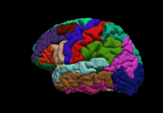
FreeSurfer is a brain imaging software package originally developed by Bruce Fischl, Anders Dale, Martin Sereno, and Doug Greve. Development and maintenance of FreeSurfer is now the primary responsibility of the Laboratory for Computational Neuroimaging at the Athinoula A. Martinos Center for Biomedical Imaging. FreeSurfer contains a set of programs with a common focus of analyzing magnetic resonance imaging (MRI) scans of brain tissue. It is an important tool in functional brain mapping and contains tools to conduct both volume based and surface based analysis. FreeSurfer includes tools for the reconstruction of topologically correct and geometrically accurate models of both the gray/white and pial surfaces, for measuring cortical thickness, surface area and folding, and for computing inter-subject registration based on the pattern of cortical folds.

Talairach coordinates, also known as Talairach space, is a 3-dimensional coordinate system of the human brain, which is used to map the location of brain structures independent from individual differences in the size and overall shape of the brain. It is still common to use Talairach coordinates in functional brain imaging studies and to target transcranial stimulation of brain regions. However, alternative methods such as the MNI Coordinate System have largely replaced Talairach for stereotaxy and other procedures.

The FMRIB Software Library, abbreviated FSL, is a software library containing image analysis and statistical tools for functional, structural and diffusion MRI brain imaging data.
Cambridge Brain Analysis (CamBA), is a software repository developed at the Brain Mapping Unit, Department of Psychiatry, University of Cambridge, UK and contains software pipelines for functional magnetic resonance imaging (fMRI) analysis. It is designed for batch processing and its main graphical user interface offers a spreadsheet-like look-and-feel.

3D Slicer (Slicer) is a free and open source software package for image analysis and scientific visualization. Slicer is used in a variety of medical applications, including autism, multiple sclerosis, systemic lupus erythematosus, prostate cancer, lung cancer, breast cancer, schizophrenia, orthopedic biomechanics, COPD, cardiovascular disease and neurosurgery.
Psychophysiological interaction (PPI) is a brain connectivity analysis method for functional brain imaging data, mainly functional magnetic resonance imaging (fMRI). It estimates context-dependent changes in effective connectivity (coupling) between brain regions. Thus, PPI analysis identifies brain regions whose activity depends on an interaction between psychological context and physiological state of the seed region.
The Human Connectome Project (HCP) is a five-year project sponsored by sixteen components of the National Institutes of Health, split between two consortia of research institutions. The project was launched in July 2009 as the first of three Grand Challenges of the NIH's Blueprint for Neuroscience Research. On September 15, 2010, the NIH announced that it would award two grants: $30 million over five years to a consortium led by Washington University in St. Louis and the University of Minnesota, with strong contributions from Oxford University (FMRIB) and $8.5 million over three years to a consortium led by Harvard University, Massachusetts General Hospital and the University of California Los Angeles.
Medical image computing (MIC) is an interdisciplinary field at the intersection of computer science, information engineering, electrical engineering, physics, mathematics and medicine. This field develops computational and mathematical methods for solving problems pertaining to medical images and their use for biomedical research and clinical care.

Resting state fMRI is a method of functional magnetic resonance imaging (fMRI) that is used in brain mapping to evaluate regional interactions that occur in a resting or task-negative state, when an explicit task is not being performed. A number of resting-state brain networks have been identified, one of which is the default mode network. These brain networks are observed through changes in blood flow in the brain which creates what is referred to as a blood-oxygen-level dependent (BOLD) signal that can be measured using fMRI.
The following outline is provided as an overview of and topical guide to brain mapping:

The Neuroimaging Tools and Resources Collaboratory is a neuroimaging informatics knowledge environment for MR, PET/SPECT, CT, EEG/MEG, optical imaging, clinical neuroinformatics, imaging genomics, and computational neuroscience tools and resources.

CONN is a Matlab-based cross-platform imaging software for the computation, display, and analysis of functional connectivity in fMRI in the resting state and during task.
Spinal Cord Toolbox (SCT) is a suite of analysis tools optimized for spinal cord images acquired with magnetic resonance imaging. Main features include segmentation, registration and calculation of anatomical metrics.
Dynamic causal modeling (DCM) is a framework for specifying models, fitting them to data and comparing their evidence using Bayesian model comparison. It uses nonlinear state-space models in continuous time, specified using stochastic or ordinary differential equations. DCM was initially developed for testing hypotheses about neural dynamics. In this setting, differential equations describe the interaction of neural populations, which directly or indirectly give rise to functional neuroimaging data e.g., functional magnetic resonance imaging (fMRI), magnetoencephalography (MEG) or electroencephalography (EEG). Parameters in these models quantify the directed influences or effective connectivity among neuronal populations, which are estimated from the data using Bayesian statistical methods.
Brain–body interactions are patterns of neural activity in the central nervous system to coordinate the activity between the brain and body. The nervous system consists of central and peripheral nervous systems and coordinates the actions of an animal by transmitting signals to and from different parts of its body. The brain and spinal cord are interwoven with the body and interact with other organ systems through the somatic, autonomic and enteric nervous systems. Neural pathways regulate brain–body interactions and allow to sense and control its body and interact with the environment.
References
- ↑ Garyfallidis, Eleftherios; Brett, Matthew; Amirbekian, Bagrat; Rokem, Ariel; van der Walt, Stefan; Descoteaux, Maxime; Nimmo-Smith, Ian; Dipy, Contributors (2014). "Dipy, a library for the analysis of diffusion MRI data". Frontiers in Neuroinformatics. 8: 8. doi: 10.3389/fninf.2014.00008 . ISSN 1662-5196. PMC 3931231 . PMID 24600385.
{{cite journal}}:|first8=has generic name (help) - ↑ Rebsamen, Michael; Rummel, Christian; Reyes, Mauricio; Wiest, Roland; McKinley, Richard (December 2020). "Direct cortical thickness estimation using deep learning‐based anatomy segmentation and cortex parcellation". Human Brain Mapping. 41 (17): 4804–4814. doi:10.1002/hbm.25159. PMC 7643371 . PMID 32786059.
- ↑ Sadigh-Eteghad S, Majdi A, Farhoudi M, Talebi M, Mahmoudi J (2014). "Different patterns of brain activation in normal aging and Alzheimer's disease from cognitional sight: meta analysis using activation likelihood estimation". Journal of the Neurological Sciences. 343 (1): 159–66. doi:10.1016/j.jns.2014.05.066. PMID 24950901. S2CID 24359894.
- ↑ Cohen-Adad J, De Leener B, Benhamou M, Cadotte D, Fleet D, Cadotte A, Fehlings MG, Pelletier Paquette JP, Thong W, Taso M, Collins DL, Callot V, Fonov V. Spinal Cord Toolbox: an open-source framework for processing spinal cord MRI data. Proceedings of the 20th Annual Meeting of OHBM, Hamburg, Germany 2014:3633