Related Research Articles

The macula (/ˈmakjʊlə/) or macula lutea is an oval-shaped pigmented area in the center of the retina of the human eye and in other animals. The macula in humans has a diameter of around 5.5 mm (0.22 in) and is subdivided into the umbo, foveola, foveal avascular zone, fovea, parafovea, and perifovea areas.

The occipital lobe is one of the four major lobes of the cerebral cortex in the brain of mammals. The name derives from its position at the back of the head, from the Latin ob, "behind," and caput, "the head."
In neuroanatomy, the optic radiation are axons from the neurons in the lateral geniculate nucleus to the primary visual cortex. The optic radiation receives blood through deep branches of the middle cerebral artery and posterior cerebral artery.

A scotoma is an area of partial alteration in the field of vision consisting of a partially diminished or entirely degenerated visual acuity that is surrounded by a field of normal – or relatively well-preserved – vision.
The visual field is the "spatial array of visual sensations available to observation in introspectionist psychological experiments". Or simply, visual field can be defined as the entire area that can be seen when an eye is fixed straight at a point.
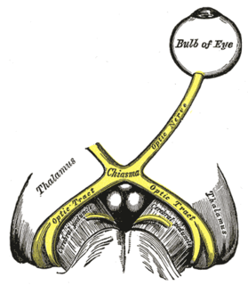
In neuroanatomy, the optic tract is a part of the visual system in the brain. It is a continuation of the optic nerve that relays information from the optic chiasm to the ipsilateral lateral geniculate nucleus (LGN), pretectal nuclei, and superior colliculus.

Bitemporal hemianopsia, is the medical description of a type of partial blindness where vision is missing in the outer half of both the right and left visual field. It is usually associated with lesions of the optic chiasm, the area where the optic nerves from the right and left eyes cross near the pituitary gland.

The Amsler grid, used since 1945, is a grid of horizontal and vertical lines used to monitor a person's central visual field. The grid was developed by Marc Amsler, a Swiss ophthalmologist. It is a diagnostic tool that aids in the detection of visual disturbances caused by changes in the retina, particularly the macula, as well as the optic nerve and the visual pathway to the brain. Amsler grid usually help detecting defects in central 20 degrees of the visual field.

The middle cerebral artery (MCA) is one of the three major paired cerebral arteries that supply blood to the cerebrum. The MCA arises from the internal carotid artery and continues into the lateral sulcus where it then branches and projects to many parts of the lateral cerebral cortex. It also supplies blood to the anterior temporal lobes and the insular cortices.
Cortical blindness is the total or partial loss of vision in a normal-appearing eye caused by damage to the brain's occipital cortex. Cortical blindness can be acquired or congenital, and may also be transient in certain instances. Acquired cortical blindness is most often caused by loss of blood flow to the occipital cortex from either unilateral or bilateral posterior cerebral artery blockage and by cardiac surgery. In most cases, the complete loss of vision is not permanent and the patient may recover some of their vision. Congenital cortical blindness is most often caused by perinatal ischemic stroke, encephalitis, and meningitis. Rarely, a patient with acquired cortical blindness may have little or no insight that they have lost vision, a phenomenon known as Anton–Babinski syndrome.
The posterior cerebral artery (PCA) is one of a pair of cerebral arteries that supply oxygenated blood to the occipital lobe, part of the back of the human brain. The two arteries originate from the distal end of the basilar artery, where it bifurcates into the left and right posterior cerebral arteries. These anastomose with the middle cerebral arteries and internal carotid arteries via the posterior communicating arteries.

Hemianopsia, or hemianopia, is a loss of vision or blindness (anopsia) in half the visual field, usually on one side of the vertical midline. The most common causes of this damage are stroke, brain tumor, and trauma.
Optic neuropathy is damage to the optic nerve from any cause. The optic nerve is a bundle of millions of fibers in the retina that sends visual signals to the brain. [1].

Hemianopsia, or hemianopia, is a visual field loss on the left or right side of the vertical midline. It can affect one eye but usually affects both eyes.

Chiasmal syndrome is the set of signs and symptoms that are associated with lesions of the optic chiasm, manifesting as various impairments of the affected's visual field according to the location of the lesion along the optic nerve. Pituitary adenomas are the most common cause; however, chiasmal syndrome may be caused by cancer, or associated with other medical conditions such as multiple sclerosis and neurofibromatosis.

Quadrantanopia,quadrantanopsia, refers to an anopia affecting a quarter of the visual field.
Posterior cerebral artery syndrome is a condition whereby the blood supply from the posterior cerebral artery (PCA) is restricted, leading to a reduction of the function of the portions of the brain supplied by that vessel: the occipital lobe, the inferomedial temporal lobe, a large portion of the thalamus, and the upper brainstem and midbrain.
Middle cerebral artery syndrome is a condition whereby the blood supply from the middle cerebral artery (MCA) is restricted, leading to a reduction of the function of the portions of the brain supplied by that vessel: the lateral aspects of frontal, temporal and parietal lobes, the corona radiata, globus pallidus, caudate and putamen. The MCA is the most common site for the occurrence of ischemic stroke.

Bonnet–Dechaume–Blanc syndrome, also known as Wyburn-Mason syndrome, is a rare congenital disorder characterized by arteriovenous malformations of the brain, retina or facial nevi. The syndrome has a number of possible symptoms and can, more rarely, affect the skin, bones, kidneys, muscles, and gastrointestinal tract. When the syndrome affects the brain, people can experience severe headaches, seizures, acute stroke, meningism, and progressive neurological deficits due to acute or chronic ischaemia caused by arteriovenous shunting.

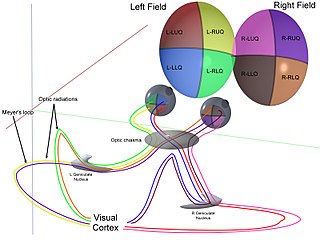
The visual pathway consists of structures that carry visual information from the retina to the brain. Lesions in that pathway cause a variety of visual field defects. In the visual system of human eye, the visual information processed by retinal photoreceptor cells travel in the following way:
Retina→Optic nerve→Optic chiasm →Optic tract→Lateral geniculate nucleus→Optic radiation→Primary and secondary visual cortices.
References
- 1 2 3 4 Whishaw, I. Q., & Kolb, B. (2015). Fundamentals of Human Neuropsychology (7th ed.). New York, NY: Worth Custom Publishing.
- 1 2 Windsor, R. L. (n.d.). Visual Fields in Brain Injury - Hemianopsia.net Everything you need to know about Hemianopsia. Retrieved from http://www.hemianopsia.net/visual-fields-in-brain-injury/
- ↑ Carroll, J. N., & Johnson, C. A. (2013, August 22). Visual Field Testing: From One Medical Student to Another. Retrieved from http://eyerounds.org/tutorials/VF-testing/
- ↑ Remington, L.A. (2012). Clinical Anatomy and Physiology of the Visual System (3rd ed.). Oxford: Elsevier Butterworth-Heinemann.
- ↑ Schiller, J., Dietrich, T.J., Lorch, L., Skalej, M., Braun, C., Schiefer, U. (1998/1999). Homonymous Visual Field Defects Perimetric findings and corresponding neuro-imaging results. The Hague, the Netherlands: Kugler Publications.
- ↑ Zhang, X., Kedar, S., Lynn, M., Newman, N., & Biousse, V. (2006). Homonymous hemianopias: Clinical-anatomic correlations in 904 cases. American Journal of Ophthalmology, 142 (2), 365-366. doi:10.1016/j.ajo.2006.06.022