Malachite green

Last updated
Malachite green
Malachite green structure.svg
Malachite green oxalate.jpg
Names
Preferred IUPAC name
4-{[4-(Dimethylamino)phenyl](phenyl)methylidene}-N,N-dimethylcyclohexa-2,5-dien-1-iminium chloride
Other names
Aniline green; Basic green 4; Diamond green B; Victoria green B
Identifiers
3D model (JSmol)
ChEBI
ChEMBL
ChemSpider
ECHA InfoCard 100.008.476 OOjs UI icon edit-ltr-progressive.svg
KEGG
PubChem CID
UNII
  • InChI=1S/C23H25N2.ClH/c1-24(2)21-14-10-19(11-15-21)23(18-8-6-5-7-9-18)20-12-16-22(17-13-20)25(3)4;/h5-17H,1-4H3;1H/q+1;/p-1 Yes check.svgY
    Key: FDZZZRQASAIRJF-UHFFFAOYSA-M Yes check.svgY
  • InChI=1/C23H25N2.ClH/c1-24(2)21-14-10-19(11-15-21)23(18-8-6-5-7-9-18)20-12-16-22(17-13-20)25(3)4;/h5-17H,1-4H3;1H/q+1;/p-1
    Key: FDZZZRQASAIRJF-REWHXWOFAA
  • CN(C)c1ccc(cc1)C(=C2C=CC(=[N+](C)C)C=C2)c3ccccc3.[Cl-]
Properties
C23H25ClN2 (chloride)
Molar mass 364.911 g/mol (chloride)
Pharmacology
QP53AX16 ( WHO )
Hazards
Occupational safety and health (OHS/OSH):
Main hazards
Moderately toxic, Extreme irritant
GHS labelling: [1]
GHS-pictogram-acid.svg GHS-pictogram-exclam.svg GHS-pictogram-silhouette.svg GHS-pictogram-pollu.svg
Danger
H302, H318, H361d, H410
P264, P270, P280, P301+P312, P305+P351+P338, P310, P330, P501
Lethal dose or concentration (LD, LC):
80mg/kg (oral, mouse)
Except where otherwise noted, data are given for materials in their standard state (at 25 °C [77 °F], 100 kPa).
Yes check.svgY  verify  (what is  Yes check.svgYX mark.svgN ?)

Malachite green is an organic compound that is used as a dyestuff and controversially as an antimicrobial in aquaculture. Malachite green is traditionally used as a dye for materials such as silk, leather, and paper. Despite its name the dye is not prepared from the mineral malachite; the name just comes from the similarity of color.

Contents

Structures and properties

Malachite green is classified in the dyestuff industry as a triarylmethane dye and also using in pigment industry. Formally, malachite green refers to the chloride salt [C6H5C(C6H4N(CH3)2)2]Cl, although the term malachite green is used loosely and often just refers to the colored cation. The oxalate salt is also marketed. The anions have no effect on the color. The intense green color of the cation results from a strong absorption band at 621 nm (extinction coefficient of 105 M−1 cm−1).

Malachite green (second transition)(pH indicator)
below pH 11.5above pH 13.2
11.513.2
Malachite green (first transition)(pH indicator)
below pH 0.2above pH 1.8
0.21.8

Malachite green is prepared by the condensation of benzaldehyde and dimethylaniline to give leuco malachite green (LMG):

Second, this colorless leuco compound, a relative of triphenylmethane, is oxidized to the cation that is MG:

C6H5CH(C6H4N(CH3)2)2 + HCl + 12 O2 → [C6H5C(C6H4N(CH3)2)2]Cl + H2O

A typical oxidizing agent is manganese dioxide.

Hydrolysis of MG gives an alcohol: [2]

[C6H5C(C6H4N(CH3)2)2]Cl + H2O → C6H5C(OH)(C6H4N(CH3)2)2 + HCl

This alcohol is important because it, not MG, traverses cell membranes. Once inside the cell, it is metabolized into LMG. Only the cation MG is deeply colored, whereas the leuco and alcohol derivatives are not. This difference arises because only the cationic form has extended pi-delocalization, which allows the molecule to absorb visible light.

On the left is leuco-malachite Green (LMG) and on the right are the two equivalent resonance structures of the MG cation. The alcohol derivative of MG is derived from LMG by replacement of the unique C-H by C-OH. MalachiteGr&Leuco.png
On the left is leuco-malachite Green (LMG) and on the right are the two equivalent resonance structures of the MG cation. The alcohol derivative of MG is derived from LMG by replacement of the unique C–H by C–OH.

Preparation

The leuco form of malachite green was first prepared by Hermann Fischer in 1877 by condensing benzaldehyde and dimethylaniline in the molecular ratio 1:2 in the presence of sulfuric acid. [3]

Synthesis of malachite green Malachite green preparation1.tif
Synthesis of malachite green

Uses

Malachite green is traditionally used as a dye. Kilotonnes of MG and related triarylmethane dyes are produced annually for this purpose. [4]

MG is active against the oomycete Saprolegnia , which infects fish eggs in commercial aquaculture, MG has been used to treat Saprolegnia and is used as an antibacterial. [5] It is a very popular treatment against Ichthyophthirius multifiliis in freshwater aquaria. The principal metabolite, leuco-malachite green (LMG), is found in fish treated with malachite green, and this finding is the basis of controversy and government regulation. See also Antimicrobials in aquaculture.

MG has frequently been used to catch thieves and pilferers. The bait, usually money, is sprinkled with the anhydrous powder. Anyone handling the contaminated money will find that on upon washing the hands, a green stain on the skin that lasts for several days will result.[ citation needed ]

Niche uses

A preparation of Bacillus subtilis showing endospores stained with malachite green (vegetative cells stained pink with safranin counterstain) Bacillus subtilis Spore.jpg
A preparation of Bacillus subtilis showing endospores stained with malachite green (vegetative cells stained pink with safranin counterstain)

Numerous niche applications exploit the intense color of MG. It is used as a biological stain for microscopic analysis of cell biology and tissue samples. In the Gimenez staining method, basic fuchsin stains bacteria red or magenta, and malachite green is used as a blue-green counterstain. Malachite green is also used in endospore staining, since it can directly stain endospores within bacterial cells; here a safranin counterstain is often used. Malachite green is a part of Alexander's pollen stain. Malachite green can also be used as a saturable absorber in dye lasers, or as a pH indicator between pH 0.2–1.8. However, this use is relatively rare. Leuco-malachite green (LMG) is used as a detection method for latent blood in forensic science. Hemoglobin catalyzes the reaction between LMG and hydrogen peroxide, converting the colorless LMG into malachite green. Therefore, the appearance of a green color indicates the presence of blood. [6]

A set of malachite green derivatives is also a key component in a fluorescence microscopy tool called the fluorogen activating protein/fluorogen system. Malachite green is in a class of molecules called fluorophores. When malachite green's rotational freedom is restricted, it transforms from a non fluorescent molecule to a highly fluorescent molecule. [7] In the fluorogen activating protein tool, established by a group at Carnegie Mellon University, Malachite green binds a specific fluorogen activating protein to become highly fluorescent. Expression of the fluorogen activating protein as fusions of targeting domains can impart subcellular localization. Its use is similar to that of GFP but has the added benefit of having a 'dark state' before the malachite green fluorophore is added. This is especially useful for FRET studies.

Regulation

In 1992, Canadian authorities determined that eating fish contaminated with malachite green posed a significant health risk. [8] Malachite green was classified a Class II Health Hazard. Due to its low manufacturing cost, malachite green is still used in certain countries with less restrictive laws for non aquaculture purposes. In 2005, analysts in Hong Kong found traces of malachite green in eels and fish imported from China. In 2006, the United States Food and Drug Administration (FDA) detected malachite green in seafood from China, among others, where the substance is also banned for use in aquaculture. [9] In June 2007, the FDA blocked the importation of several varieties of seafood due to continued malachite green contamination. [10]

Malachite green has been banned in the United States since 1983 in food-related applications. The substance is also banned in the United Kingdom. [11] It is prohibited from the use in food in Macao. [12]

Animals metabolize malachite green to its leuco form. Being lipophillic (the leuco form has a log P of 5.70), the metabolite is retained in catfish muscle longer (HL = 10 days) than is the parent molecule (HL = 2.8 days).

Toxicity

The LD50 (oral, mouse) is 80 mg/kg.[ citation needed ] Rats fed malachite green experience "a dose-related increase in liver DNA adducts" along with lung adenomas. Leucomalachite green causes an "increase in the number and severity of changes". As leucomalachite green is the primary metabolite of malachite green and is retained in fish muscle much longer, most human dietary intake of malachite green from eating fish would be in the leuco form. During the experiment, rats were fed up to 543 ppm of leucomalachite green, an extreme amount compared to the average 5 ppb discovered in fish. After a period of two years, an increase in lung adenomas in male rats was discovered but no incidences of liver tumors. Therefore, it could be concluded that malachite green caused carcinogenic symptoms, but a direct link between malachite green and liver tumor was not established. [13]

Detection

Although malachite green has almost no fluorescence in aqueous solution (quantum yield 7.9x10⁻⁵), [14] several research groups have developed technologies to detect malachite green. For example, Zhao et al., demonstrated the use of malachite green aptamer in microcantilever based sensors to detect low concentration of malachite green. [15]

Related Research Articles

<span class="mw-page-title-main">Dye</span> Soluble chemical substance or natural material which can impart color to other materials

A dye is a colored substance that chemically bonds to the substrate to which it is being applied. This distinguishes dyes from pigments which do not chemically bind to the material they color. Dye is generally applied in an aqueous solution and may require a mordant to improve the fastness of the dye on the fiber.

<span class="mw-page-title-main">Endospore</span> Protective structure formed by bacteria

An endospore is a dormant, tough, and non-reproductive structure produced by some bacteria in the phylum Bacillota. The name "endospore" is suggestive of a spore or seed-like form, but it is not a true spore. It is a stripped-down, dormant form to which the bacterium can reduce itself. Endospore formation is usually triggered by a lack of nutrients, and usually occurs in gram-positive bacteria. In endospore formation, the bacterium divides within its cell wall, and one side then engulfs the other. Endospores enable bacteria to lie dormant for extended periods, even centuries. There are many reports of spores remaining viable over 10,000 years, and revival of spores millions of years old has been claimed. There is one report of viable spores of Bacillus marismortui in salt crystals approximately 25 million years old. When the environment becomes more favorable, the endospore can reactivate itself into a vegetative state. Most types of bacteria cannot change to the endospore form. Examples of bacterial species that can form endospores include Bacillus cereus, Bacillus anthracis, Bacillus thuringiensis, Clostridium botulinum, and Clostridium tetani. Endospore formation is not found among Archaea.

<span class="mw-page-title-main">Acridine</span> Chemical compound

Acridine is an organic compound and a nitrogen heterocycle with the formula C13H9N. Acridines are substituted derivatives of the parent ring. It is a planar molecule that is structurally related to anthracene with one of the central CH groups replaced by nitrogen. Like the related molecules pyridine and quinoline, acridine is mildly basic. It is an almost colorless solid, which crystallizes in needles. There are few commercial applications of acridines; at one time acridine dyes were popular, but they are now relegated to niche applications, such as with acridine orange. The name is a reference to the acrid odour and acrid skin-irritating effect of the compound.

<span class="mw-page-title-main">Acetophenone</span> Chemical compound

Acetophenone is the organic compound with the formula C6H5C(O)CH3. It is the simplest aromatic ketone. This colorless, viscous liquid is a precursor to useful resins and fragrances.

<span class="mw-page-title-main">Dimethylaniline</span> Chemical compound

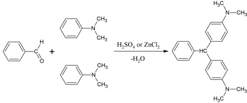
N,N-Dimethylaniline (DMA) is an organic chemical compound, a substituted derivative of aniline. It consists of a tertiary amine, featuring dimethylamino group attached to a phenyl group. This oily liquid is colourless when pure, but commercial samples are often yellow. It is an important precursor to dyes such as crystal violet.

<span class="mw-page-title-main">Fluorescent tag</span>

In molecular biology and biotechnology, a fluorescent tag, also known as a fluorescent label or fluorescent probe, is a molecule that is attached chemically to aid in the detection of a biomolecule such as a protein, antibody, or amino acid. Generally, fluorescent tagging, or labeling, uses a reactive derivative of a fluorescent molecule known as a fluorophore. The fluorophore selectively binds to a specific region or functional group on the target molecule and can be attached chemically or biologically. Various labeling techniques such as enzymatic labeling, protein labeling, and genetic labeling are widely utilized. Ethidium bromide, fluorescein and green fluorescent protein are common tags. The most commonly labelled molecules are antibodies, proteins, amino acids and peptides which are then used as specific probes for detection of a particular target.

<span class="mw-page-title-main">Staining</span> Technique used to enhance visual contrast of specimens observed under a microscope

Staining is a technique used to enhance contrast in samples, generally at the microscopic level. Stains and dyes are frequently used in histology, in cytology, and in the medical fields of histopathology, hematology, and cytopathology that focus on the study and diagnoses of diseases at the microscopic level. Stains may be used to define biological tissues, cell populations, or organelles within individual cells.

<span class="mw-page-title-main">Coomassie brilliant blue</span> Chemical compound

Coomassie brilliant blue is the name of two similar triphenylmethane dyes that were developed for use in the textile industry but are now commonly used for staining proteins in analytical biochemistry. Coomassie brilliant blue G-250 differs from Coomassie brilliant blue R-250 by the addition of two methyl groups. The name "Coomassie" is a registered trademark of Imperial Chemical Industries.

<span class="mw-page-title-main">Astaxanthin</span> Chemical compound

Astaxanthin is a keto-carotenoid within a group of chemical compounds known as terpenes. Astaxanthin is a metabolite of zeaxanthin and canthaxanthin, containing both hydroxyl and ketone functional groups. It is a lipid-soluble pigment with red coloring properties, which result from the extended chain of conjugated double bonds at the center of the compound. The presence of the hydroxyl functional groups and the hydrophobic hydrocarbons render the molecule amphiphilic.

<span class="mw-page-title-main">Rhodamine</span> Family of derivatives of xanthene used as dyes, indicators and fluorescent tracers

Rhodamine is a family of related dyes, a subset of the triarylmethane dyes. They are derivatives of xanthene. Important members of the rhodamine family are Rhodamine 6G, Rhodamine 123, and Rhodamine B. They are mainly used to dye paper and inks, but they lack the lightfastness for fabric dyeing.

<span class="mw-page-title-main">Triphenylmethane</span> Chemical compound

Triphenylmethane, or triphenyl methane, is the hydrocarbon with the formula (C6H5)3CH. This colorless solid is soluble in nonpolar organic solvents and not in water. Triphenylmethane is the basic skeleton of many synthetic dyes called triarylmethane dyes, many of them are pH indicators, and some display fluorescence. A trityl group in organic chemistry is a triphenylmethyl group Ph3C, e.g. triphenylmethyl chloride (trityl chloride) and the triphenylmethyl radical (trityl radical).

<span class="mw-page-title-main">New fuchsine</span> Chemical compound

New fuchsine is an organic compound with the formula [(H2N(CH3)C6H3)3C]Cl. It is a green-colored solid that is used as a dye of the triarylmethane class. It is one of the four components of basic fuchsine, and one of the two that are available as single dyes. The other is pararosaniline. It is prepared by condensation of ortho-toluidine with formaldehyde. This process initially gives the benzhydrol 4,4'-bis(dimethylamino)benzhydrol, which is further condensed to give the leuco (colorless) tertiary alcohol [(H2N(CH3)C6H3)3COH, which is oxidized in acid to give the dye.

<span class="mw-page-title-main">Crystal violet</span> Triarylmethane dye used as a histological stain and in Grams method of classifying bacteria

Crystal violet or gentian violet, also known as methyl violet 10B or hexamethyl pararosaniline chloride, is a triarylmethane dye used as a histological stain and in Gram's method of classifying bacteria. Crystal violet has antibacterial, antifungal, and anthelmintic (vermicide) properties and was formerly important as a topical antiseptic. The medical use of the dye has been largely superseded by more modern drugs, although it is still listed by the World Health Organization.

<span class="mw-page-title-main">Acid dye</span>

An acid dye is a dye that is typically applied to a textile at low pH. They are mainly used to dye wool, not cotton fabrics. Some acid dyes are used as food colorants, and some can also be used to stain organelles in the medical field.

<span class="mw-page-title-main">Brilliant blue FCF</span> Chemical compound

Brilliant blue FCF is a synthetic organic compound used primarily as a blue colorant for processed foods, medications, dietary supplements, and cosmetics. It is classified as a triarylmethane dye and is known under various names, such as FD&C Blue No. 1 or acid blue 9. It is denoted by E number E133 and has a color index of 42090. It has the appearance of a blue powder and is soluble in water and glycerol, with a maximum absorption at about 628 nanometers. It is one of the oldest FDA-approved color additives and is generally considered nontoxic and safe.

<span class="mw-page-title-main">Fast Green FCF</span> Chemical compound

Fast Green FCF, also called Food green 3, FD&C Green No. 3, Green 1724, Solid Green FCF, and C.I. 42053, is a turquoise triarylmethane food dye. Its E number is E143.

Triarylmethane dyes are synthetic organic compounds containing triphenylmethane backbones. As dyes, these compounds are intensely colored. They are produced industrially as dyes.

<span class="mw-page-title-main">Brilliant green (dye)</span> Chemical compound

Brilliant green is one of the triarylmethane dyes. It is closely related to malachite green.

<span class="mw-page-title-main">Endospore staining</span>

Endospore staining is a technique used in bacteriology to identify the presence of endospores in a bacterial sample. Within bacteria, endospores are protective structures used to survive extreme conditions, including high temperatures making them highly resistant to chemicals. Endospores contain little or no ATP which indicates how dormant they can be. Endospores contain a tough outer coating made up of keratin which protects them from nucleic DNA as well as other adaptations. Endospores are able to regerminate into vegetative cells, which provides a protective nature that makes them difficult to stain using normal techniques such as simple staining and gram staining. Special techniques for endospore staining include the Schaeffer–Fulton stain and the Moeller stain.

Antimicrobials destroy bacteria, viruses, fungi, algae, and other microbes. The cells of bacteria (prokaryotes), such as salmonella, differ from those of higher-level organisms (eukaryotes), such as fish. Antibiotics are chemicals designed to either kill or inhibit the growth of pathogenic bacteria while exploiting the differences between prokaryotes and eukaryotes in order to make them relatively harmless in higher-level organisms. Antibiotics are constructed to act in one of three ways: by disrupting cell membranes of bacteria, by impeding DNA or protein synthesis, or by hampering the activity of certain enzymes unique to bacteria.

References

  1. "C&L Inventory". echa.europa.eu. Retrieved 27 December 2021.
  2. Adina Raducan, Alexandra Olteanu, Mihaela Puiu, Dumitru Oancea "Influence of surfactants on the fading of malachite green" Central European Journal of Chemistry, 2008, Volume 6, pp. 1895–1066 (Print) 1644–3624 (Online). doi : 10.2478/s11532-007-0066-0
  3. Dr. M Vishwanathan. Principles of organic chemistry. Jai Sai Publications. pp. 2/37.
  4. Thomas Gessner and Udo Mayer "Triarylmethane and Diarylmethane Dyes" in Ullmann's Encyclopedia of Industrial Chemistry 2002, Wiley-VCH, Weinheim. doi : 10.1002/14356007.a27_179
  5. Srivastava, S; Sinha, R; Roy, D (2004). "Toxicological effects of malachite green". Aquatic Toxicology . 66 (3): 319–29. doi:10.1016/j.aquatox.2003.09.008. PMID   15129773.
  6. "DNA Analyst Training Laboratory Training Manual Protocol 2.18 Leucomalachite Green Presumptive Test for Blood" (PDF). National Forensic Science Technology Center. Retrieved 8 January 2018.
  7. Szent-Gyorgyi, Christopher (2007). "Fluorogen-activating single-chain antibodies for imaging cell surface proteins". Nature Biotechnology. 26 (2): 235–240. doi:10.1038/nbt1368. PMID   18157118. S2CID   21815631.
  8. Wendy C. Andersen, Sherri B. Turnipseed, and José E. Roybal "Quantitative and Confirmatory Analyses of Malachite Green and Leucomalachite Green Residues in Fish and Shrimp" J. Agric. Food Chem. 2006, volume 54, pp. 4517–4523. doi : 10.1021/jf0532258 and references therein
  9. Poopal, Rama-Krishnan; Ashwini, Rajan; Ramesh, Mathan; Li, Bin; Ren, Zongming (2023-03-01). "Triphenylmethane dye (C52H54N4O12) is potentially a hazardous substance in edible freshwater fish at trace level: toxicity, hematology, biochemistry, antioxidants, and molecular docking evaluation study". Environmental Science and Pollution Research. 30 (11): 28759–28779. doi:10.1007/s11356-022-24206-y. ISSN   1614-7499.
  10. Chinese fish crisis shows seafood safety challenges, USA Today, 7/1/2007
  11. Veterinary Residues Committee. Annual Report on Surveillance for Veterinary Residues in Food in the UK for 2001, 2002, and 2003 Archived 2012-02-11 at the Wayback Machine .
  12. "Food Safety Information - Prohibited Substances in Food".
  13. Culp, S J; Beland, FA; Heflich, R H; et al. (2002). "Mutagenicity and carcinogenicity in relation to DNA adduct formation in rats fed leucomalachite green". Mutation Research . 506–507: 55–63. doi:10.1016/S0027-5107(02)00152-5. PMID   12351145.
  14. Babendure, Jeremy R.; Adams, Stephen R.; Tsien, Roger Y. (2003). "Aptamers Switch on Fluorescence of Triphenylmethane Dyes". Journal of the American Chemical Society. American Chemical Society (ACS). 125 (48): 14716–14717. doi:10.1021/ja037994o. ISSN   0002-7863. PMID   14640641.
  15. Effect of Receptor Attachment on Sensitivity of Label Free Microcantilever Based Biosensor Using Malachite Green Aptamer https://doi.org/10.1016/j.snb.2019.126963

Further reading