Related Research Articles
Cell biology is a branch of biology that studies the structure, function, and behavior of cells. All living organisms are made of cells. A cell is the basic unit of life that is responsible for the living and functioning of organisms. Cell biology is the study of the structural and functional units of cells. Cell biology encompasses both prokaryotic and eukaryotic cells and has many subtopics which may include the study of cell metabolism, cell communication, cell cycle, biochemistry, and cell composition. The study of cells is performed using several microscopy techniques, cell culture, and cell fractionation. These have allowed for and are currently being used for discoveries and research pertaining to how cells function, ultimately giving insight into understanding larger organisms. Knowing the components of cells and how cells work is fundamental to all biological sciences while also being essential for research in biomedical fields such as cancer, and other diseases. Research in cell biology is interconnected to other fields such as genetics, molecular genetics, molecular biology, medical microbiology, immunology, and cytochemistry.

A microscope is a laboratory instrument used to examine objects that are too small to be seen by the naked eye. Microscopy is the science of investigating small objects and structures using a microscope. Microscopic means being invisible to the eye unless aided by a microscope.
In cell biology, an organelle is a specialized subunit, usually within a cell, that has a specific function. The name organelle comes from the idea that these structures are parts of cells, as organs are to the body, hence organelle, the suffix -elle being a diminutive. Organelles are either separately enclosed within their own lipid bilayers or are spatially distinct functional units without a surrounding lipid bilayer. Although most organelles are functional units within cells, some function units that extend outside of cells are often termed organelles, such as cilia, the flagellum and archaellum, and the trichocyst.

A flagellum is a hairlike appendage that protrudes from certain plant and animal sperm cells, from fungal spores (zoospores), and from a wide range of microorganisms to provide motility. Many protists with flagella are known as flagellates.
The evolution of flagella is of great interest to biologists because the three known varieties of flagella – each represent a sophisticated cellular structure that requires the interaction of many different systems.

Euglena is a genus of single cell flagellate eukaryotes. It is the best known and most widely studied member of the class Euglenoidea, a diverse group containing some 54 genera and at least 200 species. Species of Euglena are found in fresh water and salt water. They are often abundant in quiet inland waters where they may bloom in numbers sufficient to color the surface of ponds and ditches green (E. viridis) or red (E. sanguinea).

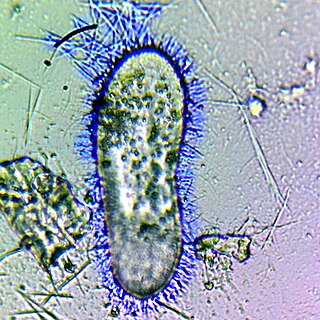
Trichomonas is a genus of anaerobic excavate parasites of vertebrates. It was first discovered by Alfred François Donné in 1836 when he found these parasites in the pus of a patient suffering from vaginitis, an inflammation of the vagina. Donné named the genus from its morphological characteristics. The prefix tricho- originates from the Ancient Greek word θρίξ (thrix) meaning hair, describing Trichomonas’s flagella. The suffix -monas, describes its similarity to unicellular organisms from the genus Monas.

In molecular biology, an axoneme, also called an axial filament, is the microtubule-based cytoskeletal structure that forms the core of a cilium or flagellum. Cilia and flagella are found on many cells, organisms, and microorganisms, to provide motility. The axoneme serves as the "skeleton" of these organelles, both giving support to the structure and, in some cases, the ability to bend. Though distinctions of function and length may be made between cilia and flagella, the internal structure of the axoneme is common to both.

A cytostome or cell mouth is a part of a cell specialized for phagocytosis, usually in the form of a microtubule-supported funnel or groove. Food is directed into the cytostome, and sealed into vacuoles. Only certain groups of protozoa, such as the Ciliophora and Excavata, have cytostomes. An example is Balantidium coli, a ciliate. In other protozoa, and in cells from multicellular organisms, phagocytosis takes place at any point on the cell or feeding takes place by absorption.
A trichocyst is an organelle found in certain ciliates and dinoflagellates.
Treponema denticola is a Gram-negative, obligate anaerobic, motile and highly proteolytic spirochete bacterium. It is one of four species of oral spirochetes to be reliably cultured, the others being Treponema pectinovorum, Treponema socranskii and Treponema vincentii. T. denticola dwells in a complex and diverse microbial community within the oral cavity and is highly specialized to survive in this environment. T. denticola is associated with the incidence and severity of human periodontal disease. Treponema denticola is one of three bacteria that form the Red Complex, the other two being Porphyromonas gingivalis and Tannerella forsythia. Together they form the major virulent pathogens that cause chronic periodontitis. Having elevated T. denticola levels in the mouth is considered one of the main etiological agents of periodontitis. T. denticola is related to the syphilis-causing obligate human pathogen, Treponema pallidum subsp. pallidum. It has also been isolated from women with bacterial vaginosis.

Malawimonadidae is a family of unicellular eukaryotes of outsize importance in understanding eukaryote phylogeny.
The genus Labyrinthula is part of the protist group Labyrinthulomycetes and contains thirteen species. The major feature of this genus is the formation of an ectoplasmic net secreted by specialized organelles called bothrosomes which surrounds the colony, which is also used by Labyrinthula for moving. The protist reproduces by zoosporulation as it sets some flagellated spores free from a sporangium. One of the flagella of the zoospores has stiff tripartite hairs (mastigonemes) - the defining characteristic of the stramenopiles.

Mastigamoeba is a genus of pelobionts, and treated by some as members of the Archamoebae group of protists. Mastigamoeba are characterized as anaerobic, amitochondriate organisms that are polymorphic. Their dominant life cycle stage is as an amoeboid flagellate. Species are typically free living, though endobiotic species have been described.

A polar organelle is a structure at a specialised region of the bacterial polar membrane that is associated with the flagellar apparatus. This flagellum-associated structure can easily be distinguished from the other membrane regions in ultrathin sections of embedded bacteria by electron microscopy when the cell membrane is orientated perpendicular to the viewing direction. There, the membrane appears slightly thickened with a finely frilled layer facing the inside of the cell. It is also possible to isolate these polar organelles from the bacterial cells and study them in face view in negatively stained preparations.
The archaellum is a unique structure on the cell surface of many archaea that allows for swimming motility. The archaellum consists of a rigid helical filament that is attached to the cell membrane by a molecular motor. This molecular motor – composed of cytosolic, membrane, and pseudo-periplasmic proteins – is responsible for the assembly of the filament and, once assembled, for its rotation. The rotation of the filament propels archaeal cells in liquid medium, in a manner similar to the propeller of a boat. The bacterial analog of the archaellum is the flagellum, which is also responsible for their swimming motility and can also be compared to a rotating corkscrew. Although the movement of archaella and flagella is sometimes described as "whip-like", this is incorrect, as only cilia from Eukaryotes move in this manner. Indeed, even "flagellum" is a misnomer, as bacterial flagella also work as propeller-like structures.

An amoeba, often called an amoeboid, is a type of cell or unicellular organism with the ability to alter its shape, primarily by extending and retracting pseudopods. Amoebae do not form a single taxonomic group; instead, they are found in every major lineage of eukaryotic organisms. Amoeboid cells occur not only among the protozoa, but also in fungi, algae, and animals.
Cell mechanics is a sub-field of biophysics that focuses on the mechanical properties and behavior of living cells and how it relates to cell function. It encompasses aspects of cell biophysics, biomechanics, soft matter physics and rheology, mechanobiology and cell biology.
Monocercomonas is a Parabasalian genus belonging to the order Trichomonadida. It presents four flagella, three forward-facing and one trailing, without the presence of a costa or any kind of undulating membrane. Monocercomonas is found in animal guts. and is susceptible to cause Monocercomoniasis in reptiles

Ultrastructural identity is a concept in biology. It asserts that evolutionary lineages of eukaryotes in general and protists in particular can be distinguished by complements and arrangements of cellular organelles. These ultrastructural components can be visualized by electron microscopy.
References
- ↑ Benchimol, Marlene (2010). "The Mastigont System in Trichomonads". Structures and Organelles in Pathogenic Protists. Microbiology Monographs. Vol. 17. pp. 1–26. doi:10.1007/978-3-642-12863-9_1. ISBN 978-3-642-12862-2. ISSN 1862-5576.
- ↑ McKhann, Heather I., and Lorraine Olendzenski, eds. Illustrated Glossary of Protoctista: Vocabulary of the Algae, Apicomplexa, Ciliates, Foraminifera, Microspora, Water Molds, Slime Molds, and the Other Protoctists. Jones & Bartlett Learning, 1993.