McGill Picture Anomaly Test

Last updated
McGill Picture Anomaly Test
Purposeassists in testing visual intelligence

The McGill Picture Anomaly Test (MPAT) is a scientific test that was created by Donald O. Hebb of McGill University and N.W. Morton that assists in testing visual intelligence as well as understanding human behavior. [1] The test includes a series of pictures that each show a typical situation but have something out of place in the photo and provides evidence that supports the idea that the right temporal lobe is involved in visual recognition. When patients with lesions to the right temporal lobe were given the MPAT, they were unable to point to the absurdity in the photo and perceived that nothing was out of place. The test is used to measure a cultural comprehension which allows for a basis to then estimate an individual's intelligence. [2] However, this test alone is not enough to accurately give a single score or representation of a person's overall intelligence. [3] The MPAT is not meant to be used across a variety of populations due to the fact that the social norms of varied populations can be tremendously different, causing the results of the test to be indeterminate. [4]

Contents

Background

The McGill Picture Anomaly test was created in 1937 by Donald O. Hebb and N.W. Morton, a member of the McGill Psychology Department. [5] Hebb applied for a job with Wilder Penfield, the founder of the Montreal Neurological Institute (MNI) where he, once hired, would study the psychological effects of brain operations. [6] His research included observing the different cognitive impairments that resulted from lesions of various areas of the brain. [7] He also conducted similar experiments in rats. After reviewing his observations, Hebb wanted to look for specific aspects of intelligence that were affected by a brain lesion. [8] He wanted to look at these specific changes instead of attempting to measure an overall intellectual change. In Hebb's looking into specific aspects of intelligence affected by a brain lesion, he was led to the creation of both the verbal Comprehension Test as well as the non-verbal Picture Anomaly Test which he developed alongside N.W. Morton.

Right Temporal Lobe

The temporal lobes are very important in the visual system as well as in the production and comprehension of language. The left temporal lobe contains Wernicke's area and Broca's area, which are both important in the production and comprehension of language. The right temporal lobe is a part of the ventral stream in the visual pathway. The ventral stream is involved in object perception and visual recognition such as faces and objects, and is therefore often referred to in relation to the McGill Picture Anomaly test. The ventral stream is often referred to as the "what" stream and provides a portrayal of the objects in the receptive field that ultimately aids in identifying the importance of the object. The different functions of each lobe account for the "disturbance of non-language capacities" experienced after the removal of the right temporal lobe. [9] If the ventral stream is disturbed or altered, the subject will not lose his vision but the ability to recognize "what" the object is in his receptive field. Disturbances in the ventral stream can be caused by damage or deterioration to any part of the brain involved in the ventral stream including the right temporal lobe.

Conducting the test

The McGill Picture Anomaly Test is a non-verbal test and requires the subject to simply point out the aspect of the photo or drawing that is out of place. When conducting the test, Hebb and Morton made it clear that the experimenter should use very little verbal instructions in order to eliminate the possibly of influencing the subject. Any remarks made by the participant during testing are ignored and do not contribute to the participant's overall score because the test is purely visual. Each series of the MPAT consists of 34 pictures each with an absurdity that is obvious to subjects with intact and normal functioning right temporal lobes but is more difficult for subjects with lesions or injuries of the right temporal lobe to recognize . [10] An example of the pictures that may be included in the test is if the picture had a group of people dressed in nice evening wear but one person is wearing a hula girl outfit. [11] In this picture, the absurdity would clearly be the hula girl. Donald O. Hebb and N.W. Norton combined the MPAT with the verbal situation series to create the McGill Adult Comprehension Examination which is used to measure adult intelligence. [12]

Hebb and Norton intended for the MPAT to be used alone with the idea that it would aid in diagnostic tests; they also created the test with the intention that the test could be used for other experiments trying to measuring intelligence. [13] The test is often used in experiments involving patients diagnosed with temporal lobe epilepsy because of the damage in their temporal lobe(s). [14] It has also been shown in a study that the recognition of picture absurdities, such as those shown in the MPAT, was significantly lower in a group of patients with dementia of Alzheimer type. [15] The dementia of Alzheimer type patients' results were compared to the results of patients with dementia syndrome of depression and cerebrovascular dementia. This impairment can be attributed to the reduced cognition and judgement utilizing visual data. [16]

Experiments

The McGill Picture Anomaly Test has been used in studies trying to measure intelligence and in studies trying to use it as a component of diagnosis. Experiments that used the MPAT for diagnostic value have shown no significant results that support any diagnostic value to the test. The potential diagnostic value of the MPAT would be in visual perception. [17] The MPAT has been used in diagnosing patients with visual-constructive disabilities, and if they receive a normal score on the test, then they are not considered to have any difficulties with picture-interpretation tasks. [18] An experiment conducted by William Sloan and Barbara Oblinger at Lincoln State School and Colony attempted to see if there were diagnostic values of Picture Anomaly tests and Verbal Absurdity tests in mental institution evaluations of patients. [19] Sloan and Oblinger found that the Verbal Absurdity tests had some significant findings but there were no significant findings when using the MPAT. Sloan and Oblinger believe that the sample size may have affected the findings for the MPAT but have not conducted a follow up experiment testing that hypothesis. [20]

Another experiment conducted by Denis Shalman attempted to see if the McGill Picture Anomaly Test has a diagnostic value with temporal lobe epilepsy. [21] Shalman used patients with right temporal lobe epileptic episodes and patients with left temporal lobe epileptic episodes; [22] Shalman hoped to replicate Brenda Milner's results which used the MPAT on patients with a temporal lobe lobectomy which found that certain dysfunctions in patients with disturbances of the right temporal lobe affected visual recognition. [23] Milner found that when the right temporal lobe was removed from a patient, pictures and representational drawings became unclear and lost their distinctiveness to the patient. [24] Shalman's findings did not support Milner's findings and showed that the McGill Picture Anomaly Test does not have the diagnostic value that Milner had previously claimed. [25] Although the McGill Picture Anomaly Test has had little diagnostic value, the test is still a common test that experimenters use to determine if there are any problems with visual recognition and interpretation or the right temporal lobe.

Notes

  1. Hebb, Donald O.; Morton, N.W. (Jan 1943). "The McGill Adult Comprehension Examination: "Verbal Situation" and "Picture Anomaly" Series". Journal of Educational Psychology. 34 (1): 16–25. doi:10.1037/h0061322.
  2. Hebb, Donald O.; Morton, N.W. (Jan 1943). "The McGill Adult Comprehension Examination: "Verbal Situation" and "Picture Anomaly" Series". Journal of Educational Psychology. 34 (1): 16–25. doi:10.1037/h0061322.
  3. Hebb, Donald O.; Morton, N.W. (1944). "Note on the measurement of adult intelligence". Journal of General Psychology. 30 (2): 217. doi:10.1080/00221309.1944.10544471. ProQuest   1290514131.
  4. Hebb, Donald O.; Morton, N.W. (Jan 1943). "The McGill Adult Comprehension Examination: "Verbal Situation" and "Picture Anomaly" Series". Journal of Educational Psychology. 34 (1): 16–25. doi:10.1037/h0061322.
  5. Hebb, Donald O.; Morton, N.W. (Jan 1943). "The McGill Adult Comprehension Examination: "Verbal Situation" and "Picture Anomaly" Series". Journal of Educational Psychology. 34 (1): 16–25. doi:10.1037/h0061322.
  6. Brown, Richard E.; Milner, Peter M. (December 2003). "The legacy of Donold O. Hebb: more than the Hebb Synapse". Nature Reviews Neuroscience. 4 (12): 1013–9. doi:10.1038/nrn1257. PMID   14682362. S2CID   205499831.
  7. Brown, Richard E.; Milner, Peter M. (December 2003). "The legacy of Donold O. Hebb: more than the Hebb Synapse". Nature Reviews Neuroscience. 4 (12): 1013–9. doi:10.1038/nrn1257. PMID   14682362. S2CID   205499831.
  8. Brown, Richard E.; Milner, Peter M. (December 2003). "The legacy of Donold O. Hebb: more than the Hebb Synapse". Nature Reviews Neuroscience. 4 (12): 1013–9. doi:10.1038/nrn1257. PMID   14682362. S2CID   205499831.
  9. Shalman, Denis C. (1961). "The diagnostic use of the McGill picture anomaly test in temporal lobe epilepsy". Journal of Neurology, Neurosurgery & Psychiatry. 24 (3): 220–222. doi:10.1136/jnnp.24.3.220. PMC   496697 . PMID   13750635.
  10. Hebb, Donald O. (1942). The McGill Picture Anomaly Series, M and N. Author, Yerkes Laboratories (Oxford, England).
  11. Hebb, Donald O.; N.W. Morton (January 1943). "The McGill Adult Comprehension Examination: "Verbal Situation" and "Picture Anomaly" Series". Journal of Educational Psychology. 34 (1): 16–25. doi:10.1037/h0061322.
  12. Hebb, Donald O.; N.W. Morton (January 1943). "The McGill Adult Comprehension Examination: "Verbal Situation" and "Picture Anomaly" Series". Journal of Educational Psychology. 34 (1): 16–25. doi:10.1037/h0061322.
  13. Hebb, Donald O.; N.W. Morton (January 1943). "The McGill Adult Comprehension Examination: "Verbal Situation" and "Picture Anomaly" Series". Journal of Educational Psychology. 34 (1): 16–25. doi:10.1037/h0061322.
  14. Shalman, Denis C. (1961). "The diagnostic use of the McGill picture anomaly test in temporal lobe epilepsy". Journal of Neurology, Neurosurgery & Psychiatry. 24 (3): 220–222. doi:10.1136/jnnp.24.3.220. PMC   496697 . PMID   13750635.
  15. Shuttleworth, Edwin C.; Huber, Steven J. (September 1989). "The picture absurdities test in the evaluation of dementia". Brain and Cognition. 11 (1): 50–9. doi:10.1016/0278-2626(89)90004-3. PMID   2789816. S2CID   5840759.
  16. Shuttleworth, Edwin C.; Huber, Steven J. (September 1989). "The picture absurdities test in the evaluation of dementia". Brain and Cognition. 11 (1): 50–9. doi:10.1016/0278-2626(89)90004-3. PMID   2789816. S2CID   5840759.
  17. Paterson, Andrew; O.W. Zangwill (December 1944). "Disorders of visual space perception associated with lesions of the right cerebral hemisphere". Brain. 67 (4): 331–358. doi:10.1093/brain/67.4.331.
  18. Paterson, Andrew; O.W. Zangwill (December 1944). "Disorders of visual space perception associated with lesions of the right cerebral hemisphere". Brain. 67 (4): 331–358. doi:10.1093/brain/67.4.331.
  19. Sloan, William; Brenda Oblinger (January 1947). "Diagnostic value of picture anomalies and verbal absurdities for institutional". American Journal of Mental Deficiency. 51: 532–534. Retrieved 12 November 2013.
  20. Sloan, William; Brenda Oblinger (January 1947). "Diagnostic value of picture anomalies and verbal absurdities for institutional". American Journal of Mental Deficiency. 51: 532–534. Retrieved 12 November 2013.
  21. Shalman, Denis (1961). "The Diagnostic use of the MPAT in temporal lobe epilepsy". Journal of Neurology, Neurosurgery & Psychiatry. 24 (3): 220–2. doi:10.1136/jnnp.24.3.220. PMC   496697 . PMID   13750635.
  22. Shalman, Denis (1961). "The Diagnostic use of the McGill picture anomaly test in temporal lobe epilepsy". Journal of Neurology, Neurosurgery & Psychiatry. 24 (3): 220–2. doi:10.1136/jnnp.24.3.220. PMC   496697 . PMID   13750635.
  23. Milner, Brenda (January 1958). "Psychological defects produced by temporal lobe excision". Research Publications of the Association for Research in Nervous & Mental Disease. 36: 244–257. PMID   13527787. ProQuest   615341349.
  24. Milner, Brenda (January 1958). "Psychological defects produced by temporal lobe excision". Research Publications of the Association for Research in Nervous & Mental Disease. 36: 244–257. PMID   13527787. ProQuest   615341349.
  25. Milner, Brenda (January 1958). "Psychological defects produced by temporal lobe excision". Research Publications of the Association for Research in Nervous & Mental Disease. 36: 244–257. PMID   13527787. ProQuest   615341349.

Related Research Articles

<span class="mw-page-title-main">Agnosia</span> Medical condition

Agnosia is the inability to process sensory information. Often there is a loss of ability to recognize objects, persons, sounds, shapes, or smells while the specific sense is not defective nor is there any significant memory loss. It is usually associated with brain injury or neurological illness, particularly after damage to the occipitotemporal border, which is part of the ventral stream. Agnosia only affects a single modality, such as vision or hearing. More recently, a top-down interruption is considered to cause the disturbance of handling perceptual information.

Source amnesia is the inability to remember where, when or how previously learned information has been acquired, while retaining the factual knowledge. This branch of amnesia is associated with the malfunctioning of one's explicit memory. It is likely that the disconnect between having the knowledge and remembering the context in which the knowledge was acquired is due to a dissociation between semantic and episodic memory – an individual retains the semantic knowledge, but lacks the episodic knowledge to indicate the context in which the knowledge was gained.

<span class="mw-page-title-main">Donald O. Hebb</span> Canadian neuropsychologist

Donald Olding Hebb was a Canadian psychologist who was influential in the area of neuropsychology, where he sought to understand how the function of neurons contributed to psychological processes such as learning. He is best known for his theory of Hebbian learning, which he introduced in his classic 1949 work The Organization of Behavior. He has been described as the father of neuropsychology and neural networks. A Review of General Psychology survey, published in 2002, ranked Hebb as the 19th most cited psychologist of the 20th century. His views on learning described behavior and thought in terms of brain function, explaining cognitive processes in terms of connections between neuron assemblies.

<span class="mw-page-title-main">Anomic aphasia</span> Medical condition

Anomic aphasia is a mild, fluent type of aphasia where individuals have word retrieval failures and cannot express the words they want to say. By contrast, anomia is a deficit of expressive language, and a symptom of all forms of aphasia, but patients whose primary deficit is word retrieval are diagnosed with anomic aphasia. Individuals with aphasia who display anomia can often describe an object in detail and maybe even use hand gestures to demonstrate how the object is used, but cannot find the appropriate word to name the object. Patients with anomic aphasia have relatively preserved speech fluency, repetition, comprehension, and grammatical speech.

<span class="mw-page-title-main">Henry Molaison</span> American memory disorder patient

Henry Gustav Molaison, known widely as H.M., was an American who had a bilateral medial temporal lobectomy to surgically resect the anterior two thirds of his hippocampi, parahippocampal cortices, entorhinal cortices, piriform cortices, and amygdalae in an attempt to cure his epilepsy. Although the surgery was partially successful in controlling his epilepsy, a severe side effect was that he became unable to form new memories.

<span class="mw-page-title-main">Temporal lobe</span> One of the four lobes of the mammalian brain

The temporal lobe is one of the four major lobes of the cerebral cortex in the brain of mammals. The temporal lobe is located beneath the lateral fissure on both cerebral hemispheres of the mammalian brain.

<span class="mw-page-title-main">Brenda Milner</span> British-Canadian neuroscientist and neuropsychologist

Brenda Milner is a British-Canadian neuropsychologist who has contributed extensively to the research literature on various topics in the field of clinical neuropsychology. Milner is a professor in the Department of Neurology and Neurosurgery at McGill University and a professor of Psychology at the Montreal Neurological Institute. As of 2020, she holds more than 25 honorary degrees and she continued to work in her nineties. Her current work covers many aspects of neuropsychology including her lifelong interest in the involvement of the temporal lobes in episodic memory. She is sometimes referred to as the founder of neuropsychology and has been essential in its development. She received the Balzan Prize for Cognitive Neuroscience in 2009, and the Kavli Prize in Neuroscience, together with John O'Keefe, and Marcus E. Raichle, in 2014. She turned 100 in July 2018 and at the time was still overseeing the work of researchers.

<span class="mw-page-title-main">Global aphasia</span> Medical condition

Global aphasia is a severe form of nonfluent aphasia, caused by damage to the left side of the brain, that affects receptive and expressive language skills as well as auditory and visual comprehension. Acquired impairments of communicative abilities are present across all language modalities, impacting language production, comprehension, and repetition. Patients with global aphasia may be able to verbalize a few short utterances and use non-word neologisms, but their overall production ability is limited. Their ability to repeat words, utterances, or phrases is also affected. Due to the preservation of the right hemisphere, an individual with global aphasia may still be able to express themselves through facial expressions, gestures, and intonation. This type of aphasia often results from a large lesion of the left perisylvian cortex. The lesion is caused by an occlusion of the left middle cerebral artery and is associated with damage to Broca's area, Wernicke's area, and insular regions which are associated with aspects of language.

Transcortical sensory aphasia (TSA) is a kind of aphasia that involves damage to specific areas of the temporal lobe of the brain, resulting in symptoms such as poor auditory comprehension, relatively intact repetition, and fluent speech with semantic paraphasias present. TSA is a fluent aphasia similar to Wernicke's aphasia, with the exception of a strong ability to repeat words and phrases. The person may repeat questions rather than answer them ("echolalia").

Semantic dementia (SD), also known as semantic variant primary progressive aphasia (svPPA), is a progressive neurodegenerative disorder characterized by loss of semantic memory in both the verbal and non-verbal domains. However, the most common presenting symptoms are in the verbal domain. Semantic dementia is a disorder of semantic memory that causes patients to lose the ability to match words or images to their meanings. However, it is fairly rare for patients with semantic dementia to develop category specific impairments, though there have been documented cases of it occurring. Typically, a more generalized semantic impairment results from dimmed semantic representations in the brain.

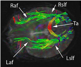
The two-streams hypothesis is a model of the neural processing of vision as well as hearing. The hypothesis, given its initial characterisation in a paper by David Milner and Melvyn A. Goodale in 1992, argues that humans possess two distinct visual systems. Recently there seems to be evidence of two distinct auditory systems as well. As visual information exits the occipital lobe, and as sound leaves the phonological network, it follows two main pathways, or "streams". The ventral stream leads to the temporal lobe, which is involved with object and visual identification and recognition. The dorsal stream leads to the parietal lobe, which is involved with processing the object's spatial location relative to the viewer and with speech repetition.

Visual agnosia is an impairment in recognition of visually presented objects. It is not due to a deficit in vision, language, memory, or intellect. While cortical blindness results from lesions to primary visual cortex, visual agnosia is often due to damage to more anterior cortex such as the posterior occipital and/or temporal lobe(s) in the brain.[2] There are two types of visual agnosia: apperceptive agnosia and associative agnosia.

Auditory verbal agnosia (AVA), also known as pure word deafness, is the inability to comprehend speech. Individuals with this disorder lose the ability to understand language, repeat words, and write from dictation. Some patients with AVA describe hearing spoken language as meaningless noise, often as though the person speaking was doing so in a foreign language. However, spontaneous speaking, reading, and writing are preserved. The maintenance of the ability to process non-speech auditory information, including music, also remains relatively more intact than spoken language comprehension. Individuals who exhibit pure word deafness are also still able to recognize non-verbal sounds. The ability to interpret language via lip reading, hand gestures, and context clues is preserved as well. Sometimes, this agnosia is preceded by cortical deafness; however, this is not always the case. Researchers have documented that in most patients exhibiting auditory verbal agnosia, the discrimination of consonants is more difficult than that of vowels, but as with most neurological disorders, there is variation among patients.

Auditory agnosia is a form of agnosia that manifests itself primarily in the inability to recognize or differentiate between sounds. It is not a defect of the ear or "hearing", but rather a neurological inability of the brain to process sound meaning. While auditory agnosia impairs the understanding of sounds, other abilities such as reading, writing, and speaking are not hindered. It is caused by bilateral damage to the anterior superior temporal gyrus, which is part of the auditory pathway responsible for sound recognition, the auditory "what" pathway.

The Cambridge Neuropsychological Test Automated Battery (CANTAB), originally developed at the University of Cambridge in the 1980s but now provided in a commercial capacity by Cambridge Cognition, is a computer-based cognitive assessment system consisting of a battery of neuropsychological tests, administered to subjects using a touch screen computer. The CANTAB tests were co-invented by Professor Trevor Robbins and Professor Barbara Sahakian. The 25 tests in CANTAB examine various areas of cognitive function, including:

The neuroanatomy of memory encompasses a wide variety of anatomical structures in the brain.

Recognition memory, a subcategory of explicit memory, is the ability to recognize previously encountered events, objects, or people. When the previously experienced event is reexperienced, this environmental content is matched to stored memory representations, eliciting matching signals. As first established by psychology experiments in the 1970s, recognition memory for pictures is quite remarkable: humans can remember thousands of images at high accuracy after seeing each only once and only for a few seconds.

Memory supports and enables social interactions in a variety of ways. In order to engage in successful social interaction, people must be able to remember how they should interact with one another, whom they have interacted with previously, and what occurred during those interactions. There are a lot of brain processes and functions that go into the application and use of memory in social interactions, as well as psychological reasoning for its importance.

The Boston Naming Test (BNT), introduced in 1983 by Edith Kaplan, Harold Goodglass and Sandra Weintraub, is a widely used neuropsychological assessment tool to measure confrontational word retrieval in individuals with aphasia or other language disturbance caused by stroke, Alzheimer's disease, or other dementing disorder. A common and debilitating feature is anomic aphasia, an impairment in the ability to name objects. The BNT contains 60 line drawings graded in difficulty. Patients with anomia often have greater difficulties with the naming of not only difficult and low frequency objects but also easy and high frequency objects. Naming difficulties may be rank ordered along a continuum. Items are rank ordered in terms of their ability to be named, which is correlated with their frequency. This type of picture-naming test is also useful in the examination of children with learning disabilities and the evaluation of brain-injured adults.

<span class="mw-page-title-main">Visual pathway lesions</span> Overview about the lesions of visual pathways

The visual pathway consists of structures that carry visual information from the retina to the brain. Lesions in that pathway cause a variety of visual field defects. In the visual system of human eye, the visual information processed by retinal photoreceptor cells travel in the following way:
Retina→Optic nerve→Optic chiasma →Optic tract→Lateral geniculate body→Optic radiation→Primary visual cortex

References