Related Research Articles

A tendon or sinew is a tough, high-tensile-strength band of dense fibrous connective tissue that connects muscle to bone. It is able to transmit the mechanical forces of muscle contraction to the skeletal system without sacrificing its ability to withstand significant amounts of tension.

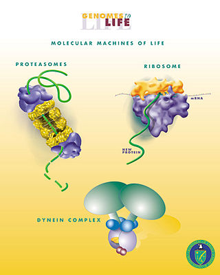
The cytoskeleton is a complex, dynamic network of interlinking protein filaments present in the cytoplasm of all cells, including those of bacteria and archaea. In eukaryotes, it extends from the cell nucleus to the cell membrane and is composed of similar proteins in the various organisms. It is composed of three main components, microfilaments, intermediate filaments and microtubules, and these are all capable of rapid growth or disassembly dependent on the cell's requirements.

Cartilage is a resilient and smooth type of connective tissue. In tetrapods, it covers and protects the ends of long bones at the joints as articular cartilage, and is a structural component of many body parts including the rib cage, the neck and the bronchial tubes, and the intervertebral discs. In other taxa, such as chondrichthyans, but also in cyclostomes, it may constitute a much greater proportion of the skeleton. It is not as hard and rigid as bone, but it is much stiffer and much less flexible than muscle. The matrix of cartilage is made up of glycosaminoglycans, proteoglycans, collagen fibers and, sometimes, elastin.

In biology, the extracellular matrix (ECM), also called intercellular matrix, is a three-dimensional network consisting of extracellular macromolecules and minerals, such as collagen, enzymes, glycoproteins and hydroxyapatite that provide structural and biochemical support to surrounding cells. Because multicellularity evolved independently in different multicellular lineages, the composition of ECM varies between multicellular structures; however, cell adhesion, cell-to-cell communication and differentiation are common functions of the ECM.

The synovial membrane is a specialized connective tissue that lines the inner surface of capsules of synovial joints and tendon sheath. It makes direct contact with the fibrous membrane on the outside surface and with the synovial fluid lubricant on the inside surface. In contact with the synovial fluid at the tissue surface are many rounded macrophage-like synovial cells and also type B cells, which are also known as fibroblast-like synoviocytes (FLS). Type A cells maintain the synovial fluid by removing wear-and-tear debris. As for the FLS, they produce hyaluronan, as well as other extracellular components in the synovial fluid.
Mechanotaxis refers to the directed movement of cell motility via mechanical cues. In response to fluidic shear stress, for example, cells have been shown to migrate in the direction of the fluid flow. Mechanotaxis is critical in many normal biological processes in animals, such as gastrulation, inflammation, and repair in response to a wound, as well as in mechanisms of diseases such as tumor metastasis.
Durotaxis is a form of cell migration in which cells are guided by rigidity gradients, which arise from differential structural properties of the extracellular matrix (ECM). Most normal cells migrate up rigidity gradients.

Hyaluronic acid, also called hyaluronan, is an anionic, nonsulfated glycosaminoglycan distributed widely throughout connective, epithelial, and neural tissues. It is unique among glycosaminoglycans as it is non-sulfated, forms in the plasma membrane instead of the Golgi apparatus, and can be very large: human synovial HA averages about 7 million Da per molecule, or about 20,000 disaccharide monomers, while other sources mention 3–4 million Da.
Chondrocytes are the only cells found in healthy cartilage. They produce and maintain the cartilaginous matrix, which consists mainly of collagen and proteoglycans. Although the word chondroblast is commonly used to describe an immature chondrocyte, the term is imprecise, since the progenitor of chondrocytes can differentiate into various cell types, including osteoblasts.

In cell biology, focal adhesions are large macromolecular assemblies through which mechanical force and regulatory signals are transmitted between the extracellular matrix (ECM) and an interacting cell. More precisely, focal adhesions are the sub-cellular structures that mediate the regulatory effects of a cell in response to ECM adhesion.
In cellular biology, mechanotransduction is any of various mechanisms by which cells convert mechanical stimulus into electrochemical activity. This form of sensory transduction is responsible for a number of senses and physiological processes in the body, including proprioception, touch, balance, and hearing. The basic mechanism of mechanotransduction involves converting mechanical signals into electrical or chemical signals.

Chondroblasts, or perichondrial cells, is the name given to mesenchymal progenitor cells in situ which, from endochondral ossification, will form chondrocytes in the growing cartilage matrix. Another name for them is subchondral cortico-spongious progenitors. They have euchromatic nuclei and stain by basic dyes.

Aggrecan (ACAN), also known as cartilage-specific proteoglycan core protein (CSPCP) or chondroitin sulfate proteoglycan 1, is a protein that in humans is encoded by the ACAN gene. This gene is a member of the lectican (chondroitin sulfate proteoglycan) family. The encoded protein is an integral part of the extracellular matrix in cartilagenous tissue and it withstands compression in cartilage.
Talin is a high-molecular-weight cytoskeletal protein concentrated at regions of cell–substratum contact and, in lymphocytes, at cell–cell contacts. Discovered in 1983 by Keith Burridge and colleagues, talin is a ubiquitous cytosolic protein that is found in high concentrations in focal adhesions. It is capable of linking integrins to the actin cytoskeleton either directly or indirectly by interacting with vinculin and α-actinin.
Mechanotherapy is a type of medical therapeutics in which treatment is given by manual or mechanical means. It was defined in 1890 as “the employment of mechanical means for the cure of disease”. Mechanotherapy employs mechanotransduction in order to stimulate tissue repair and remodelling.
Mechanosensation is the transduction of mechanical stimuli into neural signals. Mechanosensation provides the basis for the senses of light touch, hearing, proprioception, and pain. Mechanoreceptors found in the skin, called cutaneous mechanoreceptors, are responsible for the sense of touch. Tiny cells in the inner ear, called hair cells, are responsible for hearing and balance. States of neuropathic pain, such as hyperalgesia and allodynia, are also directly related to mechanosensation. A wide array of elements are involved in the process of mechanosensation, many of which are still not fully understood.
Mechanosensitive channels, mechanosensitive ion channels or stretch-gated ion channels (not to be confused with mechanoreceptors). They are present in the membranes of organisms from the three domains of life: bacteria, archaea, and eukarya. They are the sensors for a number of systems including the senses of touch, hearing and balance, as well as participating in cardiovascular regulation and osmotic homeostasis (e.g. thirst). The channels vary in selectivity for the permeating ions from nonselective between anions and cations in bacteria, to cation selective allowing passage Ca2+, K+ and Na+ in eukaryotes, and highly selective K+ channels in bacteria and eukaryotes.

Nanobiomechanics is an emerging field in nanoscience and biomechanics that combines the powerful tools of nanomechanics to explore fundamental science of biomaterials and biomechanics.
Lori Ann Setton is an American biomechanical engineer noted for her research on mechanics and mechanobiology of the intervertebral disc, articular cartilage mechanics, drug delivery, and pathomechanisms of osteoarthritis. She is currently the Department Chair as well as the Lucy and Stanley Lopata Distinguished Professor of Biomedical Engineering at Washington University in St. Louis.
Physical oncology (PO) is defined as the study of the role of mechanical signals in a cancerous tumor. The mechanical signals can be forces, pressures. If we generalize we will speak of "stress field" and "stress tensor".
References
- ↑ Wang, J. H.-C.; Thampatty, B. P. (March 2006). "An introductory review of cell mechanobiology". Biomechanics and Modeling in Mechanobiology. 5 (1): 1–16. doi:10.1007/s10237-005-0012-z. ISSN 1617-7959. PMID 16489478. S2CID 5017641.
- ↑ Smit, Theodoor H. (2020). "Adolescent idiopathic scoliosis: The mechanobiology of differential growth". JOR Spine. 3 (4): e1115. doi: 10.1002/jsp2.1115 . ISSN 2572-1143. PMC 7770204 . PMID 33392452. S2CID 225497216.
- ↑ Tracy, Lauren E.; Minasian, Raquel A.; Caterson, E.J. (March 2016). "Extracellular Matrix and Dermal Fibroblast Function in the Healing Wound". Advances in Wound Care. 5 (3): 119–136. doi:10.1089/wound.2014.0561. ISSN 2162-1918. PMC 4779293 . PMID 26989578.
- ↑ Korhonen, R.K; Laasanen, M.S; Töyräs, J; Rieppo, J; Hirvonen, J; Helminen, H.J; Jurvelin, J.S (July 2002). "Comparison of the equilibrium response of articular cartilage in unconfined compression, confined compression and indentation". Journal of Biomechanics. 35 (7): 903–909. doi:10.1016/s0021-9290(02)00052-0. ISSN 0021-9290. PMID 12052392.
- ↑ Ateshian, G.A.; Warden, W.H.; Kim, J.J.; Grelsamer, R.P.; Mow, V.C. (November 1997). "Finite deformation biphasic material properties of bovine articular cartilage from confined compression experiments". Journal of Biomechanics. 30 (11–12): 1157–1164. doi: 10.1016/s0021-9290(97)85606-0 . ISSN 0021-9290. PMID 9456384.
- ↑ Wong, M; Carter, D.R (July 2003). "Articular cartilage functional histomorphology and mechanobiology: a research perspective". Bone. 33 (1): 1–13. doi:10.1016/s8756-3282(03)00083-8. ISSN 8756-3282. PMID 12919695.
- ↑ Haapala, Jussi; Arokoski, Jari P.A.; Hyttinen, Mika M.; Lammi, Mikko; Tammi, Markku; Kovanen, Vuokko; Helminen, Heikki J.; Kiviranta, Ilkka (May 1999). "Remobilization Does Not Fully Restore Immobilization Induced Articular Cartilage Atrophy". Clinical Orthopaedics and Related Research. 362: 218–229. doi:10.1097/00003086-199905000-00031. ISSN 0009-921X.
- ↑ Stroud, Matthew J; Banerjee, Indroneal; Veevers, Jennifer; Chen, Ju (31 January 2014). "Linker of Nucleoskeleton and Cytoskeleton Complex Proteins in Cardiac Structure, Function, and Disease". Circulation Research. 114 (3): 538–548. doi: 10.1161/circresaha.114.301236 .
- ↑ Xia, Yuntao; Pfeifer, Charlotte R.; Cho, Sangkyun; Discher, Dennis E.; Irianto, Jerome (2018-12-21). del Río Hernández, Armando (ed.). "Nuclear mechanosensing". Emerging Topics in Life Sciences. 2 (5): 713–725. doi:10.1042/ETLS20180051. ISSN 2397-8554. PMC 6830732 . PMID 31693005.
- ↑ Mammoto, Akiko; Mammoto, Tadanori; Ingber, Donald E. (2012-07-01). "Mechanosensitive mechanisms in transcriptional regulation". Journal of Cell Science. 125 (13): 3061–3073. doi:10.1242/jcs.093005. ISSN 0021-9533. PMC 3434847 . PMID 22797927.
- ↑ Ou, Guangshuo; Stuurman, Nico; D’Ambrosio, Michael; Vale, Ronald D. (2010-09-30). "Polarized Myosin Produces Unequal-Size Daughters During Asymmetric Cell Division". Science. 330 (6004): 677–680. Bibcode:2010Sci...330..677O. doi:10.1126/science.1196112. ISSN 0036-8075. PMC 3032534 . PMID 20929735.
- ↑ Ingber, D. E. (October 1997). "Tensegrity: The Architectural Basis of Cellular Mechanotransduction". Annual Review of Physiology. 59 (1): 575–599. doi:10.1146/annurev.physiol.59.1.575. ISSN 0066-4278. PMID 9074778.
- ↑ Niwa, Hitoshi; Toyooka, Yayoi; Shimosato, Daisuke; Strumpf, Dan; Takahashi, Kadue; Yagi, Rika; Rossant, Janet (December 2005). "Interaction between Oct3/4 and Cdx2 Determines Trophectoderm Differentiation". Cell. 123 (5): 917–929. doi: 10.1016/j.cell.2005.08.040 . ISSN 0092-8674. PMID 16325584. S2CID 13242763.
- ↑ Buganza Tepole, A; Ploch, CJ; Wong, J; Gosain, AK; Kuhl, E (2011). "Growing skin - A computational model for skin expansion in reconstructive surgery". J. Mech. Phys. Solids. 59 (10): 2177–2190. Bibcode:2011JMPSo..59.2177B. doi:10.1016/j.jmps.2011.05.004. PMC 3212404 . PMID 22081726.
- ↑ Ingber, DE (2003). "Mechanobiology and diseases of mechanotransduction". Annals of Medicine. 35 (8): 564–77. doi:10.1080/07853890310016333. PMID 14708967. S2CID 22753025.
- ↑ Ingber, DE (1997). "Tensegrity: the architectural basis of cellular mechanotransduction". Annu. Rev. Physiol. 59: 575–599. doi:10.1146/annurev.physiol.59.1.575. PMID 9074778.
- ↑ Ingber, DE (2006). "Cellular mechanotransduction: putting all the pieces together again". FASEB J. 20 (7): 811–827. doi:10.1096/fj.05-5424rev. PMID 16675838. S2CID 21267494.