In vertebrate anatomy, ribs are the long curved bones which form the rib cage, part of the axial skeleton. In most tetrapods, ribs surround the thoracic cavity, enabling the lungs to expand and thus facilitate breathing by expanding the thoracic cavity. They serve to protect the lungs, heart, and other vital organs of the thorax. In some animals, especially snakes, ribs may provide support and protection for the entire body.

The middle ear is the portion of the ear medial to the eardrum, and distal to the oval window of the cochlea.
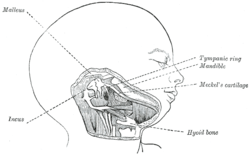
The ossicles are three bones in either middle ear that are among the smallest bones in the human body. They serve to transmit sounds from the air to the fluid-filled labyrinth (cochlea). The absence of the auditory ossicles would constitute a moderate-to-severe hearing loss. The term "ossicle" literally means "tiny bone". Though the term may refer to any small bone throughout the body, it typically refers to the malleus, incus, and stapes of the middle ear.

The skull is a bone protective cavity for the brain. The skull is composed of four types of bone i.e., cranial bones, facial bones, ear ossicles and hyoid bone, however two parts are more prominent: the cranium and the mandible. In humans, these two parts are the neurocranium (braincase) and the viscerocranium that includes the mandible as its largest bone. The skull forms the anterior-most portion of the skeleton and is a product of cephalisation—housing the brain, and several sensory structures such as the eyes, ears, nose, and mouth. In humans, these sensory structures are part of the facial skeleton.

In anatomy, the temporomandibular joints (TMJ) are the two joints connecting the jawbone to the skull. It is a bilateral synovial articulation between the temporal bone of the skull above and the mandible below; it is from these bones that its name is derived. The joints are unique in their bilateral function, being connected via the mandible.
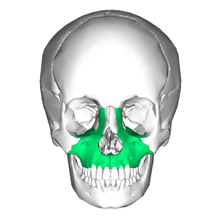
In vertebrates, the maxilla is the upper fixed bone of the jaw formed from the fusion of two maxillary bones. In humans, the upper jaw includes the hard palate in the front of the mouth. The two maxillary bones are fused at the intermaxillary suture, forming the anterior nasal spine. This is similar to the mandible, which is also a fusion of two mandibular bones at the mandibular symphysis. The mandible is the movable part of the jaw.

The jaws are a pair of opposable articulated structures at the entrance of the mouth, typically used for grasping and manipulating food. The term jaws is also broadly applied to the whole of the structures constituting the vault of the mouth and serving to open and close it and is part of the body plan of humans and most animals.

The hyoid bone is a horseshoe-shaped bone situated in the anterior midline of the neck between the chin and the thyroid cartilage. At rest, it lies between the base of the mandible and the third cervical vertebra.

The temporal bones are situated at the sides and base of the skull, and lateral to the temporal lobes of the cerebral cortex.

Fish anatomy is the study of the form or morphology of fish. It can be contrasted with fish physiology, which is the study of how the component parts of fish function together in the living fish. In practice, fish anatomy and fish physiology complement each other, the former dealing with the structure of a fish, its organs or component parts and how they are put together, such as might be observed on the dissecting table or under the microscope, and the latter dealing with how those components function together in living fish.

The quadrate bone is a skull bone in most tetrapods, including amphibians, sauropsids, and early synapsids.
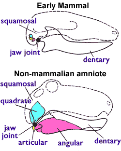
The articular bone is part of the lower jaw of most vertebrates, including most jawed fish, amphibians, birds and various kinds of reptiles, as well as ancestral mammals.

The pharyngeal arches, also known as visceral arches, are structures seen in the embryonic development of vertebrates that are recognisable precursors for many structures. In fish, the arches are known as the branchial arches, or gill arches.

The evolution of mammalian auditory ossicles was an evolutionary process that resulted in the formation of the bones of the mammalian middle ear. These bones, or ossicles, are a defining characteristic of all mammals. The event is well-documented and important as a demonstration of transitional forms and exaptation, the re-purposing of existing structures during evolution.
The splanchnocranium is the portion of the cranium that is derived from pharyngeal arches. Splanchno indicates to the gut because the face forms around the mouth, which is an end of the gut. The splanchnocranium consists of cartilage and endochondral bone. In mammals, the splanchnocranium comprises the three ear ossicles, as well as the alisphenoid, the styloid process, the hyoid apparatus, and the thyroid cartilage.

Branchial arches, or gill arches, are a series of paired bony "loops" that support the gills in fish. As gills are the primitive feature of vertebrates, all vertebrate embryos develop pharyngeal arches, though the eventual fate of these arches varies between taxa. In jawed fish, the first arch pair develops into the jaw. The second gill arches develop into the hyomandibular complex, which supports the back of the jaw and the front of the gill series. The remaining posterior arches support the gills. In amphibians and reptiles, many pharyngeal arch elements are lost, including the gill arches, resulting in only the oral jaws and a hyoid apparatus remaining. In mammals and birds, the hyoid is simplified further.

The hyomandibula, commonly referred to as hyomandibular [bone], is a set of bones that is found in the hyoid region in most fishes. It usually plays a role in suspending the jaws and/or operculum. It is commonly suggested that in tetrapods, the hyomandibula evolved into the columella (stapes).
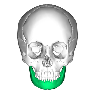
In jawed vertebrates, the mandible, lower jaw, or jawbone is a bone that makes up the lower – and typically more mobile – component of the mouth.

Most bony fishes have two sets of jaws made mainly of bone. The primary oral jaws open and close the mouth, and a second set of pharyngeal jaws are positioned at the back of the throat. The oral jaws are used to capture and manipulate prey by biting and crushing. The pharyngeal jaws, so-called because they are positioned within the pharynx, are used to further process the food and move it from the mouth to the stomach.

The malleus, or hammer, is a hammer-shaped small bone or ossicle of the middle ear. It connects with the incus, and is attached to the inner surface of the eardrum. The word is Latin for 'hammer' or 'mallet'. It transmits the sound vibrations from the eardrum to the incus (anvil).