Andries van Wezel, latinised as Andreas Vesalius, was an anatomist and physician who wrote De Humani Corporis Fabrica Libri Septem, what is considered to be one of the most influential books on human anatomy and a major advance over the long-dominant work of Galen. Vesalius is often referred to as the founder of modern human anatomy. He was born in Brussels, which was then part of the Habsburg Netherlands. He was a professor at the University of Padua (1537–1542) and later became Imperial physician at the court of Emperor Charles V.

The middle ear is the portion of the ear medial to the eardrum, and distal to the oval window of the cochlea.
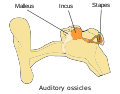
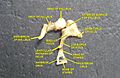
The ossicles are three bones in either middle ear that are among the smallest bones in the human body. They serve to transmit sounds from the air to the fluid-filled labyrinth (cochlea). The absence of the auditory ossicles would constitute a moderate-to-severe hearing loss. The term "ossicle" literally means "tiny bone". Though the term may refer to any small bone throughout the body, it typically refers to the malleus, incus, and stapes of the middle ear.

The stapes or stirrup is a bone in the middle ear of humans and other animals which is involved in the conduction of sound vibrations to the inner ear. This bone is connected to the oval window by its annular ligament, which allows the footplate to transmit sound energy through the oval window into the inner ear. The stapes is the smallest and lightest bone in the human body, and is so-called because of its resemblance to a stirrup.

The oval window is a connective tissue membrane-covered opening from the middle ear to the cochlea of the inner ear.

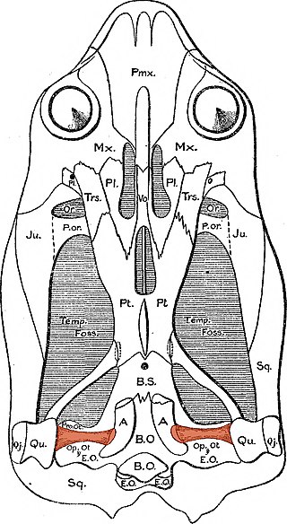
The quadrate bone is a skull bone in most tetrapods, including amphibians, sauropsids, and early synapsids.

An ear is the organ that enables hearing and body balance using the vestibular system. In mammals, the ear is usually described as having three parts: the outer ear, the middle ear and the inner ear. The outer ear consists of the pinna and the ear canal. Since the outer ear is the only visible portion of the ear in most animals, the word "ear" often refers to the external part alone. The middle ear includes the tympanic cavity and the three ossicles. The inner ear sits in the bony labyrinth, and contains structures which are key to several senses: the semicircular canals, which enable balance and eye tracking when moving; the utricle and saccule, which enable balance when stationary; and the cochlea, which enables hearing. The ear is a self cleaning organ through its relationship with earwax and the ear canals. The ears of vertebrates are placed somewhat symmetrically on either side of the head, an arrangement that aids sound localization.

De Humani Corporis Fabrica Libri Septem is a set of books on human anatomy written by Andreas Vesalius (1514–1564) and published in 1543. It was a major advance in the history of anatomy over the long-dominant work of Galen, and presented itself as such.

Tympanoplasty is the surgical operation performed to reconstruct hearing mechanism of middle ear.

The tensor tympani is a muscle within the middle ear, located in the bony canal above the bony part of the auditory tube, and connects to the malleus bone. Its role is to damp loud sounds, such as those produced from chewing, shouting, or thunder. Because its reaction time is not fast enough, the muscle cannot protect against hearing damage caused by sudden loud sounds, like explosions or gunshots.

The tympanic cavity is a small cavity surrounding the bones of the middle ear. Within it sit the ossicles, three small bones that transmit vibrations used in the detection of sound.

Jacopo Berengario da Carpi was an Italian physician. His book "Isagoge breves" published in 1522 made him the most important anatomist before Andreas Vesalius.
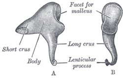
The posterior ligament of the incus is a fibrous band that connects the tip of the short crus of the incus to the fossa incudis, running to the mastoid. The posterior incudal ligament plays an important role in the vibration of the middle ear bones: together with the anterior ligament of the malleus, it forms a pivotal axis around which the ossicles rotate. This rotation conveys vibrations from the tympanum to the oval window on the bony labyrinth.

The evolution of mammalian auditory ossicles was an evolutionary process that resulted in the formation of the bones of the mammalian middle ear. These bones, or ossicles, are a defining characteristic of all mammals. The event is well-documented and important as a demonstration of transitional forms and exaptation, the re-purposing of existing structures during evolution.
The splanchnocranium is the portion of the cranium that is derived from pharyngeal arches. Splanchno indicates to the gut because the face forms around the mouth, which is an end of the gut. The splanchnocranium consists of cartilage and endochondral bone. In mammals, the splanchnocranium comprises the three ear ossicles, as well as the alisphenoid, the styloid process, the hyoid apparatus, and the thyroid cartilage.
In the auditory system, the columella contributes to hearing in amphibians, reptiles and birds. The columella form thin, bony structures in the interior of the skull and serve the purpose of transmitting sounds from the eardrum. It is an evolutionary homolog of the stapes, one of the auditory ossicles in mammals.

The incudomalleolar joint or articulatio incudomallearis is a small synovial joint between the malleus (hammer) and the incus (anvil). The joint's function is to transfer vibrations between the ossicles in the middle ear, which is perceived as sound. Contrary to other synovial joints the movement is very limited. All of the ossicles move more or less as a unit, at least at low frequencies.
The incudostapedial joint is a small, synovial ball-and-socket joint between the incus (anvil) and the stapes (stirrup). The joint's function is to transfer vibrations between the two ossicles. The incudostapedial joint lies between the long leg of the incus and the head of the stapes. The long leg moves with the rest of the incus, and a small knob, the lenticular process, articulates with the head of the stapes.

The ligaments of malleus are three ligaments that attach the malleus in the middle ear. They are the anterior, lateral and superior ligaments.

The malleus, or hammer, is a hammer-shaped small bone or ossicle of the middle ear. It connects with the incus, and is attached to the inner surface of the eardrum. The word is Latin for 'hammer' or 'mallet'. It transmits the sound vibrations from the eardrum to the incus (anvil).