Patella

Last updated
Patella
Knee diagram.svg
Right knee
Details
Pronunciation /pəˈtɛlə/
OriginsPresent at the joint of femur and tibia fibula
Identifiers
Latin patella
MeSH D010329
TA98 A02.5.05.001
TA2 1390
FMA 24485
Anatomical terms of bone

The patella (pl.: patellae or patellas), also known as the kneecap, is a flat, rounded triangular bone which articulates with the femur (thigh bone) and covers and protects the anterior articular surface of the knee joint. The patella is found in many tetrapods, such as mice, cats, birds and dogs, but not in whales, or most reptiles.

Contents

In humans, the patella is the largest sesamoid bone (i.e., embedded within a tendon or a muscle) in the body. Babies are born with a patella of soft cartilage which begins to ossify into bone at about four years of age.

Structure

The patella is a sesamoid bone roughly triangular in shape, with the apex of the patella facing downwards. The apex is the most inferior (lowest) part of the patella. It is pointed in shape, and gives attachment to the patellar ligament.

The front and back surfaces are joined by a thin margin and towards centre by a thicker margin. [1] The tendon of the quadriceps femoris muscle attaches to the base of the patella., [1] with the vastus intermedius muscle attaching to the base itself, and the vastus lateralis and vastus medialis are attached to outer lateral and medial borders of patella respectively.

The upper third of the front of the patella is coarse, flattened, and rough, and serves for the attachment of the tendon of the quadriceps and often has exostoses. The middle third has numerous vascular canaliculi. The lower third culminates in the apex which serves as the origin of the patellar ligament. [1] The posterior surface is divided into two parts. [1]

The upper three-quarters of the patella articulates with the femur and is subdivided into a medial and a lateral facet by a vertical ledge which varies in shape.

In the adult the articular surface is about 12 cm2 (1.9 sq in) and covered by cartilage, which can reach a maximal thickness of 6 mm (0.24 in) in the centre at about 30 years of age. Owing to the great stress on the patellofemoral joint during resisted knee flexion, the articular cartilage of the patella is among the thickest in the human body.

The lower part of the posterior surface has vascular canaliculi filled and is filled by fatty tissue, the infrapatellar fat pad.

Variation

In this X-ray, an anatomical variation of the patella can be seen - the bipartate patella, in which the patella is split into two parts. Patella bipartita.jpg
In this X-ray, an anatomical variation of the patella can be seen – the bipartate patella, in which the patella is split into two parts.

Emarginations (i.e. patella emarginata, a "missing piece") are common laterally on the proximal edge. [1] Bipartite patellas are the result of an ossification of a second cartilaginous layer at the location of an emargination. Previously, bipartite patellas were explained as the failure of several ossification centres to fuse, but this idea has been rejected.[ citation needed ] Partite patellas occur almost exclusively in men. Tripartite and even multipartite patellas occur.

The upper three-quarters of the patella articulates with the femur and is subdivided into a medial and a lateral facet by a vertical ledge which varies in shape. Four main types of articular surface can be distinguished:

  1. Most commonly the medial articular surface is smaller than the lateral.
  2. Sometimes both articular surfaces are virtually equal in size.
  3. Occasionally, the medial surface is hypoplastic or
  4. the central ledge is only indicated.

Development

In the patella an ossification centre develops at the age of 3–6 years. [1] The patella originates from two centres of ossification which unite when fully formed.[ citation needed ]

Function

The primary functional role of the patella is knee extension. The patella increases the leverage that the quadriceps tendon can exert on the femur by increasing the angle at which it acts.

The patella is attached to the tendon of the quadriceps femoris muscle, which contracts to extend/straighten the knee. The patella is stabilized by the insertion of the horizontal fibres of vastus medialis and by the prominence of the lateral femoral condyle, which discourages lateral dislocation during flexion. The retinacular fibres of the patella also stabilize it during exercise.

Clinical significance

Dislocation

Patellar dislocations occur with significant regularity, particularly in young female athletes. [2] It involves the patella sliding out of its position on the knee, most often laterally, and may be associated with extremely intense pain and swelling. [3] The patella can be tracked back into the groove with an extension of the knee, and therefore sometimes returns into the proper position on its own. [3]

Patella baja Patella baja.jpg
Patella baja

Vertical alignment

A patella alta is a high-riding (superiorly aligned) patella. An attenuated patella alta is an unusually small patella that develops out of and above the joint.

A patella baja is a low-riding patella. A long-standing patella baja may result in extensor dysfunction. [5]

Insall-Salvati ratio (A divided by B). Insall-Salvati ratio of patella baja.jpg
Insall-Salvati ratio (A divided by B).

The Insall-Salvati ratio helps to indicate patella baja on lateral X-rays, and is calculated as the patellar tendon length divided by the patellar bone length. An Insall-Salvati ratio of < 0.8 indicates patella baja. [6]

Fracture

The kneecap is prone to injury because of its particularly exposed location, and fractures of the patella commonly occur as a consequence of direct trauma onto the knee. These fractures usually cause swelling and pain in the region, bleeding into the joint (hemarthrosis), and an inability to extend the knee. Patella fractures are usually treated with surgery, unless the damage is minimal and the extensor mechanism is intact. [7]

Exostosis

An exostosis is the formation of new bone onto a bone, as a result of excess calcium formation. This can be the cause of chronic pain when formed on the patella.

Other animals

The patella is found in placental mammals and birds; most marsupials have only rudimentary, non-ossified patellae although a few species possess a bony patella. [8] A patella is also present in the living monotremes, the platypus and the echidna. In other tetrapods, including living amphibians and most reptiles (except some lepidosaurs), the muscle tendons from the upper leg are attached directly to the tibia, and a patella is not present. [9] In 2017 it was discovered that frogs have kneecaps, contrary to what was thought. This raises the possibility that the kneecap arose 350 million years ago when tetrapods first appeared, but that it disappeared in some animals. [10] [11]

Etymology

The word patella originated in the late 17th century from the diminutive form of Latin patina or patena or paten, meaning shallow dish. [12] [13]

See also

Related Research Articles

<span class="mw-page-title-main">Femur</span> Thigh bone

The femur, or thigh bone is the only bone in the thigh. The thigh is the region of the lower limb between the hip and the knee. In many four-legged animals the femur is the upper bone of the hindleg.

<span class="mw-page-title-main">Knee</span> Leg joint in primates

In humans and other primates, the knee joins the thigh with the leg and consists of two joints: one between the femur and tibia, and one between the femur and patella. It is the largest joint in the human body. The knee is a modified hinge joint, which permits flexion and extension as well as slight internal and external rotation. The knee is vulnerable to injury and to the development of osteoarthritis.

<span class="mw-page-title-main">Tibia</span> Leg bone in vertebrates

The tibia, also known as the shinbone or shankbone, is the larger, stronger, and anterior (frontal) of the two bones in the leg below the knee in vertebrates ; it connects the knee with the ankle. The tibia is found on the medial side of the leg next to the fibula and closer to the median plane. The tibia is connected to the fibula by the interosseous membrane of leg, forming a type of fibrous joint called a syndesmosis with very little movement. The tibia is named for the flute tibia. It is the second largest bone in the human body, after the femur. The leg bones are the strongest long bones as they support the rest of the body.

<span class="mw-page-title-main">Quadriceps</span> Group of human leg muscle

The quadriceps femoris muscle is a large muscle group that includes the four prevailing muscles on the front of the thigh. It is the sole extensor muscle of the knee, forming a large fleshy mass which covers the front and sides of the femur. The name derives from Latin four-headed muscle of the femur.

<span class="mw-page-title-main">Vastus medialis</span> Extensor muscle located medially in the thigh that extends the knee

The vastus medialis is an extensor muscle located medially in the thigh that extends the knee. The vastus medialis is part of the quadriceps muscle group.

The knee examination, in medicine and physiotherapy, is performed as part of a physical examination, or when a patient presents with knee pain or a history that suggests a pathology of the knee joint.

<span class="mw-page-title-main">Stifle joint</span>

The stifle joint is a complex joint in the hind limbs of quadruped mammals such as the sheep, horse or dog. It is the equivalent of the human knee and is often the largest synovial joint in the animal's body. The stifle joint joins three bones: the femur, patella, and tibia. The joint consists of three smaller ones: the femoropatellar joint, medial femorotibial joint, and lateral femorotibial joint.

<span class="mw-page-title-main">Lower extremity of femur</span>

The lower extremity of femur is the lower end of the femur in human and other animals, closer to the knee. It is larger than the upper extremity of femur, is somewhat cuboid in form, but its transverse diameter is greater than its antero-posterior; it consists of two oblong eminences known as the lateral condyle and medial condyle.

<span class="mw-page-title-main">Patellar tendon</span> Tendon in the human knee

The patellar tendon is the distal portion of the common tendon of the quadriceps femoris, which is continued from the patella to the tibial tuberosity. It is also sometimes called the patellar ligament as it forms a bone to bone connection when the patella is fully ossified.

<span class="mw-page-title-main">Tuberosity of the tibia</span> Slight protrusion at the top front end

The tuberosity of the tibia, tibial tuberosity or tibial tubercle is an elevation on the proximal, anterior aspect of the tibia, just below where the anterior surfaces of the lateral and medial tibial condyles end.

<span class="mw-page-title-main">Articular capsule of the knee joint</span>

The articular capsule of the knee joint is the wide and lax joint capsule of the knee. It is thin in front and at the side, and contains the patella, ligaments, menisci, and bursae of the knee. The capsule consists of an inner synovial membrane, and an outer fibrous membrane separated by fatty deposits anteriorly and posteriorly.

<span class="mw-page-title-main">Knee bursae</span>

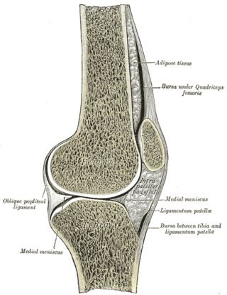
The knee bursae are the fluid-filled sacs and synovial pockets that surround and sometimes communicate with the knee joint cavity. The bursae are thin-walled, and filled with synovial fluid. They represent the weak point of the joint, but also provide enlargements to the joint space. They can be grouped into either communicating and non-communicating bursae or, after their location – frontal, lateral, or medial.

<span class="mw-page-title-main">Quadriceps tendon</span>

In human anatomy, the quadriceps tendon works with the quadriceps muscle to extend the leg. All four parts of the quadriceps muscle attach to the shin via the patella, where the quadriceps tendon becomes the patellar ligament. It attaches the quadriceps to the top of the patella, which in turn is connected to the shin from its bottom by the patellar ligament. A tendon connects muscle to bone, while a ligament connects bone to bone.

<span class="mw-page-title-main">Patellofemoral pain syndrome</span> Medical condition

Patellofemoral pain syndrome is knee pain as a result of problems between the kneecap and the femur. The pain is generally in the front of the knee and comes on gradually. Pain may worsen with sitting, excessive use, or climbing and descending stairs.

Patellar subluxation syndrome, is an injury that is concerned with the kneecap. Patellar subluxation is more common than patellar dislocation and is just as disabling.

<span class="mw-page-title-main">Patellar dislocation</span> Medical condition

A patellar dislocation is a knee injury in which the patella (kneecap) slips out of its normal position. Often the knee is partly bent, painful and swollen. The patella is also often felt and seen out of place. Complications may include a patella fracture or arthritis.

<span class="mw-page-title-main">Medial patellofemoral ligament</span> Ligament of the knee

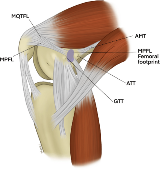
The medial patellofemoral ligament (MPFL) is one of several ligaments on the medial aspect of the knee. It originates in the superomedial aspect of the patella and inserts in the space between the adductor tubercle and the medial femoral epicondyle. The ligament itself extends from the femur to the superomedial patella, and its shape is similar to a trapezoid. It keeps the patella in place, but its main function is to prevent lateral displacement of the patella.

<span class="mw-page-title-main">Vastus muscles</span> Human thigh muscles

The vastus muscles are three of the four muscles that make up the quadriceps femoris muscle of the thigh. The three muscles are the vastus intermedius, the vastus lateralis, and the vastus medialis located in the middle, on the outside, and inside of the thigh, respectively. The fourth muscle is the rectus femoris muscle a large fleshy muscle which covers the front and sides of the femur.

In the skeleton of humans and other animals, a tubercle, tuberosity or apophysis is a protrusion or eminence that serves as an attachment for skeletal muscles. The muscles attach by tendons, where the enthesis is the connective tissue between the tendon and bone. A tuberosity is generally a larger tubercle.

References

  1. 1 2 3 4 5 6 Platzer, Werner (2004). Color Atlas of Human Anatomy, Vol. 1: Locomotor System (5th ed.). Thieme. p. 194. ISBN   3-13-533305-1.
  2. Palmu, S.; Kallio, P.E.; Donell, S.T.; Helenius, I.; Nietosvaara, Y. (2008). "Acute patellar dislocation in children and adolescents: A randomized clinical trial". Journal of Bone and Joint Surgery. 90 (3): 463–470. doi:10.2106/JBJS.G.00072. PMID   18310694.
  3. 1 2 Dath, R.; Chakravarthy, J.; Porter, K.M. (2006). "Patella Dislocations". Trauma. 8: 5–11. doi:10.1191/1460408606ta353ra. S2CID   208269986.
  4. Melloni, Pietro; Veintemillas, Maite; Marin, Anna; Valls, Rafael (2013). "Imaging Patellar Complications After Knee Arthroplasty". Arthroplasty - Update. doi:10.5772/53666. ISBN   978-953-51-0995-2. (CC-BY-3.0)
  5. Yuranga Weerakkody and Frank Gaillard. "Patella baja". Radiopaedia . Retrieved 2018-01-16.
  6. 1 2 Douglas Dennis (2017-02-25). "TKA in Patella Baja (Infera)". Orthobullets. Retrieved 2019-02-08.
  7. Bentley, G (2014). European Surgical Orthopaedics and Traumatology: The EFORT Textbook. Springer. pp. 2766–2784. ISBN   978-3642347450.
  8. Herzmark MH (1938). "The Evolution of the Knee Joint" (PDF). J Bone Joint Surg Am. 20 (1): 77–84. Archived from the original (PDF) on 2008-12-17. Retrieved 2007-11-17.
  9. Romer, Alfred Sherwood; Parsons, Thomas S. (1977). The Vertebrate Body. Philadelphia, PA: Holt-Saunders International. p. 205. ISBN   0-03-910284-X.
  10. "Frogs have hidden, ancient kneecaps". New Scientist. Jul 15, 2017.
  11. Virginia Abdala; et al. (Jul 2017). "On the presence of the patella in frogs". The Anatomical Record. 300 (10): 1747–1755. doi: 10.1002/ar.23629 . PMID   28667673.
  12. New Shorter Oxford
  13. "patella - Origin and meaning of patella by Online Etymology Dictionary". www.etymonline.com.